Abstract
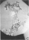
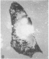
The effects of low doses of cytochalasin B (2 micrograms/ml) and cytochalasin D (0.2 microgram/ml) on the spreading of normal mouse fibroblasts in culture were investigated to find out which components of cell-substrate interactions are most sensitive to alterations of the state of actin cytoskeleton. Cytochalasin B disorganized the cortical layer of actin microfilaments and caused partial or complete disappearance of microfilament bundles; focal contacts with the substrate seen by interference-reflection microscopy also disappeared. Diffuse close contacts were apparently insensitive to cytochalasin B. Low doses of cytochalasin B did not inhibit the outgrowth and maintenance of lamellas at the cell periphery. However, in contrast to controls, these lamellas had no distal zones with convex outer edges and ruffles at the upper surfaces. The disappearance of these ruffling active edges was accompanied by loss of the ability to clear the surface of the lamellas from the concanavalin A receptors crosslinked by the corresponding ligand. The effects of cytochalasin D were similar to those of cytochalasin B. Thus, ruffling active edges and focal contacts can be regarded as specialized parts of lamellas with increased sensitivity to cytochalasins; the presence of ruffling active edges is essential for the initiation of centripetal movement of the patches of crosslinked surface receptors.
Full text
PDF


Images in this article
Selected References
These references are in PubMed. This may not be the complete list of references from this article.
- Abercrombie M., Heaysman J. E., Pegrum S. M. The locomotion of fibroblasts in culture. I. Movements of the leading edge. Exp Cell Res. 1970 Mar;59(3):393–398. doi: 10.1016/0014-4827(70)90646-4. [DOI] [PubMed] [Google Scholar]
- Atlas S. J., Lin S. Dihydrocytochalasin B. Biological effects and binding to 3T3 cells. J Cell Biol. 1978 Feb;76(2):360–370. doi: 10.1083/jcb.76.2.360. [DOI] [PMC free article] [PubMed] [Google Scholar]
- Bershadsky A. D., Tint I. S., Gelfand V. I., Rosenblat V. A., Vasiliev J. M., Gelfand I. M. Microtubular system in cultured mouse epithelial cells. Cell Biol Int Rep. 1978 Jul;2(4):345–351. doi: 10.1016/0309-1651(78)90020-6. [DOI] [PubMed] [Google Scholar]
- Bliokh Z. L., Domnina L. V., Ivanova O. Y., Pletjushkina O. Y., Svitkina T. M., Smolyaninov V. A., Vasiliev J. M., Gelfand I. M. Spreading of fibroblasts in medium containing cytochalasin B: formation of lamellar cytoplasm as a combination of several functional different processes. Proc Natl Acad Sci U S A. 1980 Oct;77(10):5919–5922. doi: 10.1073/pnas.77.10.5919. [DOI] [PMC free article] [PubMed] [Google Scholar]
- Brenner S. L., Korn E. D. Substoichiometric concentrations of cytochalasin D inhibit actin polymerization. Additional evidence for an F-actin treadmill. J Biol Chem. 1979 Oct 25;254(20):9982–9985. [PubMed] [Google Scholar]
- Brown S. S., Spudich J. A. Cytochalasin inhibits the rate of elongation of actin filament fragments. J Cell Biol. 1979 Dec;83(3):657–662. doi: 10.1083/jcb.83.3.657. [DOI] [PMC free article] [PubMed] [Google Scholar]
- Couchman J. R., Rees D. A. The behaviour of fibroblasts migrating from chick heart explants: changes in adhesion, locomotion and growth, and in the distribution of actomyosin and fibronectin. J Cell Sci. 1979 Oct;39:149–165. doi: 10.1242/jcs.39.1.149. [DOI] [PubMed] [Google Scholar]
- Domnina L. V., Ivanova O. Y., Margolis L. B., Olshevskaja L. V., Rovensky Y. A., Vasiliev J. M., Gelfand I. M. Defective formation of the lamellar cytoplasm by neoplastic fibroblasts (L cells-transformed cells-cell attachment-contact inhibition-scanning electron microscopy-microcinematography). Proc Natl Acad Sci U S A. 1972 Jan;69(1):248–252. doi: 10.1073/pnas.69.1.248. [DOI] [PMC free article] [PubMed] [Google Scholar]
- Flanagan M. D., Lin S. Cytochalasins block actin filament elongation by binding to high affinity sites associated with F-actin. J Biol Chem. 1980 Feb 10;255(3):835–838. [PubMed] [Google Scholar]
- Grumet M., Lin S. A platelet inhibitor protein with cytochalasin-like activity against actin polymerization in vitro. Cell. 1980 Sep;21(2):439–444. doi: 10.1016/0092-8674(80)90480-8. [DOI] [PubMed] [Google Scholar]
- Hartwig J. H., Stossel T. P. Cytochalasin B and the structure of actin gels. J Mol Biol. 1979 Nov 5;134(3):539–553. doi: 10.1016/0022-2836(79)90366-8. [DOI] [PubMed] [Google Scholar]
- Heath J. P., Dunn G. A. Cell to substratum contacts of chick fibroblasts and their relation to the microfilament system. A correlated interference-reflexion and high-voltage electron-microscope study. J Cell Sci. 1978 Feb;29:197–212. doi: 10.1242/jcs.29.1.197. [DOI] [PubMed] [Google Scholar]
- Höglund A. S., Karlsson R., Arro E., Fredriksson B. A., Lindberg U. Visualization of the peripheral weave of microfilaments in glia cells. J Muscle Res Cell Motil. 1980 Jun;1(2):127–146. doi: 10.1007/BF00711795. [DOI] [PubMed] [Google Scholar]
- Ivanova O. Y., Margolis L. B., Vasiliev J. M. Effect of colcemid on the spreading of fibroblasts in culture. Exp Cell Res. 1976 Aug;101(1):207–219. doi: 10.1016/0014-4827(76)90431-6. [DOI] [PubMed] [Google Scholar]
- Izzard C. S., Lochner L. R. Formation of cell-to-substrate contacts during fibroblast motility: an interference-reflexion study. J Cell Sci. 1980 Apr;42:81–116. doi: 10.1242/jcs.42.1.81. [DOI] [PubMed] [Google Scholar]
- Lin J. J., Queally S. A. A monoclonal antibody that recognizes Golgi-associated protein of cultured fibroblast cells. J Cell Biol. 1982 Jan;92(1):108–112. doi: 10.1083/jcb.92.1.108. [DOI] [PMC free article] [PubMed] [Google Scholar]
- MacLean-Fletcher S., Pollard T. D. Mechanism of action of cytochalasin B on actin. Cell. 1980 Jun;20(2):329–341. doi: 10.1016/0092-8674(80)90619-4. [DOI] [PubMed] [Google Scholar]
- Painter R. G., Whisenand J., McIntosh A. T. Effects of cytochalasin B on actin and myosin association with particle binding sites in mouse macrophages: implications with regard to the mechanism of action of the cytochalasins. J Cell Biol. 1981 Nov;91(2 Pt 1):373–384. doi: 10.1083/jcb.91.2.373. [DOI] [PMC free article] [PubMed] [Google Scholar]
- Small J. V., Isenberg G., Celis J. E. Polarity of actin at the leading edge of cultured cells. Nature. 1978 Apr 13;272(5654):638–639. doi: 10.1038/272638a0. [DOI] [PubMed] [Google Scholar]
- Vasiliev J. M., Gelfand I. M., Dominina L. V., Dorfman N. A., Pletyushkina O. Y. Active cell edge and movements of concanavalin A receptors of the surface of epithelial and fibroblastic cells. Proc Natl Acad Sci U S A. 1976 Nov;73(11):4085–4089. doi: 10.1073/pnas.73.11.4085. [DOI] [PMC free article] [PubMed] [Google Scholar]
- Vasiliev J. M., Gelfand I. M., Domnina L. V., Ivanova O. Y., Komm S. G., Olshevskaja L. V. Effect of colcemid on the locomotory behaviour of fibroblasts. J Embryol Exp Morphol. 1970 Nov;24(3):625–640. [PubMed] [Google Scholar]
- Vasiliev J. M., Gelfand I. M., Domnina L. V., Rappoport R. I. Wound healing processes in cell cultures. Exp Cell Res. 1969 Jan;54(1):83–93. doi: 10.1016/0014-4827(69)90296-1. [DOI] [PubMed] [Google Scholar]
- Vasiliev J. M., Gelfand I. M. Mechanisms of morphogenesis in cell cultures. Int Rev Cytol. 1977;50:159–274. doi: 10.1016/s0074-7696(08)60099-6. [DOI] [PubMed] [Google Scholar]
- Wehland J., Osborn M., Weber K. Cell-to-substratum contacts in living cells: a direct correlation between interference-reflexion and indirect-immunofluorescence microscopy using antibodies against actin and alpha-actinin. J Cell Sci. 1979 Jun;37:257–273. doi: 10.1242/jcs.37.1.257. [DOI] [PubMed] [Google Scholar]


