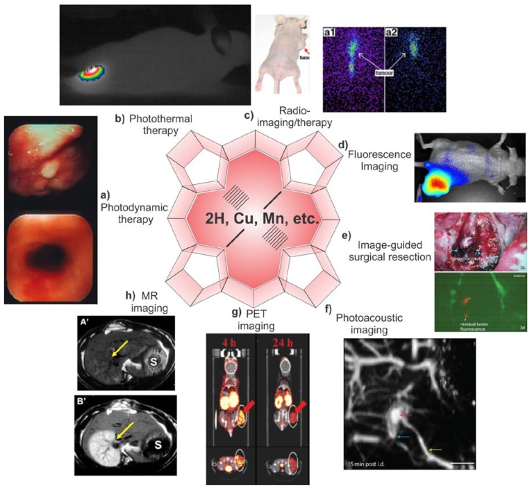
Figure 2.
Multimodal theranostic capabilities of exogenous porphyrins. a) Human esophageal cancer successfully treated with PDT 36. b) Photothermal image of a xenograft bearing mouse injected with porphysomes and then irradiated by laser for 1 min showing tumor temperature rapidly rising above 60 ºC 44. c) Radiotherapy: Melanoma imaging of 188Re-T3,4 CPP in tumor bearing mice. Scintigraphic images were collected at 8h (a1) and 24 h (a2), showing porphyrin potential in radiotherapy and imaging 85. d) Near infrared fluorescence imaging of porphysome activation in a KB xenograft bearing mouse 44. e) Fluorescence Guided Resection (FGR): the surgical cavity after white light resection of brain tumor in rabbit (top), fluorescence imaging of PpIX showing tumor margins. Additional FGR can improve the accuracy of resection 105. f) Photoacoustic image of rat lymphatics mapped following intradermal injection of porphysomes in rats 44. g) PET imaging showing clear delineation between the tumor and other tissues by PET was obtained at 4, 24 h after intravenous injection of a targeted 64Cu porphyrin 116. h) MR imaging: In the top precontrast T1 weighted image, the infarcted right liver lobe (arrow) was barely detected whereas 24 h after injection of Gadophrin-2 at 0.05 mmol/kg the infarcted liver lobe was strongly enhanced (bottom) 125. Reproduced with permission from the publishers of corresponding references.

