Abstract
Background
A problem encountered by medical examiners is that they have to assess injuries that have already been medically treated. Thus, they have to base their reports on clinical forensic examinations performed hours or days after an injury was sustained, or even base their assessment solely on information gleaned from medical files. In both scenarios, the forensic examiner has to rely heavily on the first responder’s documentation of the original injury pattern. Medical priority will be to immediately treat a patient’s injuries, and the first responder may, in addition, initially be unaware of a possibly criminal origin of an injury. As a result, the documentation of injuries is frequently of limited value for forensic purposes. This situation could be improved if photographic records were briefly made of injuries before they were treated.
Methods
German-language medicolegal, criminal, and photography journals and books were selectively searched with the help of PubMed and other databases. In addition, the authors’ experiences in creating and evaluating photographic records for clinical forensic use were assessed.
Results
This paper is an aid to creating photographic records of sufficient quality for forensic purposes. The options provided by digital photography in particular make this endeavor feasible even in a clinical setting. In addition, our paper illuminates some technical aspects of creating and archiving photographic records for forensic use, and addresses possible error sources.
Conclusion
With the requisite technical background knowledge, injuries can be photographically recorded to forensic standards during patient care.
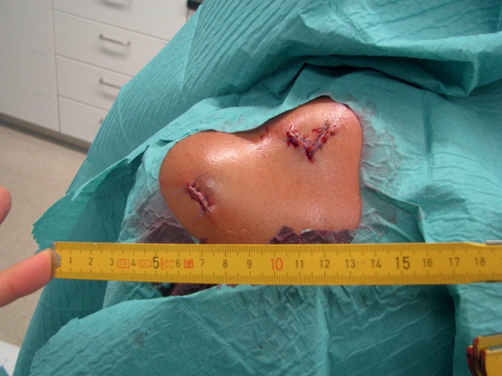
The importance of clinical forensic examination has been demonstrated in numerous publications (1– 9). By the time these examinations take place, however, the injuries will usually already have been attended to as avoiding detrimental health outcomes for the patient has priority (Figure 1). Also, a lot of forensic assessments of injuries and their possible consequences are done on reviewing the clinical and, if available, radiological documentation in the patient’s record (10).
Figure 1.
Typical findings at the time of the clinical forensic examination: The wounds are already surgically treated. A more detailed assessment of the injuries described as stab wounds is no longer possible
Any suspicion of external violence is often only raised in retrospect, so that a clinical forensic examination is only requested days after the initial emergency treatment. It can therefore be impossible to reconstruct the original clinical findings which are the forensically relevant ones.
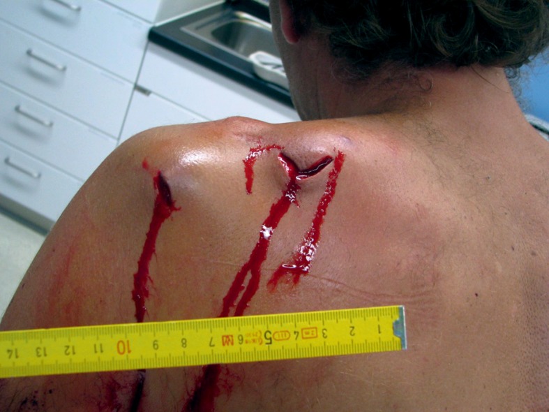
In these cases photographic evidence of the injuries prior to treatment has a crucial role to play in the forensic assessment and any subsequent criminal investigation (Figure 2). Understandably, not every injury that requires medical treatment can be documented with photographs. However, if there is a suspicious presentation (see Box), pictures of the presenting injuries should be obtained.
Figure 2.
Prior to surgical treatment of the two stab wounds (see Figure 1) the doctor in charge of the treatment took a photograph. With the help of this photo it is possible to reconstruct the injuries in retrospect far more accurately
Box. Reasons why one might suspect violence/ Indication for photo-documentation.
History: Patient or other parties report violence or inconsistent history
Type of injury: gunshot wound, stab wound, evidence of injury from blunt object (for example hematoma in the shape of the sole of a shoe after kicking, hematomas following beatings with a stick)
Position of injury: injuries on the extensor sides of joints or below the scalp line suggest accidental injury. Other positions might suggest injury by others.
Number of injuries: not explained by a singular accident
Thanks to the development of digital photography, practically everyone now has access to small digital cameras that allow economic, fast and simple production of good quality photographs. Even some mobile phones offer reasonable picture facilities. However, mobile telephones should not be used for medical documentation of injuries because the quality might be limited, there might be problems with data protection, and it might create the impression of a lack of professionalism. Other advantages of digital photography are that the picture can be checked immediately on the camera, are readily available for viewing on a computer screen, and can easily be shared and printed. One problem with the use of forensic photographs is that often a crucial picture cannot be used due to insufficient image quality. The aim of this review article is therefore to provide guidance on how to produce pictures of sufficient quality and to avoid common mistakes in order to ensure that pictures can be used as forensic evidence (11).
The following are common mistakes (12):
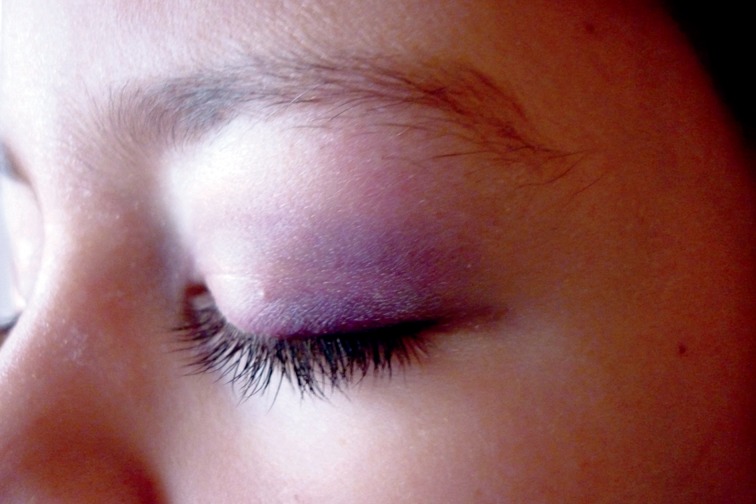
poorly focussed images (Figure 3),
blurred images due to camera shake,
over- or under-exposed images,
image noise.
Figure 3.
A boy has allegedly been involved in a physical assault. As the picture is blurred (due to poor focusing) it is not possible to assess whether the picture actually shows a bruise or for example make-up. In addition, the accuracy of color reproduction is doubtful; there is no object for comparison or standard included in the picture
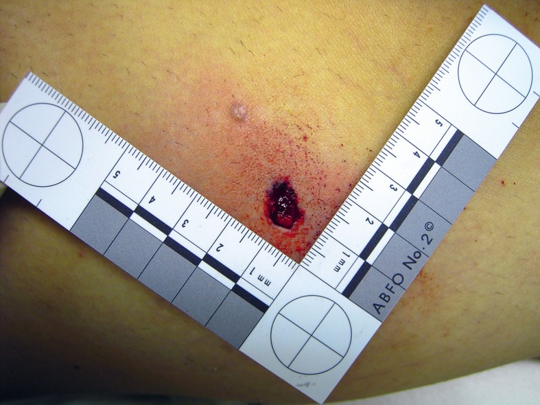
Figure 4.
A gunshot wound (entry wound) prior to wound treatment. With the help of the ruler positioned near the wound the size of the wound can be accurately judged. The yellow skin color could suggest an incorrect white balance setting. However, the pure white color of the ruler proves that this is truly jaundiced skin. Unfortunately, it is not possible to definitely locate the position of the injury as no anatomical landmarks were included in the photo and no additional overview photos were taken
From the point of view of content, perspective distortion and the choice of what details are shown in the picture are of importance (Figure 4). To understand these mistakes and to develop strategies to avoid them it is useful to learn about some technical aspects of photography as well as the technical details of the camera used. Additional information on the subject matters discussed in the following chapters can be found in summarized form in the supplementary eBox.
Key Messages.
Clinical forensic examinations of injuries are usually done only after surgical emergency treatment and closure of the wounds by suturing or similar procedures. For this reason forensic assessment of the injuries is limited compared to first presentation.
A complete reconstruction of the original injuries is usually only possible with a multidisciplinary approach of the forensic team and the physicians who first treated the patient.
Photographic documentation of the clinical findings at first presentation done by the doctors prior to any treatment can be of critical importance. It is up to the doctors to decide if they do a photo-documentation.
Pictures of injuries have to fulfil the quality criteria for forensic evidence: in focus, not blurred, overview and detail photos, detail pictures including a measuring device, indication of orientation.
The picture data have to be stored unedited as an original to allow its use as forensic evidence.
Subject—what is in the picture: the most important factor
In forensic photo-documentation it is most important to document findings exactly and in context. One should always use some means of indicating scale (Figures 1– 4). Angled rulers, standard rulers and tape measures are useful as well as, under exceptional circumstances, objects with standardized sizes such as a coin or a matchstick. The measuring device should be used in such a way that it allows accurate sizing of the injury. In order to accurately measure injuries in pictures it is important that the measuring device and the structure to be measured are in the same plane which should be as perpendicular to the optical axis as possible.
It is useful to take a series of photographs starting with an overview and then zooming in to provide more and more detail (Figure 4). It should be possible to accurately identify the anatomical location of the injury with the help of these pictures.
Focal length—lens-subject distance— proximity effect
The focal length of the lens together with the distance of the camera to the subject determine what is shown in the picture. Being very close to the subject will cause three-dimensional objects—such as faces, for example—to appear out of focus. This is called the proximity effect (13– 15). Current photographic literature also uses the word “wide angle effect” (16).
For forensic examination, macro photography (extreme close-up photography) is important. Most cameras can only achieve this very small distance to the subject by using a very short focal length (maximum wide angle). This can cause considerable distortion of the image.
Digital picture sensors
Digital single lens reflex cameras usually have better sensors than digital compact cameras and therefore produce better quality photographs.
For each picture a so-called white balance has to be set. Almost all digital cameras will do this automatically, which usually results in sufficient quality.
Alternatively, there are standardized settings a nd/or an option to set the white balance manually. To compare a picture’s color balance with ’reality’ and possibly correct its color balance, special color reference charts are available and can be included in the photograph for this purpose. Using a standardized white color in the picture can also be useful (Figure 4).
Aperture—time—sensitivity: the correct exposure
If a picture is over- or underexposed, useful information for forensic interpretation might be lost. For correct exposure the factors of aperture, time and sensitivity of the sensor need to be considered. A camera’s own exposure metering usually suggests the correct setting or selects these automatically.
Focus—autofocus
Focus describes the area of the subject of the photograph that will appear sharp.
The focussing can be done either manually or with help of an auto-focus system. Digital compact cameras usually only have a central area of focus. If a picture is taken in which the forensically relevant area is not in the centre of the photograph it is possible that the use of autofocus may result in focusing errors, which can result in focus on an unimportant part of the picture. This can be avoided if the focus is pointed at the important area, the auto-focus is activated, the measured values are stored and the camera is moved to the final chosen picture view.
Most cameras will activate the autofocus when you press the shutter release to the first level (not completely) and by holding it down the settings can be preserved.
Period of exposure
When patients, especially children, are photographed they are unlikely to hold still. Also it is important that the photographer’s hands do not shake. As a general rule, up to an exposure time of about 1/60 of a second, handheld photography will still give good quality results.
Flash photography
Using a flash can help to avoid camera shake. However, if a flash is used in an already bright environment it can lead to over-exposure due to the flash synchronisation time and can also lead to the effects of camera shake. With digital cameras, using a built-in flash or a flash which has been specifically made for the camera offers the advantage that the white balance will be easy to do.
Manual selection—automatic—picture Mode
The perfect photograph is a result of the combination of choice of subject, composition, and the above mentioned variables. In forensic photography, however, it is not the aesthetics of the picture which are important but the best possible realistic capture of the clinical findings which allow the person looking at the picture to easily grasp the type and extent of the injuries. Almost all modern cameras offer a picture mode for different photographic demands. In clinical photography, programs like “portrait” or “macro” are the most useful ones. It is important to become familiar with the camera model used and to gain an understanding of how the programmes work.
Almost all cameras offer a basic program (for example “auto”) which usually helps to achieve reliable results if a flash is also pre-selected. For macro pictures the program “macro” (flower symbol) should be chosen, and supplemented if necessary with a “soft flash”.
Photo editing
Digital pictures can be edited with the help of computer software. The camera already does some of this, usually unnoticed by the photographer, for example depending on the choice of the subject program. Some cameras offer a program that allows the picture to be edited even before it is transferred to external storage media. Pictures can also be edited in retrospect and many sophisticated photo-editing software tools are available which allow a variety of changes to be made. Shortcomings of the picture in terms of quality and content caused by mistakes during the taking of the photograph can usually only be marginally improved. However, compared to the time before digital photography existed, there are whole new dimensions of picture manipulation now available (17, 18). For forensic pictures it is mandatory that the picture information produced by the camera is left in its original state. Changes made by picture-editing software should be avoided and if the correction of technical mistakes is unavoidable it should only be done on copies of the original data.
Storage and backup of picture data
Pictures which contain patient information must be stored according to the local guidelines and rules of the medical office or hospital (for example, the hospital system OPAC). In general, if the relevant interface is not available it is better to use physical storage such as a hard drive that can be backed up. A CD should be burnt and added to the patient’s records as a form of mobile copy.
So-called metadata are important for the forensic value of digital photography as evidence (19), as this information gets stored together with the picture. These metadata consists of details about when a picture was taken, which camera model was used and other specific technical information. If the picture has been edited, the time of editing and the software used will be recorded. However, not all picture editing software does this automatically. Also, with a certain level of computer knowledge it is possible to manipulate these data. Another problem is that the date and time that document when the original picture was taken are dependent on the camera settings. Only a few cameras have an automatic switch between summer and winter time and this is a common reason for incorrect data documentation. For this reason, recording the date and time the picture was taken in the patient’s records is recommended.
Legal aspects
Like all diagnostic procedures the taking of photographs requires a patient’s consent. No special consent form is required. If a patient declines to have photographs taken, despite the doctor’s recommendation, then this should be documented in their records.
All pictures and data which are stored are protected by medical secrecy. Passing information to criminal investigations or passing information to third parties is regulated by the relevant data protection legislation for medical information.
eBOX. Supplementary technical background information.
Focal length—Distance to subject
Enlargement of the picture section with increasing distance to the subject or reducing focal length
Lens with fixed focal length: Picture detail can only be adapted by the distance to the subject, best picture quality
Zoom lens: What is in the picture can be changed without changing the position of the camera; better flexibility but loss of picture quality.
Digital picture sensor
Resolution into megapixels is not meaningful since the optical resolution is crucial; with increasing size of the image sensor the quality of the image improves. - Digital single lens reflex cameras are usually equipped with much larger sensors than compact cameras and provide better image quality.
Color definition variable, best decided by software, for each picture a so-called white balance needs to be set.
Automatic white balance or pre-selection of the standard setting for white balance, for example “artificial light”, ”direct sunlight“, “cloudy sky“ or flash. Best - practice: manual white balance with the help of measuring a neutral white surface (e.g. white paper) in the picture to allow adjustment of the color mix to this neutral value
Sensitivity of the sensor
ISO setting: ASA value first and then DIN value, for example ISO 100/21°
Example: twice the ASA value means double the sensitivity to light
Sensitivity of most digital picture sensors is in the range 100–200 ASA
I- ncreasing the sensitivity automatically or manually results in a reduction of resolution; compact cameras have smaller sensors and this usually causes deterioration of the image
Autofocus
Automatic focusing with the help of the autofocus system until a defined interference pattern is achieved
Digital compact cameras usually only have one autofocus field, in the centre of the photo
Faster and more flexible measurement with so-called multiple autofocus point systems
Focus—Depth of field
One-third of the depth of field is in front of the subject and two-thirds is behind
The depth of field depends on the way the lens has been constructed and the distance to the subject. The depth of field increases the more the aperture is closed and the further the focus is from the subject
Many digital compact cameras do not have manual aperture setting
Exposure time
Determined by the chosen aperture, the light level and the sensitivity of the sensor, to achieve ideal exposure
Risk of effects of camera shake if exposure time is longer than 1/60 of a second with a handheld camera
In macro photography, shorter exposure time is required
Camera flash and external flashes
If a flash is used exposure time is constant for example 1/100 or 1/60 of a second (so-called flash synchronization time)
How long the flash is on decides how much light effectively hits the sensor; automatic switch off of the flash when sufficient light has reached the sensor, for example after fractions of milliseconds
Built-in flash in digital compact cameras comes as standard but is often not powerful enough
With macro photos there is a risk of central overexposure
Reduction of flash intensity with the help of so-called soft flash setting
An external flash that is attached to the camera to avoid shadows from parts of the camera when taking macro photos and to improve the power of the flash
To improve the lighting in difficult situations in macro photography, a connecting wire can be attached via the hot shoe
If an external flash is acquired, assure compatibility of the systems
Data storage and backup
In digital photography, SD cards are used (so-called mass storage); they are considerably more robust and less sensitive than other storage media such as hard drives or CDs/DVDs
So-called smart card readers with USB connectors (2.0 sometimes already 3.0) to transmit the pictures from the SD card to the computer are recommended
Direct connection of the camera to the computer is almost always possible but as this method reduces the speed of transmission and uses a lot of battery power from the camera, this is not the best way of doing it
Storage of pictures is possible in almost all cameras in a compressed format; “.jpg” is the most common
Compressing the picture data to create .jpg files requires recurrent recalculation of the picture: this might have a negative impact on quality
Formats like “.bmp” or “.tif” do not compress the picture but require considerably more storage space compared to “.jpg”
Picture editing always only on the non-compressed format. Initially, create a copy of the original compressed data and then edit this copy.
Acknowledgments
Translated from the original German by Dr Ute Semrau-Boughton
Footnotes
Conflict of interest statement
Prof. Verhoff holds stock in the Leica Camera-AG company. All other authors declare that no conflict of interests exists.
References
- 1.Dlubis-Mertens K. Häusliche Gewalt gegen Frauen. Dtsch Arztebl. 2004;101(23):A 1656–A 1657. [Google Scholar]
- 2.Jacobi G, Dettmeyer R, Banaschak S, Brosig B, Herrmann B. Child abuse and neglect: diagnosis and management. Dtsch Arztebl Int. 2010;107(13):231–240. doi: 10.3238/arztebl.2010.0231. [DOI] [PMC free article] [PubMed] [Google Scholar]
- 3.Krohn J, Seifert D, Kurth H, Püschel K, Schröder AS. Gewaltdelikte mit menschlichen Bissverletzungen. Analyse von 143 Verletzungsfällen. Rechtsmedizin. 2010;20:19–24. [Google Scholar]
- 4.Matschke J, Herrmann B, Sperhake J, Körber F, Bajanowski T, Glatzel M. Shaken Baby syndrome—a common variant of nonaccidental head injury in infants. Dtsch Arztebl Int. 2009;106(13):211–217. doi: 10.3238/arztebl.2009.0211. [DOI] [PMC free article] [PubMed] [Google Scholar]
- 5.Ritz-Timme S, Graß H. Häusliche Gewalt - Werden die Opfer in der Arztpraxis optimal versorgt? Dtsch Arztebl. 2009;106(7):A 282–A 283. [Google Scholar]
- 6.Rothschild MA, Graß H. Klinische Rechtsmedizin. Aufgaben und Herausforderungen im Rahmen der medizinischen Betreuung von Opfern häuslicher Gewalt. Rechtsmedizin. 2004;14:189–193. [Google Scholar]
- 7.Seifert D, Anders S, Franke B, et al. Modellprojekt zur Implementierung eines medizinischen Kompetenzzentrums für Gewaltopfer in Hamburg. Rechtsmedizin. 2004;14:183–188. [Google Scholar]
- 8.Seifert D, Püschel K, Anders S. Selbstverletzendes Verhalten bei weiblichen Opfern von Gewalt. Vorkommen in einem rechtsmedizinischen Untersuchungskollektiv. Rechtsmedizin. 2009;19:325–330. [Google Scholar]
- 9.Siegmund-Schutze N. Vor Ort sein bei den Opfern von Gewalt. Dtsch Arztebl. 2008;105(26 A):1434–1435. [Google Scholar]
- 10.Verhoff MA, Fischer L, Alzen G, Ramsthaler F. Rekonstruktion von Einstichwunden an präoperativen CT-Daten - Befunderhebung im Rahmen einer klinisch-rechtsmedizinischen Untersuchung. Arch Kriminol. 2009;224:73–81. [PubMed] [Google Scholar]
- 11.Özkalipci Ö, Volpellier M. Photographic documentation, a practical guide for non professional forensic photography. Torture. 2010;20:45–52. [PubMed] [Google Scholar]
- 12.Verhoff MA, Gehl A, Kettner M, Kreutz K, Ramsthaler F. Digitale forensische Fotodokumentation. Rechtsmedizin. 2009;19:369–381. [Google Scholar]
- 13.Helmer R. Zugleich ein Beitrag zur Konstitutionsbiometrie und Dickenmessung der Gesichtsweichteile. Heidelberg: Kriminalistik Verlag; 1984. Schädelidentifizierung durch elektronische Bildmischung. [Google Scholar]
- 14.Verhoff MA, Witzel C, Kreutz K, Ramsthaler F. The ideal subject distance for passport pictures. Forensic Sci Int. 2008;178:153–156. doi: 10.1016/j.forsciint.2008.03.011. [DOI] [PubMed] [Google Scholar]
- 15.Verhoff MA, Witzel C, Ramsthaler F, Kreutz K. Der Einfluss von Objektabstand beziehungsweise Objektiv-Brennweite auf die Darstellung von Gesichtern. Arch Kriminol. 2007;220:36–43. [PubMed] [Google Scholar]
- 16.Osterloh G. Frankfurt am Main: Umschau Verlag; 1985. Leica M - Hohe Schule der Fotografie; pp. 201–217. [Google Scholar]
- 17.Baron C. Sleuths, Truths, and Fauxtography. Boston: Thomson Course Technology; 2008. Adobe Photoshop Forensics. [Google Scholar]
- 18.Ramsthaler F, Kettner M, Potente S, Gehl A, Kreutz K, Verhoff MA. Original oder manipuliert? Authenzität und Integrität digitaler Bildmaterialien aus forensischer Sicht. Rechtsmedizin. 2010;20:385–392. [Google Scholar]
- 19.Knopp M. Digitalfotos als Beweismittel. Z Rechtspol. 2008;41:137–168. [Google Scholar]


