Abstract
Sarcomas of the breast are relatively rare and account for 1% of all primary malignant tumors of the breast. Pure and primary chondrosarcoma of the male breast is an extremely rare tumor. It may arise either from the breast stroma itself or from underlying bone or cartilage. Differential diagnoses include cystosarcoma phyllodes and breast metaplastic carcinoma with chondroid differentiation.
Keywords: Chondrosarcoma, Male breast
Case Summary
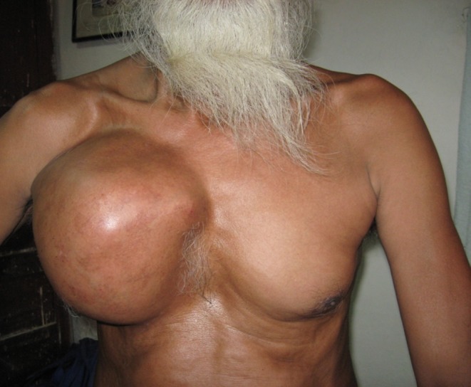
An 80-year-old man presented with a painful mass in the right breast of 9 months’ duration (Fig. 1). Physical examination revealed a firm mass occupying most of the right breast, measuring 20 cm × 10 cm in size. Rate of growth was initially slow but then progressed very rapidly in past 3 months before presentation. A chest X-ray did not reveal any involvement of underlying bony cage.
Fig. 1.

Showing a huge mass in the right breast
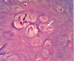
Fine needle aspiration smears showed clusters of atypical cells with abundant pale cell cytoplasm and moderately enlarged nuclei against the background of chondroid matrix. Total mastectomy with axillary sampling was performed, and subsequent histopathological examination confirmed the cytological diagnosis of primary chondrosarcoma with lymph nodes showing reactive hyperplasia. No metastasis in axillary lymph nodes was found. Pure chondrosarcoma and metaplastic cancer of the breast rarely invade axillary lymph nodes and are generally hormone receptor-negative [1–4]. Microscopically, the tumor was seen with atypical chondrocytes in single lacunae which at places were binucleated and multinucleated. The cells were having hyperchromatic nuclei with discrete anisokaryosis. Mitoses were rare (Fig. 2).
Fig. 2.
Lacunae containing atypical (black arrow head) and binucleated (white arrow head) chondrocytes against the chondroid matrix
Chondrosarcoma may occur in three different forms: as a pure chondrosarcoma, as the stromal component of a histologically malignant phyllodes tumor, or as chondrosarcomatous differentiation in a metaplastic carcinoma.
To diagnose a primary chondrosarcoma of the breast, a non-mammary site has to be excluded clinically and histologically. In the present case, underlying bony cage was not involved and only chondromatous tissue was seen without any epithelial component on histopathological examination. Differentiation from metaplastic carcinoma is possible by absence of direct transition between carcinomatous and mesenchymal components in the primary chondrosarcoma. Further, the sarcoma-like elements in metaplastic carcinoma, though acquire vimentin positivity, still retain epithelial markers [5]. Differentiation from malignant cystosarcoma phyllodes with predominant chondrosarcomatous component can be extremely difficult [6, 7]. Surgery remains the mainstay of treatment. The role of chemotherapy and radiotherapy is not yet established because of the limited number of cases reported so far. Our patient underwent a total mastectomy with axillary sampling with subsequent local radiotherapy. The patient is on regular follow-up on outpatient basis. To conclude, primary chondrosarcomas of the breast are rare tumors that must be considered in the differential diagnosis of tumors of the breast in chondrosarcomatous areas.
Acknowledgments
Funding support
Nil.
References
- 1.Pezzi CM, Patel-Parekh L, Cole K, Franko J, Himberg VS, Bland K. Characteristics and treatment of metaplastic breast cancer: analysis of 892 cases from the National Cancer Data Base. Ann Surg Oncol. 2007;14:166–173. doi: 10.1245/s10434-006-9124-7. [DOI] [PubMed] [Google Scholar]
- 2.Verfaillie G, Breucq C, Perdaens C, Bourgain C, Lamote J. Chondrosarcoma of the breast. Breast J. 2005;11:147–148. doi: 10.1111/j.1075-122X.2005.21573.x. [DOI] [PubMed] [Google Scholar]
- 3.Beatty SD, Atwood M, Tickman R, Reiner M. Metaplastic breast cancer: clinical significance. Am J Surg. 2006;191:657–664. doi: 10.1016/j.amjsurg.2006.01.038. [DOI] [PubMed] [Google Scholar]
- 4.Bhosale SJ, Kshirsagar AY, Sulhyan SR, Jagtap SV, Nikam YP. Metaplastic carcinoma with predominant chondrosarcoma of the right breast. Case Rep Oncol. 2010;3(2):277–281. doi: 10.1159/000319423. [DOI] [PMC free article] [PubMed] [Google Scholar]
- 5.Vandenhaute B, Validire P, Veilleux C, Voelh P, Zafrani B. Breast carcinoma with chondroid metaplasia. Ann Pathol. 1995;15:53–58. [PubMed] [Google Scholar]
- 6.Vera-Sempere F, García-Martínez A. Malignant phyllodes tumor of the breast with predominant chondrosarcomatous differentiation. Pathol Res Pract. 2003;199:841–845. doi: 10.1078/0344-0338-00505. [DOI] [PubMed] [Google Scholar]
- 7.Iihara H, Machinami R, Kubotas S, Itoyama S, Sato A. Malignant cystosarcoma phyllodes tumour of the breast mainly composed of chondrosarcoma—a case report. Gen Diagn Pathol. 1997;142:241–245. [PubMed] [Google Scholar]


