Abstract
Rare-earth phosphors are commonly used in display panels, security printing, and fluorescent lamps, and have potential applications in lasers and bioimaging. In the present study, Eu3+- and Dy3+-codoped uniform-shaped Y2O3 submicron particles were prepared using the urea homogeneous precipitation method. The structure and morphology of the resulting particles were characterized by X-ray diffraction, field emission scanning electron microscope, and field emission transmission electron microscope, whereas their optical properties were monitored by photoluminescence spectroscopy. The room-temperature luminescence color emission of the synthesized particles can be tuned from red to yellow by switching the excitation wavelength from 254 to 350 nm. The luminescence intensities of red and yellow emissions could be altered by varying the dopant concentration. Strong quenching was observed at high Eu3+ and Dy3+ concentrations in the Y2O3 host lattice.
Keywords: Y2O3 particles, Luminescence, Urea homogeneous precipitation, Eu3+ and Dy3+ codoped
Background
The development of novel luminescent phosphor materials with a controllable size and morphology has been a major focus in the field of photonics and optoelectronics [1]. Phosphor nanocrystals are exceptionally promising materials in many fields of technology including photonics, luminescent displays, fluorescent lamps, lasers, cathodoluminescence, and biotechnology [2]. Moreover, the emission wavelength of rare-earth-doped nanoparticles is independent of the particle size and depends only on the dopant type, leading to lower synthesis cost. They also offer excellent chemical stability as well as high quantum yield. Different methods have been used to fabricate nanocrystalline phosphor particles, such as flame spray pyrolysis [3], co-precipitation method [4], sol–gel method [5], and solvothermal method [5]. The urea homogeneous precipitation method was recognized to be a green route for the high-yield mass production of spherical ceramic submicron particles with controllable sizes. Spherical-shaped particles can improve the optical performance due to the high packing density and reduction of light scattering [6].
Yttrium oxide (Y2O3) has been investigated widely as a host material for rare-earth (RE) ion doping in optical applications [3,4,6,7] on account of its excellent chemical stability, broad transparency range (0.2 to 8 μm) with a band gap of 5.6 eV, high refractive index, and low phonon energy [1]. Furthermore, the similarities in the chemical properties and ionic radius of RE ions and Y2O3 make it an attractive choice as a host material [6,8].
The color tunability of yttria-based phosphors can be achieved by codoping the host material with some specific rare-earth elements. For example, in our previous report we investigated the color-tunability effect of Eu3+- and Tb3+-codoped Y2O3 submicron particles [8]. We showed that the color emission of synthesized particles could be tuned precisely from red to green by a simple variation of the Tb/Eu ratio and excitation wavelength. Strong energy transfer (ET) from Tb to Eu ions was observed in Tb/Eu-codoped Y2O3 submicron particles, but back ET from Eu3+ to Tb3+ was not significant. Ishiwada et al. investigated the Tb/Tm-codoped Y2O3 phosphor for high-temperature thermometry application [9]. The synthesized Tb/Tm-codoped Y2O3 phosphor showed a distinct change of visible emission colors from green to blue with increasing temperature. Therefore, research into Y2O3 codoped with other different RE activators is important because the color-tunable properties can be used in a wide range of applications. Although many studies have examined the optical properties of RE ion-doped Y2O3 phosphors, only a few have investigated the codoping of two or more different ions in the same yttria host material [6,8,9].
In recent years, the Eu/Dy codoping in a single host material has attracted a great deal of attention and has been extensively investigated. For example, the SrAl2O4 host material codoped with Eu3+ and Dy3+ is known as a new generation long-lasting luminescent phosphor material [10]. It is well known that Eu3+ doping into Y2O3 host material results in a red emission, whereas doping with Dy3+ results in blue-yellow emission [11]. To the best of the authors' knowledge, there are no reports of an Y2O3 phosphor codoped with Eu3+ and Dy3+. In addition, there is an optimum concentration of dopant ions for all RE phosphors, but the optimum concentration also depends on several parameters, such as the size of the phosphors and synthetic route.
In the present study, urea homogeneous precipitation synthesis method was used for the preparation of Eu3+- and Dy3+-codoped Y2O3 submicron particles. The morphology and particles characteristics were investigated by X-ray diffraction (XRD), field emission scanning electron microscopy (FESEM), field emission transmission electron microscopy (FETEM), and energy dispersive X-ray (EDX) spectroscopy. The optical properties of the synthesized particles were explored by photoluminescence spectroscopy (PL). Y2O3 submicron particles with different concentrations of codoped Eu3+ and Dy3+ were investigated, and the luminescence intensity of these particles were found to be strongly dependent on the activators concentration. The emission color of the Eu3+- and Dy3+-codoped Y2O3 particles could be switched from red to yellow by variation of the excitation wavelength.
Methods
Chemical synthesis
Analytical grade Y2O3 (99.9%), europium oxide (Eu2O3; 99.9%), dysprosium oxide (Dy2O3; 99.9%), nitric acid (HNO3; 70%), and urea (99% to 100.5%) were purchased from Sigma-Aldrich Corporation (MO, USA) and were used without further purification.
Uniform-shaped sub-micron Eu3+- and Dy3+-codoped Y2O3 particles were synthesized according to the reported protocols [6,8]. Phosphor precipitates were prepared by heating the corresponding RE nitrates (0.001 mol each sample) in aqueous solution of urea (40 ml H2O and 0.5 g urea). The concentration of Eu3+ varied between 1 to 3 mol%, whereas the Dy3+ concentration varied from 1 to 2 mol%.
Physical characterization
The structure of the prepared powders was examined by XRD using a Bruker D8 Discover diffractometer (Bruker Optics Inc., MA, USA) with Cu Kα radiation (λ = 0.15405 nm) and a 2θ scan range of 20 to 60°. The structural properties were also analyzed using Fourier transform infrared spectroscopy (Jasco FT/IR6300, JASCO Corp., Easton, MD, USA). The morphologies of the particles were characterized by FESEM (Hitachi S-4700, Hitachi, Ltd., Tokyo, Japan) and FETEM (JEOL JEM-2100F, JEOL Ltd., Tokyo, Japan). Elemental analysis was carried out by EDX (Horiba 6853-H, HORIBA Jobin Yvon Inc., Edison, NJ, USA). The PL measurements were performed with a Hitachi F-7000 spectrophotometer equipped with a 150-W xenon lamp as an excitation source. All the measurements were performed at room temperature.
Results and discussion
Morphology and structure
The luminescence intensity depends strongly on the phosphor crystallinity [6,11]. Therefore, all synthesized particles were calcinated at the temperature of 1,000°C. The morphology of the synthesized phosphor particles after calcination at 1,000°C was examined by FESEM. Figure 1a shows FESEM image of the Y2O3 phosphor particles codoped with a 1%Eu3+-1%Dy3+ composition. From the FESEM image, it is apparent that the Y2O3:1%Eu3+-1%Dy3+ phosphor particles consists of relatively uniform, spherical-shaped submicron particles, 100 ± 20 nm in size.
Figure 1.
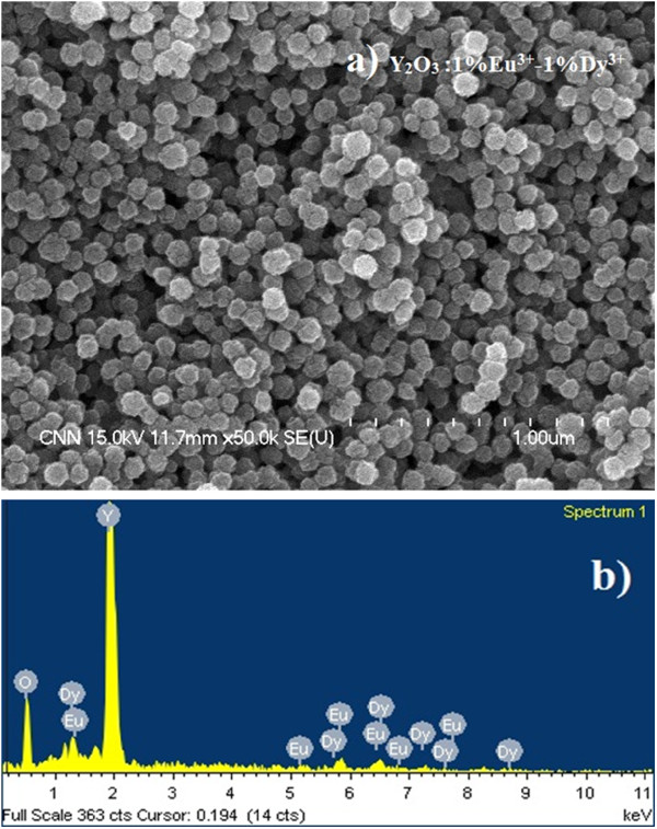
Y2O3: 1%Eu3+-1%Dy3+phosphor particles (Tcal.= 1,000°C). FESEM image (a) and EDX spectra (b).
Codoping with different Eu3+ and Dy3+ concentrations did not alter the morphology of the final phosphor product, and all particles had a spherical morphology with a size distribution of 100 ± 20 nm (not shown for other samples). On the other hand, we showed that the sizes of the final Y2O3 phosphor particles can be tuned by altering the reaction time, reaction temperature, and concentration of starting materials [8]. The EDX analysis of 1%Eu3+-1%Dy3+-codoped Y2O3 particles confirmed the presence of dysprosium and europium elements in the yttria host material (Figure 1b). Figure 2 shows the XRD patterns of Y2O3 submicron particles codoped with different Eu3+ and Dy3+ compositions, and the standard peak positions of a pure cubic Y2O3 structure (JCPDS No. 86–1107).
Figure 2.
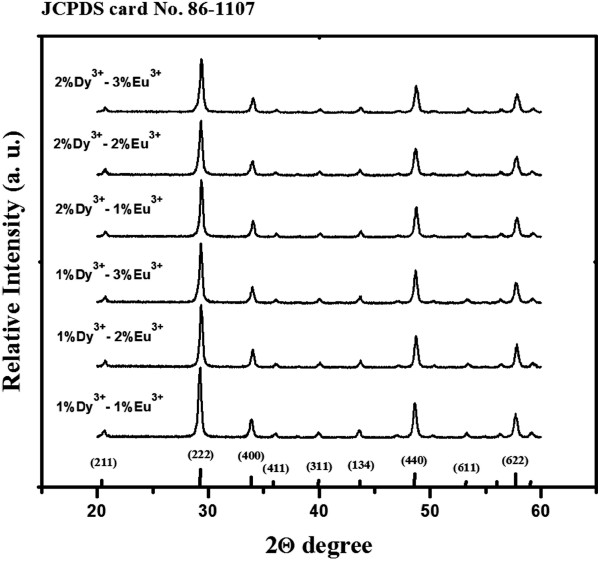
X-ray diffraction patterns of Y2O3phosphor particles. The Y2O3 particles are codoped with different RE ion (Eu3+ and Dy3+) concentrations (Tcal. = 1,000°C).
It is obvious that all the diffraction peaks could be indexed directly to the cubic Y2O3 phase with the space group Ia3 (206) according to the standard card (JCPDS No. 86–1107). No additional peaks from the doped components could be detected due to a relatively low concentration of dopant ions indicating the formation of a pure cubic Y2O3 phase. Table 1 lists the mean crystallite sizes of synthesized particles estimated from the well-known Debye-Scherrer's equation. The diffraction data of the three strongest peaks ({222}, {440}, and {622} planes) were used to calculate the mean crystallite sizes. The crystallite sizes showed a slightly increasing tendency, which can be attributed to the effect of the increased dopant concentration [8].
Table 1.
Calculated mean crystallite sizes of Eu3+- and Dy3+-codoped Y2O3particles
| 1Dy/1Eu | 1Dy/2Eu | 1Dy/3Eu | 2Dy/1Eu | 2Dy/2Eu | 2Dy/3Eu |
|---|---|---|---|---|---|
| 36.42 ± 0.11 | 36.72 ± 0.09 | 36.96 ± 0.10 | 36.67 ± 0.14 | 37.01 ± 0.16 | 37.28 ± 0.07 |
Figure 3a shows a FETEM image of a single Y2O3:1%Eu3+-1%Dy3+ phosphor particle. It is obvious that a single Y2O3:1%Eu3+-1%Dy3+ particle has a relatively spherical shape consisting of smaller crystallites (approximately 35 ± 12 nm) associated with each other, which is in good agreement with that calculated from the XRD patterns using the Debye-Scherrer's equation. The lattice fringes in the FETEM image also confirm the high crystallinity of the phosphor product. Figure 3b shows the corresponding selected area electron diffraction (SAED) image of the Y2O3:1%Eu3+-1%Dy3+ particle. The clear concentric rings from the inside to outside were indexed directly to the {211}, {222}, {400}, {440}, and {622} planes of cubic Y2O3, demonstrating the highly crystalline nature of the phosphor particles.
Figure 3.
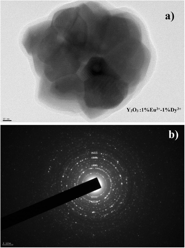
Single Y2O3:1%Eu3+-1%Dy3+phosphor particle (Tcal.= 1,000°C). FETEM image (a) and corresponding SAED image (b).
Luminescent properties
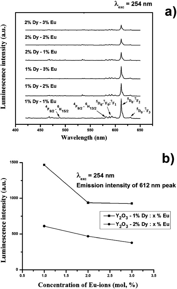
As we mention, the luminescence emission of phosphor materials depends strongly on the synthetic route, size of the phosphor materials, and concentration of dopant ions. The particle sizes were similar for all Eu3+- and Dy3+-codoped Y2O3 samples which were fabricated under identical conditions. This allows a comparison of the luminescence emission properties of Y2O3 phosphor particles codoped with different concentrations of Eu3+ and Dy3+ activators. Figure 4a shows the emission spectra of Y2O3 phosphor particles codoped with different Eu3+ and Dy3+ concentrations under ultraviolet (254 nm) irradiation. The emission spectrum showed several main groups of emission lines, which were assigned to the 5D1 → 7F1 and 5D0 → 7Fj (where j = 0, 1, 2, and 3) transitions within Eu3+[8,12]. Obviously, the emission spectrum is dominated by a red 5D0 → 7F2 (612 nm) transition within the Eu3+. Figure 4b shows the 5D0 → 7F2 (612 nm) transition peak height as a function of the codoped Eu3+ and Dy3+ concentration under ultraviolet (254 nm) irradiation. According to the recent literature, the best doping value of Eu3+ was reported to be approximately 5 mol% in Y2O3 host material [5,13]. On the other hand, the luminescence intensity of the 5D0 → 7F2 (612 nm, hypersensitive to the environment) transition decreased significantly (Figure 4a,b) with increasing codoped Eu3+ and Dy3+ concentrations in Y2O3 host.
Figure 4.
PL emission spectra and integrated luminescence emission intensity (612-nm peak). PL emission spectra of Y2O3 phosphor particles codoped with different Eu3+ and Dy3+ contents (a) and integrated luminescence emission intensity of 612-nm peak as a function of RE ion concentration (b) upon constant 254 nm excitation.
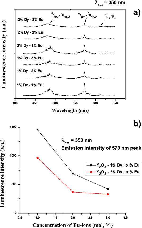
Figure 5a shows the same samples under 350-nm excitation. In this case, the emission spectrum exhibited luminescence spectra assigned to the characteristic 4F9/2 → 6H15/2 (blue region) and 4F9/2 → 6H13/2 (greenish-yellow region) transitions within Dy3+[11]. The intensity of the Dy3+ characteristic emission transitions also decreased strongly with increasing codoped Eu3+ and Dy3+ concentration, as it shown in Figure 5a,b. The strong quenching behavior at high codoped Eu3+ and Dy3+ concentrations was related to a cross-relaxation mechanism (nonradiative decay of the two ions to the ground state) within the dopant ions.
Figure 5.
PL emission spectra and integrated luminescence emission intensity (573-nm peak). PL emission spectra of Y2O3 phosphor particles codoped with different Eu3+ and Dy3+ content (a) and integrated luminescence emission intensity of 573-nm peak as a function of RE ions concentration (b) upon constant 350-nm excitation.
The mean distance between the dopant ions at high concentrations (R = 0.62/(N)1/3, where N is the concentration of ions) was much shorter. Therefore, ions can interact through an electric multipolar process leading to energy migration. The dipole-dipole quenching process is inversely proportional to the sixth power of ion-ion separation and, thus, to the square of the dopant concentration [14,15]. In Figures 4a and 5a, except for a decrease of the luminescence intensity, the emission spectrum of Y2O3 particles codoped with different Eu3+ and Dy3+ concentrations was similar due to the same f-f transitions within the specific RE ion. Therefore, the concentration of codoped RE ions plays an important role and should be strongly considered during the phosphor fabrication process.
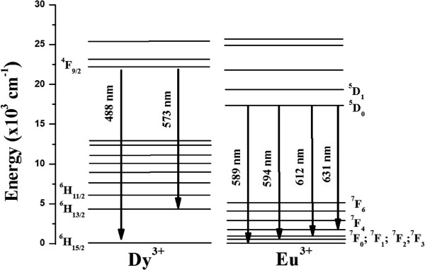
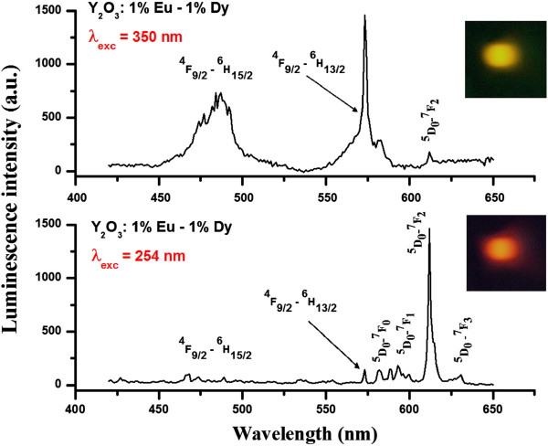
The excitation spectra of Y2O3:1% Eu3+-1% Dy3+ particles are shown in Figure 6. The excitation spectra of Y2O3:1% Eu3+-1% Dy3+ (λem. = 612 nm) consists of a band extending from approximately 230 to 260 nm, which is related to the charge-transfer band (CTB) of O2−-Eu3+ bond. Upon excitation into the CTB at 254 nm, the emission spectrum exhibited several emission lines that were assigned to the 5D1 → 7F1 and 5D0 → 7Fj (where j = 0, 1, 2, and 3) transitions within Eu3+, as shown in the Figure 7. On other hand, the weak signal at 573 nm, which was assigned to 4F9/2 → 6H13/2 (hypersensitive transition of Dy3+), was also observed, as shown in Figures 7 and 8. Figure 6 also shows the excitation spectra of Y2O3:1% Eu3+-1% Dy3+ particles measured in the 200- to 400-nm range by monitoring the yellow emission transition of Dy3+ (λem. = 573 nm). In this case, when the excitation wavelength was switched to 350 nm, the hypersensitive 6P7/2 level of Dy3+ was excited resonantly, which then quickly relaxes nonradiatively to populate the 4F9/2 level [11]. Radiative emission occurred from 4F9/2 to a lower 6H15/2 and 6H13/2, emitting at 488 and 573 nm, respectively, with feeble ET to Eu3+. The energy transferred to Eu3+ cascades rapidly via nonradiative transitions to the 5D0 state, from which luminescence associated with Eu3+ occurs. Therefore, the feeble signal of the 5D0 → 7F2 (612 nm) transition within Eu3+ was also detected in Eu3+ and Dy3+ codoped phosphor particles upon 350-nm excitation.
Figure 6.
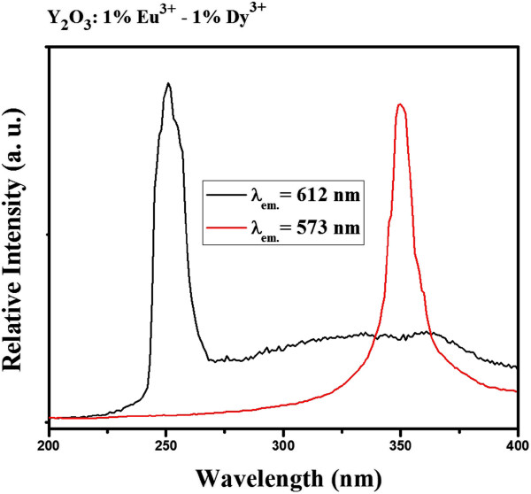
PL excitation spectra of Y2O3:1%Eu3+-1%Dy3+phosphor particles (Tcal.= 1,000°C).
Figure 7.
Schematic diagram illustrating the Dy3+/Eu3+energy level diagram and transitions between the levels.
Figure 8.
PL emission spectra of Y2O3:1%Eu3+-1%Dy3+phosphor particles (λexc.= 254 and 350 nm, respectively) with assigned transitions. Inset digital photographs showed the eye-visible red and yellow luminescence emissions.
Such behavior suggests that there is some energy transfer (ET) occurring between the two codoped ions. On the other hand, the ET observed in Eu3+- and Dy3+-codoped Y2O3 was not as strong as that observed in Tb3+-Eu3+-codoped Y2O3 phosphor [8]. Therefore, the emission wavelength and color output of the same Y2O3:1% Eu3+-1% Dy3+ particles can be adjusted by switching the radiation from 254 to 350 nm. The digital photographs of eye-visible luminescence emissions from Y2O3:1% Eu3+-1% Dy3+ particles upon 254 and 350 nm excitations are shown in the Figure 8. It is obvious that the same host material emit red or yellow color, depending on the excitation wavelength.
Conclusion
In conclusion, Eu3+- and Dy3+-codoped Y2O3 submicron spherical particles were synthesized using the urea homogeneous precipitation method. The crystal structure and morphology of synthesized particles were characterized by XRD, FESEM, EDX, and FETEM. The PL spectroscopy was used to examine luminescent properties of Eu3+- and Dy3+-codoped Y2O3 particles. PL measurements revealed strong concentration quenching at high-codoped Eu3+ and Dy3+ concentrations. The luminescence color emission could be controlled by the excitation wavelength and the incorporation of Dy3+ and Eu3+ at the appropriate concentrations into Y2O3 structure. Weak energy transfer between the codoped ions was observed. These color-tunable Y2O3:1%Eu3+-1%Dy3+ phosphor particles can be used for security printing, solid state illumination, or for optical displays.
Competing interests
The authors declare that they have no competing interests.
Authors' contributions
All the specimens used in this study and initial manuscript were prepared by TSA. YHH and HKK added a valuable discussion and coordinated the present study as principal investigators. All authors read and approved the final manuscript.
Contributor Information
Timur Sh. Atabaev, Email: atabaev@snu.ac.kr.
Yoon-Hwae Hwang, Email: yhwang@pusan.ac.kr.
Hyung-Kook Kim, Email: hkkim@pusan.ac.kr.
Acknowledgments
This work was supported by the National Research Foundation of Korea (NRF) funded by the Ministry of Education, Science and Technology (grant no.: 2010–0010575) and by the Korean government (grant no. 2012R1A1B3001357). The author would like to thank Prof. J. B. Lee for allowing the use of his equipment for the preparation and characterization of the samples.
References
- Das GK, Tan TTY. Rare-earth-doped and codoped Y2O3 nanomaterials as potential bioimaging probes. J Phys Chem C. 2008;112:11211–11217. doi: 10.1021/jp802076n. [DOI] [Google Scholar]
- Yang P, Gai S, Liu Y, Wang W, Li C, Lin J. Uniform hollow Lu2O3:Ln (Ln = Eu3+, Tb3+) spheres: facile synthesis and luminescent properties. Inorg Chem. 2011;50:2182–2190. doi: 10.1021/ic101605k. [DOI] [PubMed] [Google Scholar]
- Qin X, Yokomori T, Ju Y. Flame synthesis and characterization of rare-earth (Er3+, Ho3+, and Tm3+) doped upconversion nanophosphors. Appl Phys Lett. 2007;90:073104. doi: 10.1063/1.2561079. [DOI] [Google Scholar]
- Tu D, Liang Y, Liu R, Li D. Eu/Tb ions co-doped white light luminescence Y2O3 phosphors. J Lumin. 2011;131:2569–2573. doi: 10.1016/j.jlumin.2011.05.036. [DOI] [Google Scholar]
- Wang H, Yang J, Zhang CM, Lin J. Synthesis and characterization of monodisperse spherical SiO2@RE2O3 (RE = rare earth elements) and SiO2@Gd2O3:Ln3+ (Ln = Eu, Tb, Dy, Sm, Er, Ho) particles with core-shell structure. J Solid State Chem. 2009;182:2716–2724. doi: 10.1016/j.jssc.2009.07.033. [DOI] [Google Scholar]
- Atabaev TS, Vu H-HT, Piao Z, Kim H-K, Hwang Y-H. Tailoring the luminescent properties of codoped Gd2O3:Tb3+ phosphor particles by codoping with Al3+ ions. J Alloys Compd. 2012;541:263–268. [Google Scholar]
- Flores-Gonzales MA, Ledoux G, Roux S, Lebbou K, Perriat P, Tillement O. Preparing nanometer scaled Tb-doped Y2O3 luminescent powders by the polyol method. J Solid State Chem. 2005;178:989–997. doi: 10.1016/j.jssc.2004.10.029. [DOI] [Google Scholar]
- Atabaev TS, Lee JH, Han DW, Hwang Y-H, Kim H-K. Cytotoxicity and cell imaging potentials of submicron color-tunable yttria particles. J Biomed Mat Res A. 2012;100(9):2287–2294. doi: 10.1002/jbm.a.34168. [DOI] [PubMed] [Google Scholar]
- Ishiwada N, Fujioka S, Ueda T, Yokomori T. Co-doped Y2O3:Tb3+/Tm3+ multicolor emitting phosphors for thermometry. Opt Lett. 2011;36:5. doi: 10.1364/OL.36.000760. [DOI] [PubMed] [Google Scholar]
- Cheng B, Zhang Z, Han Z, Xiao Y, Lei S. SrAl2O4:Eu2+, Dy3+ nanobelts: synthesis by combustion and properties of long-persistent phosphorescence. J Mater Res. 2011;26:2311–2315. doi: 10.1557/jmr.2011.94. [DOI] [Google Scholar]
- Atabaev TS, Vu HHT, Kim H-K, Hwang Y-H. Synthesis and optical properties of Dy3+-doped Y2O3 nanoparticles. J Korean Phys Soc. 2012;60:244–248. doi: 10.3938/jkps.60.244. [DOI] [Google Scholar]
- Seo S, Yang H, Holloway P. Controlled shape growth of Eu- or Tb-doped luminescent Gd2O3 colloidal nanocrystals. J Colloid Interface Sci. 2009;331:236–242. doi: 10.1016/j.jcis.2008.11.016. [DOI] [PubMed] [Google Scholar]
- Liu Z, Yu L, Wang Q, Tao Y, Yang H. Effect of Eu, Tb codoping on the luminescent properties of Y2O3 nanorods. J Lumin. 2011;131:12–16. doi: 10.1016/j.jlumin.2010.08.012. [DOI] [Google Scholar]
- Atabaev TS, Vu H-HT, Kim YD, Lee JH, Kim H-K, Hwang Y-H. Synthesis and luminescence properties of Ho3+ doped Y2O3 submicron particles. J Phys Chem Solids. 2012;73:176–181. [Google Scholar]
- Patra A, Ghosh P, Chowdhury PS, Alencar MARC, Lozano W, Rakov N, Maciel GS. Red to blue tunable upconversion in Tm3+-doped ZrO2 nanocrystals. J Phys Chem B. 2005;109:10142–10146. doi: 10.1021/jp050685v. [DOI] [PubMed] [Google Scholar]


