Abstract
Modulation of P/Q-type Ca2+ currents through presynaptic voltage-gated calcium channels (CaV2.1) by binding of Ca2+/calmodulin contributes to short-term synaptic plasticity. Ca2+-binding protein-1 (CaBP1) and Visinin-like protein-2 (VILIP-2) are neurospecific calmodulin-like Ca2+ sensor proteins that differentially modulate CaV2.1 channels, but how they contribute to short-term synaptic plasticity is unknown. Here, we show that activity-dependent modulation of presynaptic CaV2.1 channels by CaBP1 and VILIP-2 has opposing effects on short-term synaptic plasticity in superior cervical ganglion neurons. Expression of CaBP1, which blocks Ca2+-dependent facilitation of P/Q-type Ca2+ current, markedly reduced facilitation of synaptic transmission. VILIP-2, which blocks Ca2+-dependent inactivation of P/Q-type Ca2+ current, reduced synaptic depression and increased facilitation under conditions of high release probability. These results demonstrate that activity-dependent regulation of presynaptic CaV2.1 channels by differentially expressed Ca2+ sensor proteins can fine-tune synaptic responses to trains of action potentials and thereby contribute to the diversity of short-term synaptic plasticity.
Neurons fire repetitively in different frequencies and patterns, and activity-dependent alterations in synaptic strength result in diverse forms of short-term synaptic plasticity that are crucial for information processing in the nervous system (1–3). Short-term synaptic plasticity on the time scale of milliseconds to seconds leads to facilitation or depression of synaptic transmission through changes in neurotransmitter release. This form of plasticity is thought to result from residual Ca2+ that builds up in synapses during repetitive action potentials and binds to a Ca2+ sensor distinct from the one that evokes neurotransmitter release (1, 2, 4, 5). However, it remains unclear how changes in residual Ca2+ cause short-term synaptic plasticity and how neurotransmitter release is regulated to generate distinct patterns of short-term plasticity.
In central neurons, voltage-gated calcium (CaV2.1) channels are localized in high density in presynaptic active zones where their P/Q-type Ca2+ current triggers neurotransmitter release (6–11). Because synaptic transmission is proportional to the third or fourth power of Ca2+ entry through presynaptic CaV2.1 channels, small changes in Ca2+ current have profound effects on synaptic transmission (2, 12). Studies at the calyx of Held synapse have provided important insights into the contribution of presynaptic Ca2+ current to short-term synaptic plasticity (13–17). CaV2.1 channels are required for synaptic facilitation, and Ca2+-dependent facilitation and inactivation of the P/Q-type Ca2+ currents are correlated temporally with synaptic facilitation and rapid synaptic depression (13–17).
Molecular interactions between Ca2+/calmodulin (CaM) and CaV2.1 channels induce sequential Ca2+-dependent facilitation and inactivation of P/Q-type Ca2+ currents in nonneuronal cells (18–21). Facilitation and inactivation of P/Q-type currents are dependent on Ca2+/CaM binding to the IQ-like motif (IM) and CaM-binding domain (CBD) of the CaV2.1 channel, respectively (20, 21). This bidirectional regulation serves to enhance channel activity in response to short bursts of depolarizations and then to decrease activity in response to long bursts. In synapses of superior cervical ganglion (SCG) neurons expressing exogenous CaV2.1 channels, synaptic facilitation is induced by repetitive action potentials, and mutation of the IM and CBD motifs prevents synaptic facilitation and inhibits the rapid phase of synaptic depression (22). Thus, in this model synapse, regulation of presynaptic CaV2.1 channels by binding of Ca2+/CaM can contribute substantially to the induction of short-term synaptic plasticity by residual Ca2+.
CaM is expressed ubiquitously, but short-term plasticity has great diversity among synapses, and the potential sources of this diversity are unknown. How could activity-dependent regulation of presynaptic CaV2.1 channels contribute to the diversity of short-term synaptic plasticity? CaM is the founding member of a large family of Ca2+ sensor (CaS) proteins that are differentially expressed in central neurons (23–25). Two CaS proteins, Ca2+-binding protein-1 (CaBP1) and Visinin-like protein-2(VILIP-2), modulate facilitation and inactivation of CaV2.1 channels in opposite directions through interaction with the bipartite regulatory site in the C-terminal domain (26, 27), and they have varied expression in different types of central neurons (23, 25, 28). CaBP1 strongly enhances inactivation and prevents facilitation of CaV2.1 channel currents, whereas VILIP-2 slows inactivation and enhances facilitation of CaV2.1 currents during trains of stimuli (26, 27). Molecular analyses show that the N-terminal myristoylation site and the properties of individual EF-hand motifs in CaBP1 and VILIP-2 determine their differential regulation of CaV2.1 channels (27, 29–31). However, the role of CaBP1 and VILIP-2 in the diversity of short-term synaptic plasticity is unknown, and the high density of Ca2+ channels and unique Ca2+ dynamics at the presynaptic active zone make extrapolation of results from studies in nonneuronal cells uncertain. We addressed this important question directly by expressing CaBP1 and VILIP-2 in presynaptic SCG neurons and analyzing their effects on synaptic plasticity. Our results show that CaM-related CaS proteins can serve as sensitive bidirectional switches that fine-tune the input–output relationships of synapses depending on their profile of activity and thereby maintain the balance of facilitation versus depression by the regulation of presynaptic CaV2.1 channels.
Results
CaBP1 Reduces Paired-Pulse Facilitation.
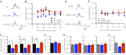
CaBP1 enhances the inactivation of CaV2.1 channels, whereas VILIP-2 reduces their inactivation and enhances facilitation (26, 27). To determine whether presynaptic expression of these CaS proteins affects short-term synaptic plasticity in SCG neurons, we designed our experiments to measure synaptic transmission driven by CaV2.1 channels expressed only on the presynaptic side of the synapse, and we blocked the endogenous N-type Ca2+ current with ω-conotoxin GVIA. Under these conditions, synaptic transmission is mediated specifically by transfected CaV2.1 channels (22, 32). We expressed CaV2.1 channels and CaBP1 or VILIP-2 by injecting cDNA into an identified SCG neuron, and we recorded excitatory postsynaptic potentials (EPSPs) from a neighboring synaptically connected but untransfected neuron, thereby isolating presynaptic effects. We first tested synaptic transmission under conditions of low release probability (1 mM extracellular Ca2+) in which facilitation is observed in control synapses. In response to paired stimuli, we observed paired-pulse facilitation (PPF) at interstimulus intervals (ISI) beginning at 50 ms, consistent with previous findings (Fig. 1 A and B) (22). In contrast, CaBP1-expressing synapses showed paired-pulse depression (PPD) rather than PPF at the same ISI (Fig. 1 B and C). At the peak of PPF for control synapses (ISI = 80 ms), the maximum paired-pulse ratio (PPR) reached in cells cotransfected with CaBP1 was reduced significantly [WT PPR80ms, 1.36 ± 0.12 (n = 23); CaBP1 PPR80ms, 0.76 ± 0.12 (n = 17), P < 0.01] (Fig. 1C). Thus, a switch from facilitation to depression was evident in CaBP1-expressing synapses at intermediate ISI. In contrast, synapses expressing VILIP-2 showed PPF that was indistinguishable from PPF in controls at all ISI (Fig. 1 B and C). To confirm that the effect of CaBP1 is caused by interaction with the channel and not by a change in initial neurotransmitter release probability, we compared the amplitude of the first EPSP in control synapses, CaBP1-expressing synapses, and VILIP-2–expressing synapses. Expression of CaBP1 and VILIP-2 did not change the amplitude of the synaptic response (Fig. 1D). Thus, CaBP1 did not affect baseline neurotransmitter release in transfected neurons. Together, our data imply that CaBP1 blocks facilitation of the Ca2+ current, resulting in reduced PPF. In contrast, coexpression of VILIP-2 with CaV2.1 channels under the conditions of our paired-pulse experiments had no effect, just as we previously observed no effect for PPF of Ca2+ currents by VILIP-2 (27).
Fig. 1.
CaBP1 blocks PPF. (A) Representative averages of 10 EPSPs evoked by paired action potentials with 50-ms ISI in the presynaptic neurons expressing CaV2.1 alone (WT) and in neurons cotransfected with CaS proteins CaBP1 (WT+CaBP1) or VILIP-2 (WT+VILIP-2) in the presence of ω-conotoxin GVIA (3 µM). (B) Average PPR plotted against ISI for WT (●), WT+CaBP1 ( ), and WT+VILIP2 (
 ) neurons in 1 mM extracellular Ca2+. (C) PPR for 50-, 80-, and 120-ms ISI for WT (black), WT+CaBP1 (blue), and WT+VILIP-2 (red) neurons. *P < 0.05, **P < 0.01, ANOVA with Bonferroni posttest. +, P < 0.05; ++, P < 0.01; ANOVA with Tukey’s posttest for differences compared with WT group (absolute P values: ISI50ms,
P = 0.006; ISI80ms,
P = 0.008; and ISI120ms,
P = 0.0002). Data are shown as mean ± SEM from 10–20 synaptic pairs. (D) Averaged initial EPSP amplitude (mV) for WT (black), WT+CaBP1 (blue), and WT+VILIP-2 (red) neurons recorded for a 50-ms interval. (E) Representative averages of 10 EPSPs evoked in neurons expressing CaV2.1IMAA-ΔCBD alone (IMCBD) or with CaS proteins CaBP1 (IMCBD+CaBP1) or VILIP-2 (IMCBD+VILIP-2) evoked by paired-action potentials with 50-ms ISI in the presynaptic neurons. (F) PPR plotted against ISI for IMCBD (
 ), IMCBD+CaBP1 (
 ), and IMCBD+VILIP2 (
 ) neurons. (G) PPR for 50-, 80-, and 120-ms ISI for IMCBD (gray), IMCBD+CaBP1 (blue), and IMCBD+VILIP-2 (red) neurons. (H) Averaged initial EPSP amplitude (mV) for WT (black), WT+CaBP1 (blue), and WT+VILIP-2 (red) neurons recorded for a 50-ms interval. Data shown in E–H are mean ± SEM from 10–15 synaptic pairs.
CaBP1 Binding to CaV2.1 Channels Is Essential for Modulation of PPF.
Immunocytochemistry studies show that CaBP1 colocalizes with CaV2.1 channels and syntaxin in the CA1 region of the hippocampus and in the molecular layer of the cerebellum, although postsynaptic staining also has been observed (26). Because CaBP1 is transfected only in the presynaptic neuron of SCG synaptic pairs, its effect is limited to the presynaptic sites involved in synaptic plasticity. However, it is possible that CaBP1 may act on other presynaptic machinery involved in neurotransmitter release rather than on CaV2.1 channels. Does CaBP1 bind directly to CaV2.1 channels to block synaptic facilitation? The interaction between CaBP1 and CaV2.1 channels requires the same intracellular domain of CaV2.1 that binds CaM. To address whether CaBP1 acts directly on CaV2.1 channels, we transfected SCG neurons with mutant CaV2.1 channels in which the IM motif was mutated to AA and the CBD was deleted (CaV2.1IM-AA/ΔCBD). Coexpression of CaBP1 or VILIP-2 had no effect on these mutant CaV2.1IM-AA/△CBD channels in paired-pulse experiments (Fig. 1 E–G). CaV2.1IM-AA/△CBD channels showed much less PPF at intermediate ISI than did CaV2.1 channels (Fig. 1 B and F). Coexpression of CaBP1 or VILIP-2 did not alter basal neurotransmission or PPF observed for any ISI (Fig. 1 G and H). These results confirm that the effect of CaBP1 in reducing PPF results from its binding to the CaS-binding site on CaV2.1 channels.
CaBP1 Reduces Synaptic Facilitation During Trains of Activity.
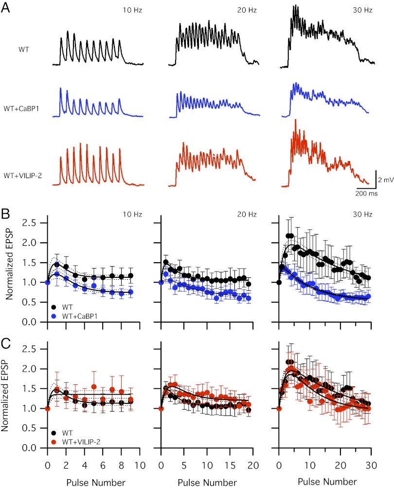
Activity-dependent increases in Ca2+ entry cause facilitation followed by inactivation of CaV2.1-channel currents (13–19). This dual regulation is caused by sequential binding of the two lobes of Ca2+/CaM to the IQ-like domain and CBD of CaV2.1 channels (18, 20, 21). To investigate the effects of CaBP1 and VILIP-2 on CaV2.1 channels during trains of activity, we stimulated synapses at varying frequencies and recorded EPSPs during each stimulation in the presence of 1 mM extracellular Ca2+ to give a low probability of neurotransmitter release (Fig. 2A). In control synapses expressing WT CaV2.1 channels alone, we observed synaptic facilitation that then decayed at all stimulus frequencies (Fig. 2B). To include all the data points in our statistical analysis, the mean synaptic responses were fit to a biexponential function, the time constant for depression was derived from this fit, and the value for maximum facilitation was estimated by extrapolation to the time of the second stimulus in the train. Errors in the fits and parameter estimates were expressed as 95% confidence limits (Fig. 2B, dotted lines). This profile of synaptic facilitation and depression resembles the regulation of CaV2.1 channels by endogenous CaM (Fig. 2B) (17, 19, 22). In contrast, synapses expressing CaBP1 showed less facilitation and increased depression at each stimulus frequency (Fig. 2B). Peak synaptic facilitation was reduced on average from 2.22 ± 0.25 to 1.53 ± 0.15 at 30 Hz (P < 0.05), and synaptic depression following the peak response also was enhanced, as indicated by the more rapid decrement in EPSP amplitude (Fig. 2B).
Fig. 2.
CaBP1 blocks synaptic facilitation in trains of stimuli. (A) Representative EPSPs in 1 mM extracellular Ca2+ evoked by repetitive action potentials at 10, 20, and 30 Hz for 1s in the presynaptic neurons expressing CaV2.1 alone (WT, black) or cotransfected with CaBP1 (WT+CaBP1, blue) or VILIP-2 (WT+VILIP-2, red). Data from 10 sweeps repeated every 30 s at each frequency were averaged. (B and C) Mean normalized EPSP amplitude from 10–16 synaptic pairs at 10-, 20-, and 30-Hz frequency. EPSP amplitudes were normalized to the first EPSP of each train and plotted against action potential number for WT (●), WT+CaBP1 ( ), and WT+VILIP-2 (
 ) neurons. Points represent the mean ± SEM. Solid lines are best fits of a biexponential equation as described in SI Materials and Methods. Dotted lines indicate 95% confidence intervals.
Although VILIP-2 has no effect on facilitation by a single pulse, it remained possible that VILIP-2 has an important effect on synaptic transmission during trains of stimuli. Previous studies in transfected tsA-201 cells have shown that VILIP-2 reduces inactivation of Ca2+ currents during trains of repetitive depolarizations and thereby enhances and prolongs Ca2+ current facilitation (27). In contrast to our expectations from these previous results in nonneuronal cells, synapses expressing VILIP-2 showed synaptic facilitation followed by synaptic depression that was similar to that in control synapses (Fig. 2C). At 30 Hz, no difference in the rate of facilitation or depression was observed (Fig. 2C), in contrast to the marked effects of CaBP1 expression at this stimulus frequency (Fig. 2B). These results illustrate that effects of VILIP-2 on short-term synaptic plasticity are not extrapolated accurately from the effects observed on Ca2+ currents in transfected nonneuronal cells under these conditions, perhaps because of the differences in Ca2+ dynamics in presynaptic active zones versus nonneuronal cells. We conclude that CaBP1-expressing synapses have reduced synaptic facilitation during trains of activity but that VILIP-2 has no effect under conditions of low release probability.
CaBP1 Binding to CaV2.1 Channels Is Required for Its Effects on Synaptic Plasticity in Trains of Stimuli.
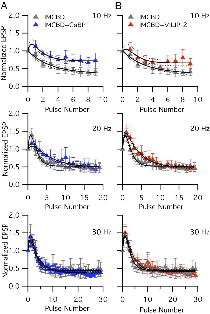
To test whether direct binding of CaBP1 to CaV2.1 channels is required for the modulation of synaptic facilitation during trains of stimuli, we expressed CaBP1 with the mutant CaV2.1IM-AA/△CBD. SCG neurons expressing CaV2.1IM-AA/△CBD show peak synaptic facilitation that is reduced from 2.22 ± 0.21 to 1.48 ± 0.12 at 30 Hz (compare Figs. 3A and 2B, 30 Hz, P < 0.05), and synaptic facilitation is reduced or completely blocked throughout the trains of stimuli at 10–30 Hz (Fig. 3A and Fig. S1). These results are consistent with the previous conclusion that normal synaptic facilitation in this model synapse requires facilitation of CaV2.1 channels via CaS proteins interacting with the IM and CBD domains (22). Coexpression of CaBP1 does not further reduce facilitation of synapses expressing the mutant CaV2.1IM-AA/△CBD channel (Fig. 3A and Fig. S1). These data indicate that mutation of CaS-binding sites eliminates regulation by CaBP1 and therefore suggest that direct binding of CaBP1 to CaV2.1 channels is sufficient to cause a switch from synaptic facilitation to depression.
Fig. 3.
Mutation of the CaS-binding site reduces modulation of short-term synaptic plasticity by CaBP1 and VILIP-2. (A) Normalized EPSP amplitudes for CaV2.1IM-AA/ΔCBD (IMCBD) alone ( ) and with coexpression of CaBP1 (IMCBD+CaBP1) (
 ) in 1 mM extracellular Ca2 at a stimulation frequency of 10, 20, and 30 Hz. (B) Normalized EPSP amplitudes for CaV2.1IMAA-ΔCBD alone (
 , IMCBD) and with coexpression of VILIP-2 (IMCBD+VILIP-2) (
 ) at a stimulation frequency of 10, 20, or 30 Hz. Mean EPSP amplitudes were normalized to the first EPSP of the train. Data shown are mean ± SEM from 8–14 synaptic pairs.
VILIP-2 was unable to increase facilitation of synaptic transmission in paired pulses or trains of stimuli under conditions of low release probability (Figs. 1 and 2). As a further control for the specificity of VILIP-2 effects, we tested VILIP-2 on SCG neurons expressing the CaV2.1IM-AA/△CBD mutant channel. As expected from the results of Figs. 1 and 2, coexpression of VILIP-2 did not significantly increase facilitation during trains of stimuli at 10, 20, or 30 Hz (Fig. 3B and Fig. S1). These results are consistent with the conclusion that the CaV2.1IM-AA/△CBD mutation completely prevents CaS protein regulation.
VILIP-2 Enhances Synaptic Facilitation at High Basal Release Probability.
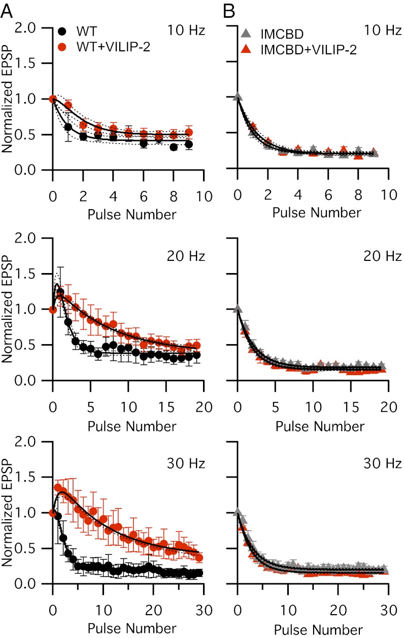
Facilitation during trains of stimuli in SCG neuron synapses is robust, approaching 2.22-fold at 30 Hz in 1 mM extracellular Ca2+ (Fig. 2). This substantial level of facilitation may represent the maximum possible at these synapses. We hypothesized that this high level of basal synaptic facilitation may occlude effects of VILIP-2. Therefore, we raised external Ca2+ from 1 mM to 2 mM to enhance release probability and increase synaptic depression. Under these conditions, control synapses show rapid synaptic depression at all three stimulus frequencies (Fig. 4A and Fig. S2), but synapses expressing VILIP-2 show significantly more facilitation and significantly less depression at 20 Hz and 30 Hz than controls (P < 0.05) (Figs. 4A and Fig. S2). At 30 Hz, VILIP-2 restores synaptic facilitation during the first five pulses followed by pulse-wise synaptic depression (1.37 ± 0.08, P < 0.01) (Fig. 4A and Fig. S2). Peak synaptic facilitation observed with VILIP-2 in 2 mM Ca2+ (Fig. 4A) was 27% less than in the corresponding control without VILIP-2 in 1 mM extracellular Ca2+ (P < 0.05) (Fig. 2B). The increase in facilitation during stimulus trains in 2 mM Ca2+ highlights the ability of VILIP-2 to facilitate neurotransmitter release specifically at high frequencies of stimulation when basal neurotransmitter release is high. This specificity of the effect of VILIP-2 for the facilitation of CaV2.1 channel activity under conditions of high release probability is unexpected from previous work in nonneuronal cells and indicates that the Ca2+-signaling environment at the active zone markedly affects the ability of CaS proteins to modulate short-term synaptic plasticity. The synaptic facilitation during trains of stimuli shows that VILIP-2 binding to CaV2.1 channels slows their cumulative Ca2+-dependent inactivation and thereby enhances their Ca2+-dependent facilitation (27).
Fig. 4.
VILIP-2 enhances synaptic facilitation via the CaS protein-binding site on CaV2.1 channels. (A) Normalized EPSPs recorded after the expression of CaV2.1 channels alone (WT) (●) and with VILIP-2 ( ) in 2 mM extracellular Ca2+ during trains of action potentials at 10, 20, and 30 Hz. (B) Normalized EPSPs recorded after the expression of the CaV2.1 channel double mutant CaV2.1IMAA-ΔCBD (IMCBD) alone (
 ) and with VILIP-2 (IMCBD+VILIP-2) (
 ) in 2 mM extracellular Ca2+ during trains of action potentials at 10, 20, and 30 Hz. The averaged first EPSP amplitude recorded in 2 mM extracellular Ca2+: WT, 6.99 ± 1.9 mV (n = 10); WT+VILIP-2, 5.60 ± 0.98 mV (n = 14); IMCBD, 7.69 ± 1.01 mV (n = 8); and IMCBD+VILIP-2, 6.77 ± 1.19 (n = 12). Data shown are mean ± SEM from 5–14 synaptic pairs.
CaS-Binding Sites of CaV2.1 Channels Are Essential for VILIP-2 Modulation.
To confirm that the effect of VILIP-2 in 2 mM Ca2+ is caused by the direct interaction with the CaM-binding site on the channel, we coexpressed VILIP-2 with CaV2.1IM-AA/△CBD mutant channels. Under conditions of high release probability in 2 mM Ca2+, CaV2.1IM-AA/△CBD channels undergo rapid synaptic depression, as seen at other synapses when external Ca2+ is elevated (Fig. 4B) (22). Coexpression of VILIP-2 has no effect on depression or facilitation of CaV2.1IM-AA/△CBD channels (Fig. 4B and Fig. S2). This result is consistent with the conclusion that, when external Ca2+ and basal release probability are high, VILIP-2 prevents rapid Ca2+-dependent channel inactivation and enhances Ca2+-dependent facilitation by interaction with the CaS protein regulatory site on CaV2.1 channels. Evidently, when bound in place of CaM, VILIP-2 can switch SCG synapses from synaptic depression to facilitation. Thus, CaBP1 and VILIP-2 provide bidirectional control of facilitation and depression by CaS proteins.
Discussion
Short-term synaptic plasticity converts the information encoded in the frequency and pattern of action potential firing in the presynaptic terminal into an analog signal for transmission to the postsynaptic neuron (3). Our results show that this information processing can be controlled in a bidirectional manner at the level of the presynaptic Ca2+ channel by CaS proteins, which are poised to modulate these channels and alter neurotransmitter release. Direct regulation of presynaptic CaV2.1 channels by the CaS proteins CaBP1 and VILIP-2 may serve as a bidirectional switch to control the input–output relationships of synapses in response to trains of action potentials. This conversion of synapses from depressing to facilitating and vice-versa would be expected to have profound consequences for the encoding properties of neural circuits, thereby fine-tuning the synaptic plasticity of different types of synapses (3).
Molecular Analysis of Synaptic Plasticity in SCG Neurons.
Analysis of the functional effects of presynaptic Ca2+ channel regulation in synaptic transmission is challenging because of the need to alter regulation only in the presynaptic cell. Our experiments took advantage of the unique characteristics of SCG neurons as an expression system for studies of presynaptic effects on synaptic transmission. These neurons lack CaS proteins, which are expressed specifically in the central nervous system and retina (23, 25), and they lack CaV2.1 channels, which are expressed primarily in central neurons (6, 9, 10). Thus, SCG neurons provide a null genetic background for studies of these key components of central synapses. The large cell bodies of cultured SCG neurons allow microinjection of cDNA encoding CaV2.1 channels into individual cell nuclei without damage, giving expression only in presynaptic cells, and the level of functional expression is in the same range as the endogenous CaV2.2 channels, which can be blocked specifically and completely by ω-conotoxin GVIA (32). Facilitation followed by depression is observed in these synapses (22). Both facilitation and the rapid phase of depression are dependent on the regulation of CaV2.1 channels by CaM interaction with the IM and CBD motifs in the C-terminal domain (22). These favorable characteristics have allowed us to test directly the role of CaS proteins in the modulation of synaptic plasticity that is dependent on CaV2.1 channels and by using mutant channel constructs to demonstrate the requirement for CaS protein binding to the IM and CBD motifs.
CaS Proteins Override CaM-Dependent Synaptic Plasticity in a Ca2+-Dependent Manner.
Like CaM, the CaS proteins are expressed in high levels in central neurons (23). Our results show that specific expression of CaBP1 and VILIP-2 in presynaptic neurons can compete effectively with endogenous CaM, without changing peak CaV2.1 currents (Fig. S3). CaBP1 expression blocks CaM-dependent facilitation and enhances rapid synaptic depression when studied in the presence of 1 mM extracellular Ca2+, where synaptic facilitation is prominent. In contrast, CaBP1 did not have detectable effects on synaptic plasticity at 2 mM Ca2+, where synaptic depression is rapid (n = 6; P > 0.05). Thus, CaBP1 is able to compete with CaM for binding to their common regulatory site and can induce changes in short-term synaptic plasticity under physiological conditions, depending on the level of Ca2+ entry and the resulting level of release probability.
In contrast to CaBP1, VILIP-2 has no effect on synaptic plasticity in SCG neurons at 1 mM extracellular Ca2+, but it reduces synaptic depression and enhances synaptic facilitation at 2 mM Ca2+, where synaptic depression is rapid. These results show that VILIP-2 also can override the synaptic plasticity induced by CaM under appropriate conditions of Ca2+ entry. It is noteworthy that the effects of CaBP1 and VILIP-2 on synaptic plasticity respond in opposite ways to changes in the level of release probability—CaBP1 reduces facilitation and increases depression more effectively at low release probability in the presence of low extracellular Ca2+, whereas VILIP-2 reduces depression and enhances facilitation more effectively at high release probability in the presence of higher extracellular Ca2+. This difference in the effects of CaBP1 and VILIP-2 under different physiological conditions highlights the importance of the functional state of the synapse in dictating the response of short-term synaptic plasticity to these CaS proteins. Taken together, our results show that the CaS proteins CaBP1 and VILIP-2 can serve as a bidirectional switch controlling synaptic facilitation and depression in a push–pull manner through opposing regulation of CaV2.1 channels.
CaS Proteins and Short-Term Synaptic Plasticity.
It is becoming increasingly apparent that CaS proteins play a significant role in coupling changes in intracellular Ca2+ to modulation of different types of ion channels and other regulatory proteins. Ca2+ and CaM have long been implicated in mechanisms of synaptic plasticity (33–35). In addition, the CaS proteins frequenin, neuronal Ca2+ sensor-1 (NCS-1), and KChIP modulate a broad range of voltage-gated and ligand-gated channels (36–40). NCS-1, which is closely related to VILIP-2, enhances P/Q-type Ca2+ currents in the calyx of Held and can facilitate synaptic transmission at the calyx of Held and in hippocampal synapses (39, 40), but modulation of CaV2.1 channels by direct binding of NCS-1 has not been reported. Our results indicate that CaBP1 and VILIP-2 can regulate short-term synaptic plasticity in SCG neurons directly in opposing ways by binding to the same regulatory site as CaM and differentially regulating CaV2.1 channels. The fact that these effects of CaS proteins on synaptic transmission are largely eliminated when mutant CaV2.1IM-AA/ΔCBD channels mediate Ca2+ entry demonstrates the regulation of CaV2.1 channels by direct binding of CaS proteins as opposed to other possible presynaptic targets. Thus, regulated expression of these CaS proteins in different classes of synapses could work as a binary switch to change short-term synaptic plasticity from depressing to facilitating or vice versa through direct modulation of CaV2.1 channels.
Distinct Expression of CaS Proteins in the Central Nervous System.
CaS proteins are expressed in a cell-specific manner in the central nervous system (23–25, 28). VILIP-2 is expressed mainly in the caudate-putamen, neocortex, and hippocampus (28, 38). Expression of VILIP-2 is notably absent in the cerebellum and pons (28, 41). CaBP1 is expressed primarily in cerebral cortex, retina, and hippocampus, whereas CaM is expressed ubiquitously (25). Additionally, CaBP1 and CaV2.1 channels coexist in the CA1 region of the hippocampus and in presynaptic nerve terminals in the molecular layer of the cerebellum (26). Neuronal CaS proteins have Ca2+-binding affinities considerably higher than those of CaM (42). Therefore, the diversity of this family of CaS proteins and their Ca2+-binding properties raises the possibility that they may override the ubiquitous effects of CaM and contribute broadly to the diversity of Ca2+ channel regulation and synaptic plasticity.
CaS Proteins as Effectors of the Diversity of Synaptic Transmission.
Different synapses show diverse patterns of facilitation and depression, and the underlying mechanism for this diversity is unknown (2, 3). CaS proteins contribute substantially to the diversity of neuronal Ca2+ signaling in the postsynaptic cell (23). Our results suggest a key role for CaS proteins in synaptic plasticity by showing that CaBP1 and VILIP-2 can exert opposing influences on facilitation and depression of synaptic transmission through the regulation of presynaptic CaV2.1 channels. This perspective opens the way for a broader analysis of the potential roles of other CaS proteins in control of short-term synaptic plasticity by regulation of presynaptic Ca2+ channels. Given the widespread distribution of CaV2.1 channels in the central nervous system, cell-specific modulation of presynaptic CaV2.1 channels by CaBP1, VILIP-2, and other CaS proteins may act as a molecular switch linking CaS-dependent facilitation and inactivation of CaV2.1 channels to the encoding of information in the frequency domain at synapses.
Materials and Methods
SCG neurons were cultured as described to allow synapse formation (43). cDNAs encoding α12.1 subunit, α12.1IMAA/ΔCBD, CaBP1, VILIP-2, and eGFP were microinjected into nuclei, and synaptic transmission was measured as described previously (22). More detailed descriptions of the experimental conditions can be found in SI Materials and Methods.
Supplementary Material
Footnotes
The authors declare no conflict of interest.
This article contains supporting information online at www.pnas.org/lookup/suppl/doi:10.1073/pnas.1215172109/-/DCSupplemental.
References
- 1.Katz B, Miledi R. The role of calcium in neuromuscular facilitation. J Physiol. 1968;195:481–492. doi: 10.1113/jphysiol.1968.sp008469. [DOI] [PMC free article] [PubMed] [Google Scholar]
- 2.Zucker RS, Regehr WG. Short-term synaptic plasticity. Annu Rev Physiol. 2002;64:355–405. doi: 10.1146/annurev.physiol.64.092501.114547. [DOI] [PubMed] [Google Scholar]
- 3.Abbott LF, Regehr WG. Synaptic computation. Nature. 2004;431:796–803. doi: 10.1038/nature03010. [DOI] [PubMed] [Google Scholar]
- 4.Dittman JS, Kreitzer AC, Regehr WG. Interplay between facilitation, depression, and residual calcium at three presynaptic terminals. J Neurosci. 2000;20:1374–1385. doi: 10.1523/JNEUROSCI.20-04-01374.2000. [DOI] [PMC free article] [PubMed] [Google Scholar]
- 5.Catterall WA, Few AP. Calcium channel regulation and presynaptic plasticity. Neuron. 2008;59:882–901. doi: 10.1016/j.neuron.2008.09.005. [DOI] [PubMed] [Google Scholar]
- 6.Uchitel OD, et al. P-type voltage-dependent calcium channel mediates presynaptic calcium influx and transmitter release in mammalian synapses. Proc Natl Acad Sci USA. 1992;89:3330–3333. doi: 10.1073/pnas.89.8.3330. [DOI] [PMC free article] [PubMed] [Google Scholar]
- 7.Regehr WG, Mintz IM. Participation of multiple calcium channel types in transmission at single climbing fiber to Purkinje cell synapses. Neuron. 1994;12:605–613. doi: 10.1016/0896-6273(94)90216-x. [DOI] [PubMed] [Google Scholar]
- 8.Luebke JI, Dunlap K, Turner TJ. Multiple calcium channel types control glutamatergic synaptic transmission in the hippocampus. Neuron. 1993;11:895–902. doi: 10.1016/0896-6273(93)90119-c. [DOI] [PubMed] [Google Scholar]
- 9.Wheeler DB, Randall A, Tsien RW. Roles of N-type and Q-type Ca2+ channels in supporting hippocampal synaptic transmission. Science. 1994;264:107–111. doi: 10.1126/science.7832825. [DOI] [PubMed] [Google Scholar]
- 10.Westenbroek RE, et al. Immunochemical identification and subcellular distribution of the alpha 1A subunits of brain calcium channels. J Neurosci. 1995;15:6403–6418. doi: 10.1523/JNEUROSCI.15-10-06403.1995. [DOI] [PMC free article] [PubMed] [Google Scholar]
- 11.Wu LG, Westenbroek RE, Borst JG, Catterall WA, Sakmann B. Calcium channel types with distinct presynaptic localization couple differentially to transmitter release in single calyx-type synapses. J Neurosci. 1999;19:726–736. doi: 10.1523/JNEUROSCI.19-02-00726.1999. [DOI] [PMC free article] [PubMed] [Google Scholar]
- 12.Dodge FA, Jr, Rahamimoff R. Co-operative action a calcium ions in transmitter release at the neuromuscular junction. J Physiol. 1967;193:419–432. doi: 10.1113/jphysiol.1967.sp008367. [DOI] [PMC free article] [PubMed] [Google Scholar]
- 13.Forsythe ID, Tsujimoto T, Barnes-Davies M, Cuttle MF, Takahashi T. Inactivation of presynaptic calcium current contributes to synaptic depression at a fast central synapse. Neuron. 1998;20:797–807. doi: 10.1016/s0896-6273(00)81017-x. [DOI] [PubMed] [Google Scholar]
- 14.Borst JG, Sakmann B. Facilitation of presynaptic calcium currents in the rat brainstem. J Physiol. 1998;513:149–155. doi: 10.1111/j.1469-7793.1998.149by.x. [DOI] [PMC free article] [PubMed] [Google Scholar]
- 15.Inchauspe CG, Martini FJ, Forsythe ID, Uchitel OD. Functional compensation of P/Q by N-type channels blocks short-term plasticity at the calyx of Held presynaptic terminal. J Neurosci. 2004;24:10379–10383. doi: 10.1523/JNEUROSCI.2104-04.2004. [DOI] [PMC free article] [PubMed] [Google Scholar]
- 16.Ishikawa T, Kaneko M, Shin HS, Takahashi T. Presynaptic N-type and P/Q-type Ca2+ channels mediating synaptic transmission at the calyx of Held of mice. J Physiol. 2005;568:199–209. doi: 10.1113/jphysiol.2005.089912. [DOI] [PMC free article] [PubMed] [Google Scholar]
- 17.Xu J, Wu LG. The decrease in the presynaptic calcium current is a major cause of short-term depression at a calyx-type synapse. Neuron. 2005;46:633–645. doi: 10.1016/j.neuron.2005.03.024. [DOI] [PubMed] [Google Scholar]
- 18.Lee A, et al. Ca2+/calmodulin binds to and modulates P/Q-type calcium channels. Nature. 1999;399:155–159. doi: 10.1038/20194. [DOI] [PubMed] [Google Scholar]
- 19.Lee A, Scheuer T, Catterall WA. Ca2+/calmodulin-dependent facilitation and inactivation of P/Q-type Ca2+ channels. J Neurosci. 2000;20:6830–6838. doi: 10.1523/JNEUROSCI.20-18-06830.2000. [DOI] [PMC free article] [PubMed] [Google Scholar]
- 20.DeMaria CD, Soong TW, Alseikhan BA, Alvania RS, Yue DT. Calmodulin bifurcates the local Ca2+ signal that modulates P/Q-type Ca2+ channels. Nature. 2001;411:484–489. doi: 10.1038/35078091. [DOI] [PubMed] [Google Scholar]
- 21.Lee A, Zhou H, Scheuer T, Catterall WA. Molecular determinants of Ca2+/calmodulin-dependent regulation of CaV2.1 channels. Proc Natl Acad Sci USA. 2003;100:16059–16064. doi: 10.1073/pnas.2237000100. [DOI] [PMC free article] [PubMed] [Google Scholar]
- 22.Mochida S, Few AP, Scheuer T, Catterall WA. Regulation of presynaptic CaV2.1 channels by Ca2+ sensor proteins mediates short-term synaptic plasticity. Neuron. 2008;57:210–216. doi: 10.1016/j.neuron.2007.11.036. [DOI] [PubMed] [Google Scholar]
- 23.Burgoyne RD. Neuronal calcium sensor proteins: Generating diversity in neuronal Ca2+ signalling. Nat Rev Neurosci. 2007;8:182–193. doi: 10.1038/nrn2093. [DOI] [PMC free article] [PubMed] [Google Scholar]
- 24.Burgoyne RD, Weiss JL. The neuronal calcium sensor family of Ca2+-binding proteins. Biochem J. 2001;353:1–12. [PMC free article] [PubMed] [Google Scholar]
- 25.Haeseleer F, Imanishi Y, Sokal I, Filipek S, Palczewski K. Calcium-binding proteins: Intracellular sensors from the calmodulin superfamily. Biochem Biophys Res Commun. 2002;290:615–623. doi: 10.1006/bbrc.2001.6228. [DOI] [PubMed] [Google Scholar]
- 26.Lee A, et al. Differential modulation of CaV2.1 channels by calmodulin and Ca2+-binding protein 1. Nat Neurosci. 2002;5:210–217. doi: 10.1038/nn805. [DOI] [PMC free article] [PubMed] [Google Scholar]
- 27.Lautermilch NJ, Few AP, Scheuer T, Catterall WA. Modulation of CaV2.1 channels by the neuronal calcium-binding protein visinin-like protein-2. J Neurosci. 2005;25:7062–7070. doi: 10.1523/JNEUROSCI.0447-05.2005. [DOI] [PMC free article] [PubMed] [Google Scholar]
- 28.Paterlini M, Revilla V, Grant AL, Wisden W. Expression of the neuronal calcium sensor protein family in the rat brain. Neuroscience. 2000;99:205–216. doi: 10.1016/s0306-4522(00)00201-3. [DOI] [PubMed] [Google Scholar]
- 29.Few AP, Lautermilch NJ, Westenbroek RE, Scheuer T, Catterall WA. Differential regulation of CaV2.1 channels by calcium-binding protein 1 and visinin-like protein-2 requires N-terminal myristoylation. J Neurosci. 2005;25:7071–7080. doi: 10.1523/JNEUROSCI.0452-05.2005. [DOI] [PMC free article] [PubMed] [Google Scholar]
- 30.Few AP, Nanou E, Scheuer T, Catterall WA. Molecular determinants of CaV2.1 channel regulation by calcium-binding protein-1. J Biol Chem. 2011;286:41917–41923. doi: 10.1074/jbc.M111.292417. [DOI] [PMC free article] [PubMed] [Google Scholar]
- 31.Nanou E, Martinez GQ, Scheuer T, Catterall WA. Molecular determinants of modulation of CaV2.1 channels by visinin-like protein 2. J Biol Chem. 2012;287:504–513. doi: 10.1074/jbc.M111.292581. [DOI] [PMC free article] [PubMed] [Google Scholar]
- 32.Mochida S, Westenbroek RE, Yokoyama CT, Itoh K, Catterall WA. Subtype-selective reconstitution of synaptic transmission in sympathetic ganglion neurons by expression of exogenous calcium channels. Proc Natl Acad Sci USA. 2003;100:2813–2818. doi: 10.1073/pnas.262787299. [DOI] [PMC free article] [PubMed] [Google Scholar]
- 33.Pang ZP, Cao P, Xu W, Südhof TC. Calmodulin controls synaptic strength via presynaptic activation of calmodulin kinase II. J Neurosci. 2010;30:4132–4142. doi: 10.1523/JNEUROSCI.3129-09.2010. [DOI] [PMC free article] [PubMed] [Google Scholar]
- 34.Chin D, Means AR. Calmodulin: A prototypical calcium sensor. Trends Cell Biol. 2000;10:322–328. doi: 10.1016/s0962-8924(00)01800-6. [DOI] [PubMed] [Google Scholar]
- 35.Lisman J, Schulman H, Cline H. The molecular basis of CaMKII function in synaptic and behavioural memory. Nat Rev Neurosci. 2002;3:175–190. doi: 10.1038/nrn753. [DOI] [PubMed] [Google Scholar]
- 36.An WF, et al. Modulation of A-type potassium channels by a family of calcium sensors. Nature. 2000;403:553–556. doi: 10.1038/35000592. [DOI] [PubMed] [Google Scholar]
- 37.Nakamura TY, et al. A role for frequenin, a Ca2+-binding protein, as a regulator of Kv4 K+-currents. Proc Natl Acad Sci USA. 2001;98:12808–12813. doi: 10.1073/pnas.221168498. [DOI] [PMC free article] [PubMed] [Google Scholar]
- 38.Braunewell KH, Gundelfinger ED. Intracellular neuronal calcium sensor proteins: A family of EF-hand calcium-binding proteins in search of a function. Cell Tissue Res. 1999;295:1–12. doi: 10.1007/s004410051207. [DOI] [PubMed] [Google Scholar]
- 39.Tsujimoto T, Jeromin A, Saitoh N, Roder JC, Takahashi T. Neuronal calcium sensor 1 and activity-dependent facilitation of P/Q-type calcium currents at presynaptic nerve terminals. Science. 2002;295:2276–2279. doi: 10.1126/science.1068278. [DOI] [PubMed] [Google Scholar]
- 40.Sippy T, Cruz-Martín A, Jeromin A, Schweizer FE. Acute changes in short-term plasticity at synapses with elevated levels of neuronal calcium sensor-1. Nat Neurosci. 2003;6:1031–1038. doi: 10.1038/nn1117. [DOI] [PMC free article] [PubMed] [Google Scholar]
- 41.Kajimoto Y, Shirai Y, Mukai H, Kuno T, Tanaka C. Molecular cloning of two additional members of the neural visinin-like Ca2+-binding protein gene family. J Neurochem. 1993;61:1091–1096. doi: 10.1111/j.1471-4159.1993.tb03624.x. [DOI] [PubMed] [Google Scholar]
- 42.Mikhaylova M, Hradsky J, Kreutz MR. Between promiscuity and specificity: Novel roles of EF-hand calcium sensors in neuronal Ca2+ signalling. J Neurochem. 2011;118:695–713. doi: 10.1111/j.1471-4159.2011.07372.x. [DOI] [PubMed] [Google Scholar]
- 43.Mochida S, Sheng ZH, Baker C, Kobayashi H, Catterall WA. Inhibition of neurotransmission by peptides containing the synaptic protein interaction site of N-type Ca2+ channels. Neuron. 1996;17:781–788. doi: 10.1016/s0896-6273(00)80209-3. [DOI] [PubMed] [Google Scholar]
Associated Data
This section collects any data citations, data availability statements, or supplementary materials included in this article.


