Abstract
Background
The amplification of murine double minutes (MDM2) is the primary feature of well-differentiated liposarcomas (WDLPS) and dedifferentiated liposarcomas (DDLPS), while DDIT3 rearrangement is the main one of myxoid liposarcomas (MLPS). Our aim was to evaluate the added value of MDM2 amplification and DDIT3 rearrangement in making a diagnosis and classifying lipogenic tumors.
Methods
Eighty-two cases of liposarcoma and 60 lipomas diagnosed between 1995 and 2010 were analysed for MDM2 amplification and DDIT3 rearrangement using a fluorescence in situ hybridization (FISH). The subtypes of liposarcoma were reclassified according to the molecular results, whose results were reviewed with an analysis of the relevant histologic and immunohistochemical findings.
Results
One case of lipoma (1.67%) was reclassified as a WDLPS. Of the liposarcomas, 13.4% (16/82) were reclassified after the molecular testing. Five cases of MLPS were reclassified as four cases of DDLPS and one case of myxoid lipoma. Two cases of WDLPS were reclassified as one case of spindle cell lipoma and another case of myxofibrosarcoma. Four cases of DDLPS were reclassified as two cases of leiomyosarcoma, one case of angiomyolipoma and another case of fibroinflammatory lesion. Of the six cases of pleomorphic liposarcoma, five were reclassified as DDLPS.
Conclusions
In our series, a critical revision of diagnosis was found at a rate of 3.5% (5/142) after a review of the lipomatous lesions. The uses of molecular testing by MDM2 and DDIT3 FISH were valuable to make an accurate subtyping of liposarcomas as well as to differentiate WDLPS from benign lipomatous tumor.
Keywords: Liposarcoma; MDM2; DDIT3; In situ hybridization, fluorescence
Liposarcomas (LPSs) are the most frequent soft tissue sarcomas that occur in adults. LPSs are divided into four categories according to diagnostic criteria in histopathology: atypical lipomatous tumour/well differentiated liposarcoma (WDLPS), dedifferentiated liposarcomas (DDLPS), myxoid liposarcoma (MLPS) and pleomorphic liposarcoma (PLPS).1 WDLPS includes a variety of histologic subtypes: lipoma-like, sclerosing, inflammatory and spindle cell subtypes. DDLPS is found in up to 10% of WDLPS of any type and it has a more aggressive course than WDLPS or MLPS. MLPS can progress to the round cell liposarcoma. It would be mandatory to make a differential diagnosis between lipomas and LPS, followed by the classification into subtypes, which is essential for providing patients with tailored medical services and predicting a prognosis of them. Unlike lipomas, a wide excision should be done for LPS to avoid its recurrence. MLPS is highly sensitive to the radioactivity and they show favourable responses to specific agents such as trabectedin.2 In some cases, however, LPS may not have notable findings on histopathology. This poses a diagnostic challenge to pathologists.
Molecular characteristics are useful to classify LPS into the subtypes. In cases of WDLPS, fluorescence in situ hybridization (FISH) studies have revealed the amplification of materials from 12q14-15, and these include the genes murine double minutes (MDM) 2 and cyclin-dependent kinase (CDK) 4.3-5 But the amplification of materials from 12q14-15 is not detected in lipomas. This feature can be used to make a differential diagnosis of WDLPS from benign lipomas.6 As shown in cases of WDLPS, the DDLPS is characterized by the amplification of materials from the 12q13-21 region. But the amplification of MDM2 gene is not consistently seen in cases of PLPS.7
In nearly all cases of MLPS, there is a mutation in a specific gene, i.e., a translocation mutation t(12;16)(q13;p11), leading to the fusion of the CHOP (DDIT3) gene located on 12q13 and the TLS (FUS) gene on 16p11, or t(12;22)(q13;q12), leading to the fusion of CHOP (DDIT3) and the EWSR1. These mutations are not detected in other cases of LPS.7
The aim of this study is to evaluate the added value of MDM2 amplification and DDIT3 rearrangement on the FISH in making a diagnosis and a classification of liposarcoma.
MATERIALS AND METHODS
A retrospective analysis of the pathology reports was performed for all the patients who were diagnosed with LPS and then treated at our medical institution during a period from 1995 to 2010. Among the 153 collected cases, 82 where paraffin-embedded blocks were available were enrolled in the current study. These 82 cases of LPS had been classified into 42 cases of WDLPS, 14 cases of DDLPS, 20 cases of MLPS and six cases of PLPS. Medical data of these patients were collected. Sixty cases of lipoma of more than 10 cm in size were served as normal controls. All the pathologic specimens were independently reviewed by three pathologists (JC, SEL, and YLC).
According to the manufacturer's instructions, the FISH was performed on interphase nuclei present on formalin-fixed, paraffin-embedded tissue sections. Unstained 4-µm sections were placed on electrostatically charged slides (SuperFrost, Fisher Scientific, Hampton, NH, USA) and then evaluated using MDM2 (12q15) dual-colour probe and DDIT3 (12q13) dual-colour, break-apart probe (Vysis, Downers Grove, IL, USA). The hybridized slides were reviewed on an Olympus IX-50 microscope (Olympus, Tokyo, Japan) at 100× magnification with oil immersion using a DAPI/Green/Red triple band pass filter set. The tissues were scored by evaluating a minimum of 100 tumour nuclei per sample. The amplification of the MDM2 was defined as an MDM2/CEP12 ratio of ≥2.2 in 100 tumour cells. The interpretation of intact and split signals of DDIT3 was done based on generally accepted guidelines recommended by the Vysis, for which the width of space between the two signals should be larger than that of one signal and this is essential for determining whether there is a split signal. Positive results for DDIT3 were defined when more than 10% of tumor nuclei had evidences demonstrating that rearrangement of the DDIT3 rearrangement. To avoid false-positive results originating from the nuclear truncation that occurs in a subset of cells in paraffin-embedded samples, we excluded overlapping cells indistinguishable as separate nuclei from the current analysis.
Our cases of LPS were diagnosed and classified into the subtypes based on histopathologic findings and molecular status. If necessary, we performed an immunohistochemical staining (IHC) for MDM2 (1:100, Invitrogen, Carlsbad, CA, USA), CD34 (1:400, Dako, Carpintertia, CA, USA), smooth muscle actin (1:100, Dako) and human melanoma black-45 (HMB-45; 1:50, Dako). In addition, we retrieved the clinical data through a retrospective analysis of the medical records.
A chi-square test was used to identify the correlations between the qualitative clinicopathological variables. Besides, a Mann-Whitney U-test was performed to identify the correlations between the variables including patient age, tumor size and the length of follow-up period. Both a disease-free survival (DFS) and an overall survival (OS) were estimated based on a Kaplan-Meier analysis, whose results were compared using the log rank test based on the original and revised diagnoses. Statistical analysis was done using SPSS ver. 18 (SPSS Inc., Chicago, IL, USA) and R.2.12.1. A p<0.05 was considered statistically significant.
RESULTS
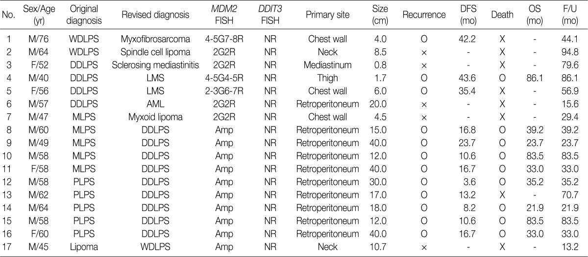
After molecular analysis, the original diagnosis was revised in one case of lipoma and 16 cases of LPS (Table 1). In addition, the classification was revised to DDLPS in four cases of MLPS following a revision analysis of the slides and positive results for MDM2 on immunohistochemistry in non-lipogenic area. Based on the revised classification, the population was reduced to 36 cases of WDLPS, 23 cases of DDLPS, 15 cases of MLPS and one case of PLPS. In Table 2, the clinicopathologic data of these cases are summarized. The mean and median age of the patients was 53 and 56 years (range, 23 to 76 years), respectively. The mean follow-up period was 59.1±40.1 months (range, 2.0 to 175.3 months). There was an almost equal number of male and female patients with a male-to-female ratio of 1.22:1. The most frequent site of occurrence was the retroperitoneum (41 cases), which was followed by the thigh (16 cases) and mediastinum (six cases).
Table 1.
Patients whose diagnosis was revised based on the FISH analysis of MDM2 and DDIT3
FISH, fluorescence in situ hybridization; MDM2, murine double minutes; DFS, disease-free survival; OS, overall survival; F/U, follow-up period; M, male; WDLPS, well differentiated liposarcoma; G, green signal of CEP12 probe; R, red signal of MDM; NR, no rearrangement; F, female; DDLPS, dedifferentiated liposarcoma; LMS, leiomyosarcoma; AML, angiomyolipoma; MLPS, myxoid liposarcoma; Amp, amplification; PLPS, pleomorphic liposarcomas.
Table 2.
Clinical data of 75 patients with LPS after the classification was revised on the FISH analysis of MDM2 and DDIT3
LPS, liposarcomas; FISH, fluorescence in situ hybridization; MDM2, murine double minutes; WDLPS, well-differentiated liposarcoma; DDLPS, dedifferentiated liposarcoma; MLPS, myxoid liposarcoma; PLPS, pleomorphic liposarcoma; F/U, follow-up.
aPLPS is not included in analysis; bValues are presented as mean±standard deviation (range).
There were 43 cases of tumor recurrence during the follow-up period. The recurrence rates of WDLPS, DDLPS, and MLPS were 41.7% (15/36), 87.0% (20/23), and 53.3% (8/15), respectively.
The median value of a ratio of MDM2/CEP12 was 13.4 (range, 3.0 to 25.5) in WDLPS, 24.2 (range 13.3 to 50.5) in DDLPS, 2.2 (range, 0.8 to 2.5) in MLPS and 0.6 in PLPS. In most amplified cases, there was a large homogeneously staining region. This indicates a high level of amplification in more than 20 copies. All the 15 cases of myxoid liposarcoma showed rearrangements involving the DDIT3 gene at a mean proportion of positive cells per case of 85% (range, 68 to 95%).
Of the 42 cases which had been previously diagnosed as WDLPS, two had no amplification of MDM2. These two cases occurred in the neck (8.5 cm) and chest wall (4.0 cm), whose classification was revised to spindle cell lipoma and myxofibrosarcoma, respectively. In case of myxofibrosarcoma, there was an MDM2 polysomy on the FISH analysis. The spindle cell lipoma was strongly positive for CD34 on immunohistochemistry.
Of a total of 14 cases of DDLPS, four had no amplification of the MDM2 gene on the on the FISH analysis. Of these, two occurring in the thigh (1.7 cm) or the chest wall (6.0 cm) had an MDM2 polysomy without amplification. After reviewing hematoxylin and eosin (H&E) slides and additional IHC property, the diagnosis was revised to leiomyosarcoma in these two cases because both cases were diffusely positive for smooth muscle actin. In another case in which there was a retroperitoneal mass of 20 cm in diameter, the classification was revised to angiomyolipoma. This was confirmed based on the positive staining results for HMB-45. The other case that occurred in the mediastinum (0.8 cm) turned out to be sclerosing mediastinitis on histologic re-examination.
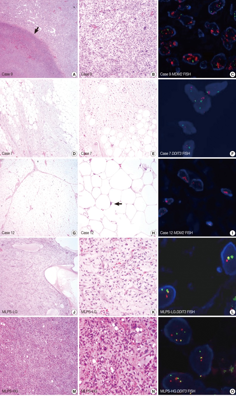
Of the 20 cases which had originally been diagnosed as MLPS, four had an amplification of the MDM2 gene. All of these cases occurred in the retroperitoneum with relatively larger sizes, and their classification was revised to DDLPS after histologic review (Fig. 1A-C). All the remaining cases but one had a break-apart rearrangement of CHOP (DDIT3) on the FISH analysis. In one case, the classification was revised to myxoid lipoma of the chest (Fig. 1D-F). Of the 60 cases of benign lipoma, one had an amplification of the MDM2 gene (MDM2/CEP12 ratio, 3.0) and its classification was revised to WDLPS (Fig. 1G-I). In addition to the presence of a break-apart rearrangement of DDIT3 in typical MLPS (Figs. 1J-O), some cases of high-grade MLPS showed a polysomy for DDIT3 (Fig. 1O). Five of six cases of PLPS had an amplification of the MDM2 gene, and the classification was revised to DDLPS.
Fig. 1.
(A-C) Case 9. A 49-year-old man with a 40-cm sized retroperitoneal mass. Original diagnosis, myxoid liposarcoma (MLPS); revised diagnosis, dedifferentiated liposarcoma. (A) The low-power view reveals a tumour necrosis (arrow) which causes a myxoid change in the surrounding tumor. (B) In this case, there are pleomorphic tumour cells with high mitotic activity. (C) A high amplification of the murine double minutes (MDM2) gene (red signal) is detected on the fluorescence in situ hybridization (FISH) analysis. (D-F) Case 7. A 47-year-old man with a 4.5-cm sized mass on the chest wall. Original diagnosis, MLPS; revised diagnosis, myxoid lipoma. (D, E) Adipocytes with mild focal atypia are intermingled with fibromyxoid stroma. (F) No rearrangement is identified on the FISH analysis of the DDIT3 gene (a break-apart probe). (G-I) Case 12. A 45-year-old man with a 10.7-cm sized mass in the neck. Original diagnosis, lipoma; revised diagnosis, well-differentiated liposarcoma. (G, H) Mature adipocytes with mild atypical nuclei (arrow). (I) The amplification of the MDM2 gene (red signal) is detected on the FISH analysis. (J-L) Low-grade MLPS (MLPS-LG). (J, K) Loose myxoid tumour showing cystic changes with arborization of the capillary vessels. (L) A break-apart rearrangement of the DDIT3 gene is detected on the FISH analysis. (M-O) High-grade MLPS (MLPS-HG). (M, N) Tumor cells with a round-to-oval shape showing a nuclear pleomorphism in the myxoid stroma. (O) Multiple break-apart rearrangements of the DDIT3 gene are detected on the FISH analysis.
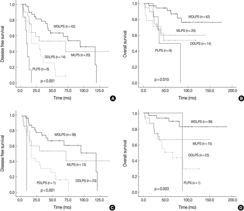
A Kaplan-Meier survival analysis was performed to estimate the results of different cumulative DFS and OS in cases for which the original diagnosis and classification were revised. This showed that the p-value for the OS decreased from 0.010 to 0.003. But there were no significant differences in the DFS or OS between the two diagnosis groups (Fig. 2).
Fig. 2.
The Kaplan-Meier survival curve of the disease-free survival (DFS) and overall survival (OS) based on the original (A, B) and revised (C, D) diagnosis. The DFS (A) and OS (B) for original diagnosis. The DFS (C) and OS (D) for revised diagnosis. WDLPS, well-differentiated liposarcoma; PLPS, pleomorphic liposarcoma; MLPS, myxoid liposarcoma; DDLPS, dedifferentiated liposarcoma.
DISCUSSION
Most sarcomas are diagnosed based on their histologic features and immunohistochemical profiles. Recently, however, a molecular analysis of the specific alterations in some types of sarcoma has been stressed as helpful for diagnosis. Due to the histologic heterogeneity, LPS have a high frequency of discordant histopathological classification. MDM2 enhances the tumorigenic potential of cells by inactivating p53 in the cell cycle.8 Because MDM2 amplification is found in some cases of LPS, several testing modalities have been used to determine if there are underlying mutations in MDM2.
An IHC of MDM2 has been widely performed in adipocytic and non-adipocytic tumours. An IHC is a valuable diagnostic modality to make a differential diagnosis of WDLPS or DDLPS from benign lipomas and other mesenchymal tumors. In many non-adipocytic sarcomas, such as malignant peripheral nerve sheath tumours, myxofibrosarcomas and rhabdomyosarcomas, however, the MDM2 is expressed.9,10 In addition to the IHC, molecular methods such as quantitative polymerase chain reaction (Q-PCR) and FISH have also been used to determine the amplification of MDM2 in adipose tissue tumors. The Q-PCR and FISH showed highly concordant results, although their concordance with those of IHC was relatively lower.11
The fusions between the genes, FUS-DDIT3 (TLS-CHOP) and EWSR1-DDIT3 (EWS-CHOP), are the characteristics of myxoid and round cell LPS but these characteristics are rarely seen in other neoplasms.12 Our results showed that a diagnosis of LPS could be confirmed based on the amplification of the MDM2 gene and a break-apart rearrangement of the DDIT3 gene on the FISH analysis. In particular, these diagnostic clues could also be applied to accurately making a differential diagnosis of WDLPS or DDLPS from benign lipomas and other sarcomas and of MLPS from other myxoid lesions. In the current study, we evaluated cases that had been diagnosed as LPS on conventional histologic examination.
Of a total of 42 cases which had been originally diagnosed as WDLPS, two had no MDM2 amplification on the FISH analysis. In cases whose diagnosis was revised to spindle cell lipoma, there were no recurrence or metastasis during a period of 94.8 months despite a lack of the adjuvant therapy. In other case whose diagnosis was revised to myxofibrosarcoma, however, the recurrence occurred at 42.2 months after the initial operation.
Of 36 cases of WDLPS, 22 occurred in the retroperitoneum. All these cases had a MDM2 amplification on the FISH analysis. According to a previous study, however, none of the 19 cases of retroperitoneal lipomatous tumor without cytologic atypia had an MDM2 amplification on the FISH analysis.13 We therefore recommend that a diagnosis of retroperitoneal lipomatous tumor be made only after a careful review of the morphologic and genetic results.
On the H&E slides of cases of WDLPS or DDLPS, whose diagnoses were revised to non-lipogenic lesions, the adipocytes were distributed between the spindle cells with variable nuclear atypia. It is probable that these adipocytes were mistaken for malignant neoplastic cells that are seen in LPS. In making a differential diagnosis, it should be considered that there is a possibility of entrapped non-neoplastic adipose tissue or benign neoplasm containing adipose cellular components. We therefore do not recommend that a differential diagnosis of WDLPS/DDLPS be made from other lipomatous lesions solely based on histologic examination. The six non-lipomatous lesions occurred in the chest wall (three cases), neck (one case), mediastinum (one case) and thigh (one case), none of which were uncommon sites for LPS.1 It would therefore mandatory to perform a molecular analysis including MDM2 FISH, as well as to evaluate the clinical data and histopathologic findings, in ruling out diverse lesions mimicking WDLPS or DDLPS.
Of the 20 cases which had originally been diagnosed as MLPS, four had an amplification of the MDM2 gene on the FISH analysis. Because there is a difference in the genetic mechanism of tumorigenesis between MLPS and WDLPS/DDLPS, the MDM2 amplification is not detected in cases of MLPS.14 Furthermore, de Vreeze et al.15 reported that the primary retroperitoneal MLPS is not a real disease entity based on immunohistochemical and molecular biological analyses. In these four cases, there were stromal myxoid features on histopathologic examination, in the surrounding area of necrosis in particular, which led to the misdiagnosis of myxoid liposarcoma as shown in cases 8 and 9 (Fig. 1A-F). The stromal myxoid change alone should not be considered a pathognomonic feature of the MLPS, and molecular tests for FUS-DDIT3 or EWSR1-DDIT3 gene fusions are essential for making an accurate diagnosis of myxoid lesion.
Of 60 cases of benign lipoma of more than 10 cm in diameter, occurring in the neck, one has been revised to WDLPS on the FISH analysis of MDM2. In this case, the ratio for MDM2/CEP12 was 3.0 and this corresponds to the lowest value of all of our collected cases. On histopathologic examination of the H&E slides, there were no notable nuclear atypia that might have been overlooked in the original diagnosis. A closer examination of the tumor showed, however, that there were both a marked proliferation of the lobules of mature adipocytes and a mild nuclear atypism. Lipomatous tumors of more than 5 cm in diameter may be of malignant nature. In any lipomatous lesions of more than 5 cm in size, where there is a marked proliferation, it would be mandatory to perform the MDM2 FISH in making a differential diagnosis from WDLPS. De Vreeze et al.16 reported that a molecular analysis was helpful for making a diagnosis of LPS based on their single-institution experience. These authors also noted that the classification of lipomas is frequently revised to WDLPS. In addition, they also reported that the original diagnosis of MLPS was largely correct with the exception of retroperitoneal tumors with morphological features mimicking those of MLPS. This is in agreement with our results.
PLPS is a high-grade sarcoma containing lipoblasts and it is mainly classified based on histology. Our results showed that there was an amplification of the MDM2 gene on the FISH analysis in five of six cases which had originally been diagnosed as PLPS. It has been argued that MDM2 gene amplification is not a consistent feature of PLPS.7 Recently, Mariño-Enríquez et al.2 showed that DDLPS can show a lipoblastic differentiation in the high-grade component and this is a clue for the differential diagnosis from PLPS. Thus, these authors proposed criteria for revising the diagnosis in cases of DDLPS.2 It is possible that the incidence of histologic diagnosis of PLPS will continue to decrease due to the ambiguity of this particular disease entity. In cases of PLPS with an amplification of the MDM2 gene, there were mixed histopathologic features of WDLPS. This indicates that these tumours could have originated from antecedent WDLPS or DDLPS. Our results showed that the classification was revised to DDLPS in five cases with an amplification of the MDM2 gene. There were no significant differences in the DFS or OS between the two diagnosis groups, although the P-value for the OS decreased from 0.010 to 0.003 in the group where the diagnosis was revised.
To summarize, our results indicate that the FISH analysis of MDM2 and DDIT3 FISH, as well as histopathological findings, is helpful for making a differential diagnosis of lipomatous tumors.
Acknowledgments
This work was supported by a grant of the Korea Healthcare technology R&D project, Ministry for Health & Welfare Affairs, Republic of Korea. (A092255).
Footnotes
No potential conflict of interest relevant to this article was reported.
References
- 1.Fletcher CD, Unni KK, Mertens F World Health Organization classification of tumours. Pathology and genetics of tumours of soft tissue and bone. Lyon: IARC Press; 2002. [Google Scholar]
- 2.Mariño-Enríquez A, Fletcher CD, Dal Cin P, Hornick JL. Dedifferentiated liposarcoma with "homologous" lipoblastic (pleomorphic liposarcoma-like) differentiation: clinicopathologic and molecular analysis of a series suggesting revised diagnostic criteria. Am J Surg Pathol. 2010;34:1122–1131. doi: 10.1097/PAS.0b013e3181e5dc49. [DOI] [PubMed] [Google Scholar]
- 3.Dal Cin P, Kools P, Sciot R, et al. Cytogenetic and fluorescence in situ hybridization investigation of ring chromosomes characterizing a specific pathologic subgroup of adipose tissue tumors. Cancer Genet Cytogenet. 1993;68:85–90. doi: 10.1016/0165-4608(93)90001-3. [DOI] [PubMed] [Google Scholar]
- 4.Pedeutour F, Suijkerbuijk RF, Forus A, et al. Complex composition and co-amplification of SAS and MDM2 in ring and giant rod marker chromosomes in well-differentiated liposarcoma. Genes Chromosomes Cancer. 1994;10:85–94. doi: 10.1002/gcc.2870100203. [DOI] [PubMed] [Google Scholar]
- 5.Pedeutour F, Forus A, Coindre JM, et al. Structure of the supernumerary ring and giant rod chromosomes in adipose tissue tumors. Genes Chromosomes Cancer. 1999;24:30–41. [PubMed] [Google Scholar]
- 6.Dei Tos AP, Doglioni C, Piccinin S, et al. Coordinated expression and amplification of the MDM2, CDK4, and HMGI-C genes in atypical lipomatous tumours. J Pathol. 2000;190:531–536. doi: 10.1002/(SICI)1096-9896(200004)190:5<531::AID-PATH579>3.0.CO;2-W. [DOI] [PubMed] [Google Scholar]
- 7.Sandberg AA. Updates on the cytogenetics and molecular genetics of bone and soft tissue tumors: liposarcoma. Cancer Genet Cytogenet. 2004;155:1–24. doi: 10.1016/j.cancergencyto.2004.08.005. [DOI] [PubMed] [Google Scholar]
- 8.Momand J, Zambetti GP, Olson DC, George D, Levine AJ. The mdm-2 oncogene product forms a complex with the p53 protein and inhibits p53-mediated transactivation. Cell. 1992;69:1237–1245. doi: 10.1016/0092-8674(92)90644-r. [DOI] [PubMed] [Google Scholar]
- 9.Aleixo PB, Hartmann AA, Menezes IC, Meurer RT, Oliveira AM. Can MDM2 and CDK4 make the diagnosis of well differentiated/dedifferentiated liposarcoma? An immunohistochemical study on 129 soft tissue tumours. J Clin Pathol. 2009;62:1127–1135. doi: 10.1136/jcp.2009.070201. [DOI] [PubMed] [Google Scholar]
- 10.Binh MB, Sastre-Garau X, Guillou L, et al. MDM2 and CDK4 immunostainings are useful adjuncts in diagnosing well-differentiated and dedifferentiated liposarcoma subtypes: a comparative analysis of 559 soft tissue neoplasms with genetic data. Am J Surg Pathol. 2005;29:1340–1347. doi: 10.1097/01.pas.0000170343.09562.39. [DOI] [PubMed] [Google Scholar]
- 11.Sirvent N, Coindre JM, Maire G, et al. Detection of MDM2-CDK4 amplification by fluorescence in situ hybridization in 200 paraffin-embedded tumor samples: utility in diagnosing adipocytic lesions and comparison with immunohistochemistry and real-time PCR. Am J Surg Pathol. 2007;31:1476–1489. doi: 10.1097/PAS.0b013e3180581fff. [DOI] [PubMed] [Google Scholar]
- 12.Willmore-Payne C, Holden J, Turner KC, Proia A, Layfield LJ. Translocations and amplifications of chromosome 12 in liposarcoma demonstrated by the LSI CHOP breakapart rearrangement probe. Arch Pathol Lab Med. 2008;132:952–957. doi: 10.5858/2008-132-952-TAAOCI. [DOI] [PubMed] [Google Scholar]
- 13.Macarenco RS, Erickson-Johnson M, Wang X, et al. Retroperitoneal lipomatous tumors without cytologic atypia: are they lipomas? A clinicopathologic and molecular study of 19 cases. Am J Surg Pathol. 2009;33:1470–1476. doi: 10.1097/PAS.0b013e3181b278bf. [DOI] [PubMed] [Google Scholar]
- 14.Pilotti S, Della Torre G, Lavarino C, et al. Molecular abnormalities in liposarcoma: role of MDM2 and CDK4-containing amplicons at 12q13-22. J Pathol. 1998;185:188–190. doi: 10.1002/(SICI)1096-9896(199806)185:2<188::AID-PATH53>3.0.CO;2-2. [DOI] [PubMed] [Google Scholar]
- 15.de Vreeze RS, de Jong D, Tielen IH, et al. Primary retroperitoneal myxoid/round cell liposarcoma is a nonexisting disease: an immunohistochemical and molecular biological analysis. Mod Pathol. 2009;22:223–231. doi: 10.1038/modpathol.2008.164. [DOI] [PubMed] [Google Scholar]
- 16.de Vreeze RS, de Jong D, Nederlof PM, et al. Added value of molecular biological analysis in diagnosis and clinical management of liposarcoma: a 30-year single-institution experience. Ann Surg Oncol. 2010;17:686–693. doi: 10.1245/s10434-009-0806-9. [DOI] [PMC free article] [PubMed] [Google Scholar]


