
Figure 1.
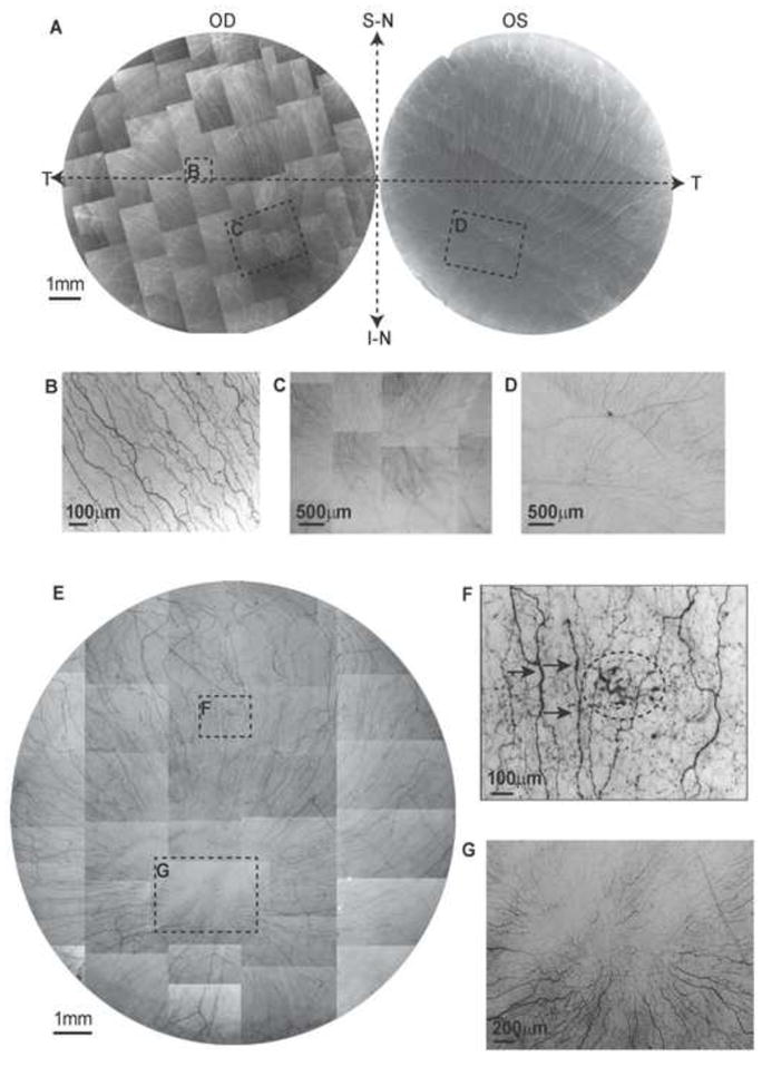
Whole mount view of corneal epithelial nerve distribution in normal and diabetic corneas at early stage of the disease. A) Images were acquired in a time-lapse mode with a Nikon Eclipse TE200 and with a 5X lens in compliance with the natural shape of the cornea from the two eyes of a 64-year-old normal male donor. OD: oculus dexter (right eye); OS: oculus sinister (left eye); S-N: superior nasal quadrant; I-N: inferior nasal quadrant; T: temporal quadrant. The epithelial nerve bundles ran in a radial pattern from the periphery to merge in an area within the inferior-nasal quadrant beyond the corneal apex. Highlighted images showed the detailed architecture of the epithelial nerve distribution in the central (B), and merging areas (C, D). E) Images were taken from the right eye of a 45-year-old male with insulin-dependent diabetes mellitus (IDDM) for two years. Highlighted images show the detailed nerve distribution in the converging area (G). F) Image F, outlined with the small frame, shows the increased thickness in parts of the nerves (arrows) and a sub-epithelial neuropathy (circle).