Introduction
The objective of CBTRUS Statistical Report: Primary Brain and Central Nervous System Tumors Diagnosed in the United States in 2005–2009 is to provide a current and comprehensive review of the descriptive epidemiology of primary brain and central nervous system (CNS) tumors in the United States population. CBTRUS has obtained data on all primary brain and CNS tumors from the Centers for Disease Control and Prevention, National Program of Cancer Registries (NPCR) and the National Cancer Institute, Surveillance, Epidemiology and End Results (SEER) program for diagnosis years 2005–2009. Incidence counts and rates of primary malignant and non-malignant brain and CNS tumors are documented by histology, gender, age, race, and Hispanic ethnicity.
Background
The Central Brain Tumor Registry of the United States (CBTRUS) is a unique professional research organization that focuses exclusively on providing quality statistical data on population-based primary brain and CNS incident tumors in the United States. CBTRUS is currently the only population-based site-specific registry in the United States that works in partnership with a public surveillance organization, the National Program of Central Registries (NPCR), and from which data are directly received under a special agreement. This agreement permits transfer of data through the NPCR-CSS Submission Specifications mechanism,1 the system utilized for collection of central (state) cancer data as mandated in 1992 by Public Law 102-515, the Cancer Registries Amendment Act.2 CBTRUS combines the NPCR data with data from the SEER program3 which was established for national cancer surveillance in the early 1970s. Working with these premier surveillance organizations enables CBTRUS to report high quality data on brain and CNS tumors that are useful to the communities it serves.
Since 1995, CBTRUS has self-published fourteen reports that have contributed to the surveillance of brain and CNS tumors in the United States. As a result of partnering with the Society for Neuro-Oncology (SNO)4, this fifteenth CBTRUS report is the first to be published as a supplement to Neuro-Oncology, the official journal of SNO and marks an historic milestone for both organizations.
CBTRUS was incorporated as a nonprofit 501(c)3 organization with a founding and sustaining grant from the Pediatric Brain Tumor Foundation in 1992 following a two–year study conducted by the American Brain Tumor Association to determine the feasibility of a central registry for all primary brain and CNS tumor cases in the United States. Until that time, standard data reporting in the United States had been limited to only malignant cases. Non–malignant brain tumors, those classified as having a benign or uncertain behavior, however, may, and often do, impose similar costs to society in terms of medical care, case fatality, and lost productivity as do malignant brain tumors. In addition, as molecular markers have been discovered, it has become clear that certain non–malignant brain tumors may become malignant over time.
Passed in 2002, the Benign Brain Tumor Cancer Registries Amendment Act (Public Law 107–260)5 expanded the collection of primary brain and CNS tumor incidence data by the NPCR to include non-malignant brain and CNS tumors having International Classification of Diseases for Oncology Third Edition (ICD-O-3)6 codes beginning with the 2004 diagnosis year. All central (state) cancer registries now include these data in their collection practices. Starting in 2004, Uniform Data Standards (UDS) as directed by the North American Association of Cancer Registries (NAACCR)7, an umbrella organization for tumor registries, governmental agencies, professional associations and private groups, guide the collection of required information on non-malignant brain and CNS tumors; in 2005, the UDS for the collection of malignant brain and CNS tumors were revised. The Multiple Primary and Histology Coding Rules for malignant and non-malignant brain and CNS tumors have been undergoing revision in 2012 under the leadership of SEER.
The CBTRUS database contains the largest aggregation of population–based data on the incidence of all primary brain and CNS tumors in the United States. This report represents a dramatic increase in population coverage (approximately 97% from the initial CBTRUS Reports). The central cancer registries receive population-based, standardized data from all healthcare data sources primarily through certified tumor registrars. Along with the UDS, there are quality control checks and a system for rating each central registry to further insure that these data are reported as accurately and completely as possible. These individuals and organizations provide the high quality data that are the foundation of the CBTRUS statistical reports and scientific activities.
This statistical report continues the past efforts that CBTRUS has made to provide population–based incidence rates for all primary brain and CNS tumors by histology, age, gender, race, and Hispanic ethnicity. As in previous reports, these data have been organized by clinically relevant histological groupings that are useful for surveillance. The information is important for allocation and planning of specialty health–care services, in the planning of disease prevention and control programs, and in research activities. These data may lead to clues that will stimulate research into the causes of this terrible disease.
In 2012, the CBTRUS staff in collaboration with three neuropathologists, Drs. Janet Bruner (University of Texas M.D. Anderson Medical Center), Roger McLendon (Duke University) and Tarik Tihan (University of California at San Francisco) revised the CBTRUS Histology Grouping Scheme to reflect the 2007 WHO Classification of Tumours of the Central Nervous System.8 CBTRUS will continue to update its grouping scheme to reflect state-of-the-art classification for brain and CNS tumors mindful that any future revisions will incorporate accepted ICD-O coding. CBTRUS will continue to share its expertise and to work cooperatively with other surveillance organizations as well as brain tumor clinicians and researchers to assure that primary brain and CNS tumors are collected and reported as accurately and completely as possible.
Technical Notes
Data Collection
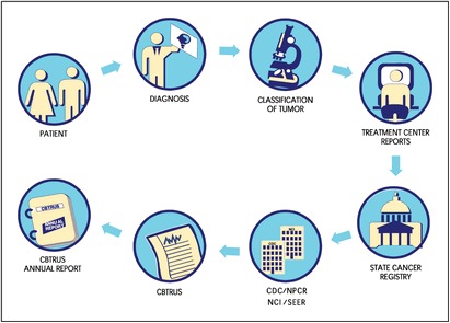
CBTRUS does not collect data directly from patient's medical records. As noted, data for CBTRUS analyses come from NPCR and SEER programs. By law, cancer and benign brain tumors are reportable diseases, and central cancer registries in states are mandated to collect pertinent information on their residents, collate these data, and provide data files to NPCR and SEER. State central cancer registries (including the District of Columbia) play an essential role in the collection process, diagrammatically presented as follows.
CBTRUS obtained incidence data from 49 population-based cancer registries (44 NPCR and 5 SEER) that include cases of malignant and non–malignant (benign and uncertain) primary brain and CNS tumors. It should be noted that metastatic tumors including those found in the brain and CNS are not collected by surveillance organizations in the United States. Data were requested for all newly-diagnosed primary malignant and non-malignant tumors in 2005–2009 at any of the following anatomic sites (ICD-O-3 topography codes in parentheses): brain (C71.0–C71.9), meninges (C70.0–C70.9), spinal cord, cranial nerves, and other parts of the central nervous system (C72.0–C72.9), pituitary and pineal glands (C75.1–C75.3), and olfactory tumors of the nasal cavity [C30.0 (ICD-O-3 histology codes 9522 and 9523)].6
NPCR provided data on 300,205 primary brain and CNS tumors diagnosed in 2005–2009. NPCR cancer registries had to agree to participate in the CBTRUS Statistical Report and to pass certain data quality standards required by NPCR in order for CBTRUS to receive data. From the SEER research data files, data from state central cancer registries not included in the NPCR dataset were obtained and included 12,339 primary brain and CNS tumor case records diagnosed in 2005–2009. These data were combined into a single data set for analyses. A total of 1,342 records (0.43%) were deleted from the final analytic data set because of invalid site/histology combinations based on a review by the CBTRUS consulting neuropathologist, or because of reclassification based on adjudication of multiple records. Of these, 131 cases had bilateral acoustic neuromas in which the records were consolidated. The final analytic data set included 311,202 records from 49 population-based central cancer registries.
Definitions
Measures in Surveillance Epidemiology
The incidence rate is the basic measure of disease occurrence as it expresses probability or risk of disease in a defined population over a specified period of time. Incidence Rates measure the occurrence of newly-diagnosed cases of disease per 100,000 population. Mortality Rates quantify the number of people who have died from the disease per 100,000 population in a specific time period. Prevalence Rates measure the number of people with a disease per 100,000 population at a particular point in time or during a particular period of time. Survival Rates (percentages) are the probability of surviving for a specified time period. Relative Survival Rates are defined as the observed probability of survival adjusted for the expected survival rate of the population for that age, gender and calendar year.
Incidence and mortality rates in this report are expressed per units of observation. For cancer, rates are usually expressed per 100,000 population. The unadjusted rate of disease in an entire population is the Crude Rate. Crude rates are frequently adjusted by age because as a population ages the crude rate would increase, reflecting only the aging of the population, not an actual incidence increase. Age–Adjusted Rates to a common standard population allow for comparisons of rates in populations across regions with different age structures. Incidence and mortality brain and CNS tumor rates in this report are age adjusted to the Year 2000 U.S. Standard Population. Rates for a subset of a population are termed specific rates. Age–Specific Rates that describe the rate of disease in a defined age group are presented in this report. Specific rates by gender, race, and Hispanic ethnicity are also reported. The variability around the estimates of rates is reflected in the Standard Error, which is incorporated into the formula for computing the confidence interval associated with a certain rate. A Confidence Interval (CI) is the computed interval with a given probability, eg, 95 percent, that the true value of a variable such as a mean, proportion or rate occurs within the interval. For example, the age–adjusted primary brain and CNS tumor incidence rate is 20.59 cases per 100,000. We can assume with 95 percent certainty that the actual incidence rate is within the range of 20.52 and 20.66 cases per 100,000. Statistically Significant refers to the likelihood that a result or relationship is caused by something other than mere random chance. Statistical hypothesis testing is traditionally employed to determine if a result is statistically significant or not. This provides a “p-value” representing the probability that random chance could explain the result. In general, a 5% or lower p-value is considered to be statistically significant.
In order to be able to compare incidence rates among statistical reports, agencies, or registries a number of factors should be considered such as whether the case definition, data collection, and rate calculation are similar by asking some of the following questions:
How is an incident case defined?
Are all primary malignant and non–malignant tumors included in the data set?
Or, are only malignant tumors being analyzed?
What anatomic locations (primary sites) are included?
Are lymphomas and hematopoietic neoplasms included in the incidence rates?
Are the populations comparable?
Are the incidence rates age–adjusted? And if so, to which standard population are they age–adjusted?
Classification by Behavior and Histology
Brain and CNS tumor classifications according to behavior ICD-O-3 standards for benign, uncertain and malignant behaviors are coded 0, 1, and 3, respectively. The histology groupings in CBTRUS statistical reports were initially developed in collaboration with the CBTRUS consulting neuropathologist, Dr. Janet Bruner. In 2012, Drs. Roger McLendon and Tarik Tihan joined Dr. Bruner and the CBTRUS staff to synchronize the CBTRUS histology grouping scheme with the 2007 World Health Organization (WHO) classification of tumors of the central nervous system.8,9 This report uses this most recent 2012 CBTRUS histology grouping scheme (Table 1). The classification scheme utilizes ICD-O-3 codes6 and may include morphology codes that were not previously reported to CBTRUS.10 Tables 1a and 1b list malignant only and non-malignant only histologies, respectively. In this report, incidence rates are provided by major histology grouping and detailed histology.
Table 1.
Central Brain Tumor Registry of the United States (CBTRUS), Brain and Central Nervous System Tumor Histology Groupings
| Histology | ICD-O-3† Histology Code |
|---|---|
| Tumors of Neuroepithelial Tissue | |
| Pilocytic astrocytoma | 9421 |
| Diffuse astrocytoma | 9400, 9410, 9411, 9420 |
| Anaplastic astrocytoma | 9401 |
| Unique astrocytoma variants | 9381, 9384, 9424 |
| Glioblastoma | 9440, 9441, 9442/3‡ |
| Oligodendroglioma | 9450 |
| Anaplastic oligodendroglioma | 9451, 9460 |
| Oligoastrocytic tumors | 9382 |
| Ependymal tumors | 9383, 9391, 9392, 9393, 9394 |
| Glioma malignant, NOS | 9380 |
| Choroid plexus tumors | 9390 |
| Other neuroepithelial tumors | 9363, 9423, 9430, 9444 |
| Neuronal and mixed neuronal-glial tumors | 8680, 8681, 8690, 8693, 9412, 9413, 9442/1§, |
| 9492 (excluding site C75.1), 9493, 9505, 9506, 9522, 9523 | |
| Tumors of the pineal region | 9360, 9361, 9362 |
| Embryonal tumors | 8963, 9364, 9470, 9471, 9472, 9473, 9474, |
| 9490, 9500, 9501, 9502, 9508 | |
| Tumors of Cranial and Spinal Nerves | |
| Nerve sheath tumors | 9540, 9541, 9550, 9560, 9561, 9570, 9571 |
| Other tumors of cranial and spinal nerves | 9562 |
| Tumors of Meninges | |
| Meningioma | 9530, 9531, 9532, 9533, 9534, 9537, 9538, 9539 |
| Mesenchymal tumors | 8324, 8800, 8801, 8802, 8803, 8804, 8805, 8806, 8810, 8815, 8824, 8830, |
| 8831, 8835, 8836, 8850, 8851, 8852, 8853, 8854, 8857, 8861, 8870, 8880, | |
| 8890, 8897, 8900, 8901, 8902, 8910, 8912, 8920, 8921, 8935, 8990, 9040, 9136, | |
| 9150, 9170, 9180, 9210, 9241, 9260, 9373, 9480 | |
| Primary melanocytic lesions | 8720, 8728, 8770, 8771 |
| Other neoplasms related to the meninges | 9161, 9220, 9231, 9240, 9243, 9370, 9371, 9372, 9535 |
| Lymphomas and Hemopoietic Neoplasms | |
| Lymphoma | 9590, 9591, 9596, 9650, 9651, 9652, 9653, 9654, 9655, 9659, 9661, |
| 9662, 9663, 9664, 9665, 9667, 9670, 9671, 9673, 9675, 9680, 9684, | |
| 9687, 9690, 9691, 9695, 9698, 9699, 9701, 9702, 9705, 9714, 9719, | |
| 9728, 9729 | |
| Other hemopoietic neoplasms | 9727, 9731, 9733, 9734, 9740, 9741, 9750, 9751, 9752, 9753, 9754, 9755, |
| 9756, 9757, 9758, 9760, 9766, 9823, 9826, 9827, 9832, 9837, 9860, 9861, 9866, 9930, 9970 | |
| Germ Cell Tumors and Cysts | |
| Germ cell tumors, cysts and heterotopias | 8020, 8440, 9060, 9061, 9064, 9065, 9070, 9071, 9072, 9080, 9081, 9082, |
| 9083, 9084, 9085, 9100, 9101 | |
| Tumors of Sellar Region | |
| Tumors of the pituitary | 8040, 8140, 8146, 8246, 8260, 8270, 8271, 8272, |
| 8280, 8281, 8290, 8300, 8310, 8323, 9492 (Site C75.1 only), 9582 | |
| Craniopharyngioma | 9350, 9351, 9352 |
| Unclassified Tumors | |
| Hemangioma | 9120, 9121, 9122, 9123, 9125, 9130, 9131, 9133, 9140 |
| Neoplasm, unspecified | 8000, 8001, 8002, 8003, 8004, 8005, 8010, 8021 |
| All other | 8320, 8452, 8710, 8711, 8713, 8811, 8840, 8896, 8980, 9173, 9503, 9580 |
† International Classification of Diseases for Oncology, 3rd Edition, 2000. World Health Organization, Geneva, Switzerland.
‡ Morphology 9442/3 only.
§ Morphology 9442/1 only.
CBTRUS defines the broad category of gliomas to include ICD-O-3 histology codes 9380-9384,9391-9460,9480.
Abbreviations: CBTRUS, Central Brain Tumor Registry of the United States; NOS, not otherwise specified.
Table 1a.
Central Brain Tumor Registry of the United States (CBTRUS), Brain and Central Nervous System Tumor Malignant Histologies†
| Histology | ICD-O-3‡ Histology Code |
|---|---|
| Tumors of Neuroepithelial Tissue | |
| Pilocytic astrocytoma | 9421/1 [Included with malignant tumors] |
| Diffuse astrocytoma | 9400/3, 9410/3, 9411/3, 9420/3 |
| Anaplastic astrocytoma | 9401/3 |
| Unique astrocytoma variants | 9381/3, 9424/3 |
| Glioblastoma | 9440/3, 9441/3, 9442/3 |
| Oligodendroglioma | 9450/3 |
| Anaplastic oligodendroglioma | 9451/3, 9460/3 |
| Oligoastrocytic tumors | 9382/3 |
| Ependymal tumors | 9391/3, 9392/3, 9393/3 |
| Glioma malignant, NOS | 9380/3 |
| Choroid plexus | 9390/3 |
| Other neuroepithelial tumors | 9423/3, 9430/3 |
| Neuronal and mixed neuronal- glial tumors | 8680/3, 8693/3, 9505/3, 9522/3, 9523/3 |
| Tumors of the pineal region | 9362/3 |
| Embryonal tumors | 8963/3, 9364/3, 9470/3, 9471/3, 9472/3,9473/3, 9474/3, |
| 9490/3, 9500/3, 9501/3, 9502/3, 9508/3 | |
| Tumors of Cranial and Spinal Nerves | |
| Nerve sheath tumors | 9540/3, 9560/3, 9561/3, 9571/3 |
| Tumors of Meninges | |
| Meningioma | 9530/3, 9538/3, 9539/3 |
| Mesenchymal tumors | 8800/3, 8801/3, 8802/3, 8803/3, 8804/3, 8805/3, 8806/3, 8810/3, 8815/3, 8830/3, |
| 8850/3, 8851/3, 8852/3, 8853/3, 8854/3, 8857/3, 8890/3, 8900/3, 8901/3, 8902/3, | |
| 8910/3, 8912/3, 8920/3, 8921/3, 8990/3, 9040/3, 9150/3, 9170/3, 9180/3, 9260/3, 9480/3 | |
| Primary melanocytic lesions | 8720/3, 8728/3, 8770/3, 8771/3 |
| Other neoplasms related to the meninges | 9220/3, 9231/3, 9240/3, 9243/3, 9370/3, 9371/3, 9372/3 |
| Lymphomas and Hemopoietic Neoplasms | |
| Lymphoma | 9590/3, 9591/3, 9596/3, 9650/3, 9651/3, 9652/3, 9653/3, 9654/3, 9655/3, 9659/3, |
| 9661/3, 9662/3, 9663/3, 9664/3, 9665/3, 9667/3, 9670/3, 9671/3, 9673/3, 9675/3, | |
| 9680/3, 9684/3, 9687/3, 9690/3, 9691/3, 9695/3, 9698/3, 9699/3, 9701/3, 9702/3, | |
| 9705/3, 9714/3, 9719/3, 9728/3, 9729/3 | |
| Other hemopoietic neoplasms | 9727/3, 9731/3, 9733/3, 9734/3, 9740/3, 9741/3, 9750/3, 9754/3, 9755/3, 9756/3, 9757/3, 9758/3, |
| 9760/3, 9823/3, 9826/3, 9827/3, 9832/3, 9837/3, 9860/3, 9861/3, 9866/3, 9930/3 | |
| Germ Cell Tumors and Cysts | |
| Germ cell tumors, cysts and | 8020/3, 8440/3, 9060/3, 9061/3, 9064/3, 9065/3, 9070/3, 9071/3, 9072/3, 9080/3, |
| heterotopias | 9081/3, 9082/3, 9083/3, 9084/3, 9085/3, 9100/3, 9101/3 |
| Tumors of Sellar Region | |
| Tumors of the pituitary | 8140/3, 8246/3, 8260/3, 8270/3, 8272/3, 8280/3, 8281/3, 8290/3, 8300/3, 8310/3, 8323/3 |
| Unclassified Tumors | |
| Hemangioma | 9120/3, 9130/3, 9133/3, 9140/3 |
| Neoplasm, unspecified | 8000/3, 8001/3, 8002/3, 8003/3, 8004/3, 8005/3, 8010/3, 8021/3 |
| All other | 8320/3, 8710/3, 8711/3, 8811/3, 8840/3, 8896/3, 8980/3, 9503/3, 9580/3 |
† Includes all the histologies listed in the standard definition of reportable brain tumors from the Consensus Conference10 on Brain Tumor Definitions.
‡ International Classification of Diseases for Oncology, 3rd Edition, 2000. World Health Organization, Geneva, Switzerland.
Abbreviations: CBTRUS, Central Brain Tumor Registry of the United States; NOS, not otherwise specified.
Table 1b.
Central Brain Tumor Registry of the United States (CBTRUS), Brain and Central Nervous System Tumor Non-Malignant Histologies†
| Histology | ICD-O-3‡ Histology Code |
|---|---|
| Tumors of Neuroepithelial Tissue | |
| Pilocytic astrocytoma | 9421/1 [Included with malignant tumors] |
| Unique astrocytoma variants | 9384/1 |
| Ependymal tumors | 9383/1; 9394/1 |
| Choroid plexus | 9390/0,1 |
| Other neuroepithelial tumors | 9363/0; 9444/1 |
| Neuronal and mixed neuronalglial tumors | 8680/0,1; 8681/1; 8690/1; 8693/1; 9412/1; 9413/0; 9442/1; |
| 9492/0 (excluding site C75.1); 9493/0; 9505/1; 9506/1 | |
| Tumors of the pineal region | 9360/1; 9361/1 |
| Embryonal tumors | 9490/0 |
| Tumors of Cranial and Spinal Nerves | |
| Nerve sheath tumors | 9540/0,1; 9541/0, 9550/0; 9560/0,1; 9570/0; 9571/0 |
| Other tumors of cranial and spinal nerves | 9562/0 |
| Tumors of Meninges | |
| Meningioma | 9530/0,1; 9531/0; 9532/0; 9533/0; 9534/0; 9537/0; 9538/1; 9539/1 |
| Mesenchymal tumors | 8324/0; 8800/0; 8810/0; 8815/0; 8824/0,1; 8830/0,1; 8831/0; 8835/1; 8836/1; |
| 8850/0,1; 8851/0; 8852/0, 8854/0; 8857/0; 8861/0; 8870/0; 8880/0, 8890/0,1; 8897/1; | |
| 8900/0; 8920/1; 8935/0,1; 8990/0,1; 9040/0; 9136/1, 9150/0,1; 9170/0; 9180/0; 9210/0; 9241/0; 9373/0 | |
| Primary melanocytic lesions | 8728/0,1; 8770/0; 8771/0 |
| Other neoplasms related to the meninges | 9161/1; 9220/0,1; 9535/0 |
| Lymphomas and Hemopoietic Neoplasms | |
| Other hemopoietic neoplasms | 9740/1; 9751/1; 9752/1; 9753/1; 9766/1; 9970/1 |
| Germ Cell Tumors and Cysts | |
| Germ cell tumors, cysts and heterotopias | 8440/0; 9080/0,1; 9084/0 |
| Tumors of Sellar Region | |
| Tumors of the pituitary | 8040/0,1; 8140/0,1; 8146/0; 8260/0; 8270/0; 8271/0; 8272/0; |
| 8280/0; 8281/0; 8290/0; 8300/0; 8310/0; 8323/0; 9492/0 (site C75.1 only); 9582/0 | |
| Craniopharyngioma | 9350/1; 9351/1; 9352/1 |
| Unclassified Tumors | |
| Hemangioma | 9120/0; 9121/0; 9122/0; 9123/0; 9125/0; 9130/0,1; 9131/0; 9133/1 |
| Neoplasm, unspecified | 8000/0,1; 8001/0,1; 8005/0; 8010/0 |
| All other | 8452/1; 8711/0; 8713/0; 8811/0; 8840/0; 9173/0; 9580/0 |
† Includes all the histologies listed in the standard definition of reportable brain tumors from the Consensus Conference10 on Brain Tumor Definition.
‡ International Classification of Diseases for Oncology, 3rd Edition, 2000. World Health Organization, Geneva, Switzerland.
Abbreviations: CBTRUS, Central Brain Tumor Registry of the United States; NOS, not otherwise specified.
Gliomas are tumors that arise from glial cells, and include astrocytoma, glioblastoma, oligodendroglioma, ependymoma, mixed glioma, malignant glioma, not otherwise specified (NOS), and a few more rare histologies. Whereas there is no standard definition, CBTRUS defines glioma as ICD-O-3 histology codes 9380-9384, 9391-9460, and 9480. It is also important to note that the statistics for lymphomas and hematopoietic neoplasms contained in this report refer only to those lymphomas and hematopoietic neoplasms that arise in the brain and CNS.
Anatomic Location of Tumor Sites
Various terms are used to describe the regions of the brain and central nervous system. The sites referred to in this report are broadly based on the categories and site codes defined in the SEER Site/Histology Validation List.11 Tumors include olfactory tumors of the nasal cavity in addition to brain tumors located in sites included in the standard definition from the Consensus Conference on Brain Tumor Definition for Registration.10 According to the standard definition from the Consensus Conference, reportable primary brain–related tumors (intracranial and central nervous system tumors) are all primary tumors, irrespective of histology and behavior, occurring in the following sites: brain; meninges; pineal gland; pituitary gland and craniopharyngeal duct; and spinal cord, cranial nerves, and other parts of the central nervous system. As per the site definition outlined by the Consensus Conference, brain lymphomas coded to any of the brain or CNS site codes listed above are included in the CBTRUS report. The group of tumors known as spinal cord tumors is coded to the following sites: spinal meninges, spinal cord, and cauda equina, and is highlighted in this report.
Statistics by ICD-O-3 primary sites are grouped in the following manner: the frontal lobe (C71.1); temporal lobe (C71.2); parietal lobe (C71.3); and occipital lobe (C71.4) are grouped together. Cerebrum (C71.0), ventricle (C71.5), cerebellum (C71.6), and brain stem (C71.7) are each grouped independently. Overlapping lesions of the brain, as well as brain sites not otherwise specified (NOS), are defined by ICD-O site codes C71.8–C71.9. The cranial nerve category (C72.2–C72.5) includes the olfactory nerve, optic nerve, acoustic nerve, and other cranial nerves. The spinal cord (C72.0) and cauda equina (C72.1) are grouped together. Overlapping lesions of the brain and central nervous system, as well as nervous system sites not otherwise specified (NOS), are defined by ICD-O site codes C72.8–C72.9. The meninges (C70.0–C70.9) include the cerebral meninges and spinal meninges. Pituitary tumors (C75.1–C75.2) include tumors located in the pituitary gland and craniopharyngeal duct. Pineal tumors (C75.3) include tumors located in the pineal gland. In this report, tumors located in the nasal cavity (C30.0) are olfactory tumors (defined by ICD-O-3 histology codes 9522 and 9523).
Measurement Methods
Counts, means, rates, ratios, proportions, and other relevant statistics were calculated using SPSS and/or SEER*Stat statistical software.12,13 Statistics are suppressed when counts are fewer than 16 within a cell. However, the data in the suppressed cells are included in the counts and rates for the totals.
Population data for each geographic region were obtained from the SEER program website14 for the purpose of rate calculation. The estimates adjusted for the impact of the Katrina and Rita hurricanes on affected state populations were used in the data analyses for the statistics presented in this report.
Age-adjusted incidence rates and 95% confidence intervals15 for malignant and non–malignant tumors and for selected histology groupings by gender, race, Hispanic ethnicity, and pediatric, young adult, and adult age groups were estimated. Age–adjustment was based on five–year age groupings and standardized to the Year 2000 U.S. standard population. Age-specific incidence rates by five-year age groups were also calculated. The age distribution of the 2000 U.S. standard population is shown in Appendix A. Combined populations for the regions included in this report are shown in Appendix B and Appendix C.
CBTRUS presents statistics on the pediatric age group 0-19 years in order to include and describe specific brain and CNS tumor patterns in age groups 0-4, 5-9, 10-14 as well as 15-19 years. However, the 0-14 year age group is a standard age category for childhood cancer used by other cancer surveillance organizations and has been included in this report for consistency and comparison purposes. Race categories in this report are all races, white, black, American Indian/Alaskan Native (AIAN), and Asian Pacific Islander (API). Other race: unspecified and unknown race are included in all race statistics. Hispanic ethnicity was defined using the NAACCR Hispanic Identification Algorithm, version 2, data element, which utilizes a combination of cancer registry data fields (Spanish/Hispanic Origin data element, birthplace, race, and surnames) to directly and indirectly classify cases as Hispanic or non–Hispanic.16 Trends across annual incidence rates were not estimated because a timeframe of five years for both malignant and non-malignant tumors as presented in this report is not sufficient to detect a real change in the rate pattern with any degree of confidence.
Brain Tumor Definition Differences
It should be noted that NPCR, SEER, and NAACCR report brain tumors differently than CBTRUS. The definition of brain and CNS tumors used by these organizations (in their published incidence and mortality statistics) includes tumors located in the brain, meninges, and other central nervous system tumors (C70.0–9, C71.0–9, and C72.0–9), but excludes lymphoma and leukemia histologies (9590–9989) from all brain and CNS sites. NPCR and SEER include separate tables for malignant and non–malignant brain and CNS tumors reflecting the 2002 Consensus Conference10 definition in their respective publications. With the inclusion of non-malignant brain tumors, an increase in incidence rates may result, especially for the following histology groups and subgroups: (groups) tumors of meninges; tumors of cranial and spinal nerves; tumors of the sellar region; and (subgroups) unique astrocytoma variants; ependymal tumors; choroid plexus; neuronal and mixed neuronal-glial tumors; tumors of the pineal region; nerve sheath tumors; meningioma; mesenchymal tumors; other neoplasms related to the meninges; germ cell tumors; tumors of the pituitary; craniopharyngioma; hemangioma; neoplasm, unspecified; and all other.
In contrast, the CBTRUS reports data on all tumor morphologies located within the Consensus Conference site definition including the leukemia and lymphoma histologies (9590–9989) as well as olfactory tumors of the nasal cavity [C30.0 (9522–9523)].10 NPCR, SEER, and NAACCR include pilocytic astrocytomas [a tumor listed in the WHO Classification of Tumours of the Central Nervous System8 as having uncertain behavior (ICD-0-3 behavior code of 1)] in their malignant (ICD-0-3 behavior code of 3) brain tumor data and statistics. In support of consistency within cancer surveillance reporting, the CBTRUS categorizes pilocytic astrocytomas in the malignant tumor category to enhance comparability of rates to those reported by NPCR, SEER, and NAACCR, especially for comparison of childhood brain and CNS tumor rates. It is important to understand these differences in definition, as they influence the direct comparison of published rates.
Estimation of Expected Numbers of Brain and CNS Tumors in 2012 and 2013
Estimated numbers of expected malignant and non–malignant brain and CNS tumors were calculated for diagnosis years 2012 and 2013. The age-specific rate method was utilized to project 2012 and 2013 estimates of all primary brain and CNS tumors using the CBTRUS 2005–2009 age-sex-race-specific brain tumor incidence rates for a group by the age-sex-race-specific population projections for that group. Projected population estimates for 2012 and 2013 were derived for the 50 states and District of Columbia using the US Census Bureau 2000-2009 population data (seer.cancer.gov/popdata/index.html).14
Estimation of Mortality Rates for Underlying Cause from Brain and CNS Tumors
Age-adjusted mortality rates for deaths resulting from all brain and CNS tumors were calculated using SEER Stat 7.0.9.12,17 The underlying mortality data were provided to the SEER program by the National Center for Health Statistics (NCHS) (www.cdc.gov/nchs). In addition to total age-adjusted rate for the United States, age-adjusted rates are presented by gender and state.
Estimation of Survival Rates
SEER*Stat 7.0.9 statistical software was used to estimate one–through ten–year relative survival rates for primary malignant brain tumor cases diagnosed between 1995–2009 in eighteen SEER areas.12,18 This software utilizes life–table (actuarial) methods to compute survival estimates and accounts for current follow-up. The traditional cohort analysis of survival rates was utilized for the survival estimates presented in this report. Survival was estimated for brain (C71.0–C71.9), meninges (C70.0–C70.9), spinal cord, cranial nerves, and other parts of the central nervous system (C72.0–C72.9), pituitary and pineal glands (C75.1–C75.3), and olfactory tumors of the nasal cavity [C30.0 (9522–9523)]. Lymphomas and leukemias (morphology codes 9590–9989) and meningiomas (9530–9539) are included from all brain and CNS sites. Second or later primary tumors, cases diagnosed at autopsy, cases in which race or sex is coded as other or unknown, and cases known to be alive but for whom follow–up time could not be calculated were excluded from the SEER survival data analyses.
Data Interpretation
The CBTRUS works diligently to support the broader surveillance efforts aimed at improving the collection and reporting of primary brain and CNS tumors. The central cancer registry data provided to NPCR and SEER and, subsequently, to CBTRUS vary from year-to-year due to ongoing updates in collection and data refinement aimed to improve completeness and accuracy. The data presented in this report must be interpreted within this surveillance framework as well as taking into account the information provided in the technical notes. Therefore, it is important to note that data from previous CBTRUS Reports cannot be compared to data in CBTRUS Statistical Report: Primary Brain and Central Nervous System Tumors Diagnosed in the United States in 2005–2009.
Random fluctuations in average annual rates are usual especially for rates based on small incidence counts. The CBTRUS policy to suppress data presentation for cells with counts of less than 16 is consistent with the NPCR policy. The rationale for this policy is that rates produced with small counts are unreliable. The suppression of data with counts of fewer than 16 is more than adequate to protect confidentially given the CBTRUS data set is an aggregate of 48 states and the District of Columbia for 2005–2009.
Delays in reporting and late ascertainment are a reality and a known issue influencing registry completeness and, consequently, rate underestimations - especially for more recent data collection years.19 CBTRUS also recognizes that the problem may be even more likely to occur in the reporting of non-malignant brain and CNS tumors, where reporting often comes from non-hospital based sources and mandated collection is relatively recent (2004).
Reporting from Veteran's Health Administration (VHA) hospitals, the sole source of data for cancer cases diagnosed among Veterans served by those institutions, affects completeness of data. Cancer cases from VHA facilities account for at least three percent and possibly as much as eight percent of all cancer cases diagnosed among men. VHA policy that went into effect in 2007 restricting Veterans' health data sharing has resulted in the underreporting of cancer incidence data for diagnosis years 2005 through 2007. Since late 2008, VHA facilities and states with central cancer registries have been working to establish data transfer agreements that correct the problem to assure more complete ascertainment of national cancer incidence including brain and CNS tumor incidence data used in CBTRUS statistical reports.20
CBTRUS editing practices conducted yearly aim to refine the data for accuracy and clinical relevance should also be recognized in interpreting these report data. Exclusion of site and histology combinations considered to be invalid by the consulting neuropathologists may have the impact of conservatively underestimating the incidence of brain and CNS tumors. Editing changes also incorporate updates to the cancer registration coding rules that influence case ascertainment and data collection. For example, beginning in 2004, some brain and CNS site codes were reconsidered as paired sites, that is having a tumor on the left and right hemispheres, would result in multiple tumors being reported rather than unpaired sites which has likely caused some increase in reported incidence. Another relevant coding tool affecting reporting was the 2007 Multiple Primary and Histology Coding Rules. These rules revised the way malignant and non-malignant brain tumors are reported and may affect incident rate changes for certain histologies.
Population estimates used for denominators affect incidence rates. CBTRUS has utilized population data estimates for years 2005–2009 available on the SEER website for rate calculations in this report. These population estimates are provided to SEER on an annual basis from the United States Bureau of the Census. It should be noted that these estimates do not reflect the 2010 decennial census which may or may not impact the rates published. Finally, because this report includes incidence for diagnosis year 2005, adjusted population estimates were used for rate calculation in this report. These estimates adjust for the impact of hurricanes Katrina and Rita on the displacement of populations along the Gulf Coast of Louisiana, Alabama, Mississippi, and Texas in the fall of 2005.
Results
Primary Brain and CNS Tumors: Distributions and Incidence by Histology Group, Histology, Gender, Race, Hispanic Ethnicity, Age Group, Cancer Registry and Behavior
Counts of the 311,202 incident tumors (109,695 malignant; 201,507 non-malignant) reported during 2005–2009 by histology and demographic characteristics for all ages and for children ages 0-19 are presented in Tables 2–4. Approximately seven percent of the cases were in individuals less than 20 years of age at the time of diagnosis, and 93% were in individuals 20 years of age or older. Approximately 42% of all brain and CNS tumors occurred in males and 58% in females. The overall number of all reported tumors is listed by central cancer registry in Table 5. The average annual combined 2005–2009 population of 293,011,631 represents approximately 97% of the U.S. population for those years. The overall percent of non–malignant tumors varied considerably by cancer registry (range: 53-73%). About 65% of all tumors had a histologically confirmed diagnosis, with substantial regional variation (see range: 53-97% in Table 5).
Table 2.
Number of Brain and Central Nervous System Tumors by Major Histology Groupings, Histology, Gender, Race and Hispanic Ethnicity, CBTRUS Statistical Report: NPCR and SEER, 2005–2009
| Total | Gender |
Race |
Hispanic Ethnicity† |
||||||
|---|---|---|---|---|---|---|---|---|---|
| Histology | Male | Female | White | Black | AIAN | API | Hispanic | Non-Hispanic | |
| Tumors of Neuroepithelial Tissue | 99,063 | 55,149 | 43,914 | 88,414 | 6,666 | 516 | 1,823 | 9,034 | 90,029 |
| Pilocytic astrocytoma | 4,636 | 2,374 | 2,262 | 3,870 | 491 | 39 | 104 | 649 | 3,987 |
| Diffuse astrocytoma | 8,616 | 4,811 | 3,805 | 7,616 | 591 | 67 | 170 | 814 | 7,802 |
| Anaplastic astrocytoma | 5,374 | 3,059 | 2,315 | 4,856 | 295 | 31 | 103 | 455 | 4,919 |
| Unique astrocytoma variants | 938 | 500 | 438 | 757 | 126 | – | – | 132 | 806 |
| Glioblastoma | 49,088 | 27,994 | 21,094 | 45,140 | 2,614 | 182 | 668 | 3,203 | 45,885 |
| Oligodendroglioma | 3,973 | 2,181 | 1,792 | 3,558 | 231 | 19 | 85 | 419 | 3,554 |
| Anaplastic oligodendroglioma | 1,687 | 933 | 754 | 1,507 | 88 | – | 40 | 156 | 1,531 |
| Oligoastrocytic tumors | 3,020 | 1,724 | 1,296 | 2,691 | 165 | 22 | 81 | 310 | 2,710 |
| Ependymal tumors | 6,117 | 3,371 | 2,746 | 5,311 | 473 | 41 | 144 | 707 | 5,410 |
| Glioma malignant, NOS | 6,574 | 3,329 | 3,245 | 5,569 | 638 | 33 | 164 | 770 | 5,804 |
| Choroid plexus tumors | 772 | 375 | 397 | 664 | 58 | – | 21 | 131 | 641 |
| Other neuroepithelial tumors | 95 | 35 | 60 | 80 | – | – | – | – | 82 |
| Neuronal and mixed neuronal-glial tumors | 3,887 | 2,093 | 1,794 | 3,245 | 396 | 25 | 108 | 438 | 3,449 |
| Tumors of the pineal region | 579 | 240 | 339 | 444 | 103 | – | – | 80 | 499 |
| Embryonal tumors | 3,707 | 2,130 | 1,577 | 3,106 | 384 | 24 | 111 | 757 | 2,950 |
| Tumors of Cranial and Spinal Nerves | 25,942 | 12,377 | 13,565 | 22,614 | 1,349 | 106 | 854 | 2,108 | 23,834 |
| Nerve sheath tumors | 25,926 | 12,371 | 13,555 | 22,603 | 1,348 | 106 | 852 | 2,107 | 23,819 |
| Other tumors of cranial and spinal nerves | 16 | – | – | – | – | – | – | – | – |
| Tumors of Meninges | 114,363 | 31,030 | 83,333 | 94,854 | 13,112 | 592 | 3,380 | 9,790 | 104,573 |
| Meningioma | 110,359 | 28,884 | 81,475 | 91,469 | 12,757 | 561 | 3,238 | 9,298 | 101,061 |
| Mesenchymal tumors | 1,192 | 584 | 608 | 998 | 113 | – | 38 | 127 | 1,065 |
| Primary melanocytic lesions | 106 | 65 | 41 | 95 | – | – | – | – | 91 |
| Other neoplasms related to the meninges | 2,706 | 1,497 | 1,209 | 2,292 | 236 | 19 | 101 | 350 | 2,356 |
| Lymphomas and Hemopoietic Neoplasms | 6,956 | 3,701 | 3,255 | 5,874 | 693 | 39 | 251 | 721 | 6,235 |
| Lymphoma | 6,774 | 3,607 | 3,167 | 5,731 | 667 | 36 | 245 | 694 | 6,080 |
| Other hemopoietic neoplasms | 182 | 94 | 88 | 143 | 26 | – | – | 27 | 155 |
| Germ Cell Tumors and Cysts | 1,418 | 967 | 451 | 1,126 | 146 | – | 91 | 285 | 1,133 |
| Germ cell tumors, cysts and heterotopias | 1,418 | 967 | 451 | 1,126 | 146 | – | 91 | 285 | 1,133 |
| Tumors of Sellar Region | 46,562 | 21,057 | 25,505 | 34,608 | 8,535 | 372 | 1,566 | 6,699 | 39,863 |
| Tumors of the pituitary | 43,882 | 19,728 | 24,154 | 32,590 | 8,056 | 351 | 1,473 | 6,289 | 37,593 |
| Craniopharyngioma | 2,680 | 1,329 | 1,351 | 2,018 | 479 | 21 | 93 | 410 | 2,270 |
| Unclassified Tumors | 16,898 | 7,489 | 9,409 | 14,313 | 1,733 | 108 | 337 | 1,791 | 15,107 |
| Hemangioma | 3,240 | 1,436 | 1,804 | 2,765 | 256 | 17 | 111 | 370 | 2,870 |
| Neoplasm, unspecified | 13,566 | 6,008 | 7,558 | 11,471 | 1,470 | 89 | 222 | 1,409 | 12,157 |
| All other | 92 | 45 | 47 | 77 | – | – | – | – | 80 |
| Total | 311,202 | 131,770 | 179,432 | 261,803 | 32,234 | 1,742 | 8,302 | 30,428 | 280,774 |
- Counts are not presented when fewer than 16 cases were reported for the specific histology category. The suppressed cases are included in the counts for totals. Counts for other race, unspecified and unknown race are included in the counts for totals.
† Hispanic ethnicity is not mutually exclusive of race; Classified using the North American Association of Central Cancer Registries Hispanic Identification Algorithm, version 2 (NHIA v2).
Abbreviations: CBTRUS, Central Brain Tumor Registry of the United States; NPCR, CDC's National Program of Cancer Registries; SEER, NCI's Surveillance, Epidemiology and End Results program; AIAN, American Indian/Alaskan Native; API, Asian Pacific Islander.
Table 3.
Number of Childhood (Ages 0-19) Brain and Central Nervous System Tumors by Major Histology Groupings, Histology, Gender, Race and Hispanic Ethnicity, CBTRUS Statistical Report: NPCR and SEER, 2005–2009
| Total | Gender |
Race |
Hispanic Ethnicity† |
||||||
|---|---|---|---|---|---|---|---|---|---|
| Histology | Male | Female | White | Black | AIAN | API | Hispanic | Non-Hispanic | |
| Tumors of Neuroepithelial Tissue | 14,152 | 7,631 | 6,521 | 11,484 | 1,717 | 114 | 399 | 2,449 | 11,703 |
| Pilocytic astrocytoma | 3,220 | 1,668 | 1,552 | 2,654 | 367 | 28 | 74 | 502 | 2,718 |
| Diffuse astrocytoma | 1,082 | 577 | 505 | 869 | 136 | – | 31 | 178 | 904 |
| Anaplastic astrocytoma | 320 | 187 | 133 | 258 | 40 | – | – | 61 | 259 |
| Unique astrocytoma variants | 430 | 224 | 206 | 328 | 74 | – | – | 73 | 357 |
| Glioblastoma | 562 | 324 | 238 | 425 | 101 | – | 23 | 105 | 457 |
| Oligodendroglioma | 233 | 131 | 102 | 187 | 38 | – | – | 34 | 199 |
| Anaplastic oligodendroglioma | 49 | 27 | 22 | 40 | – | – | – | – | 40 |
| Oligoastrocytic tumors | 137 | 63 | 74 | 117 | – | – | – | 18 | 119 |
| Ependymal tumors | 1,112 | 641 | 471 | 899 | 126 | 16 | 48 | 228 | 884 |
| Glioma malignant, NOS | 2,345 | 1,148 | 1,197 | 1,885 | 273 | – | 76 | 397 | 1,948 |
| Choroid plexus tumors | 394 | 221 | 173 | 339 | 26 | – | – | 86 | 308 |
| Other neuroepithelial tumors | 30 | – | 24 | 23 | – | – | – | – | 29 |
| Neuronal and mixed neuronal-glial tumors | 1,456 | 805 | 651 | 1,203 | 163 | – | 27 | 204 | 1,252 |
| Tumors of the pineal region | 165 | 80 | 85 | 103 | 51 | – | – | 34 | 131 |
| Embryonal tumors | 2,617 | 1,529 | 1,088 | 2,154 | 294 | 21 | 84 | 519 | 2,098 |
| Tumors of Cranial and Spinal Nerves | 1,100 | 558 | 542 | 884 | 134 | – | 27 | 193 | 907 |
| Nerve sheath tumors | 1,100 | 558 | 542 | 884 | 134 | – | 27 | 193 | 907 |
| Other tumors of cranial and spinal nerves | – | – | – | – | – | – | – | – | – |
| Tumors of Meninges | 857 | 409 | 448 | 687 | 107 | – | 23 | 144 | 713 |
| Meningioma | 548 | 263 | 285 | 431 | 74 | – | 17 | 71 | 477 |
| Mesenchymal tumors | 150 | 61 | 89 | 129 | – | – | – | 28 | 122 |
| Primary melanocytic lesions | 19 | – | – | 16 | – | – | – | – | – |
| Other neoplasms related to the meninges | 140 | 75 | 65 | 111 | 18 | – | – | 39 | 101 |
| Lymphomas and Hemopoietic Neoplasms | 90 | 56 | 34 | 67 | – | – | – | 27 | 63 |
| Lymphoma | 59 | 38 | 21 | 40 | – | – | – | 18 | 41 |
| Other hemopoietic neoplasms | 31 | 18 | – | 27 | – | – | – | – | 22 |
| Germ Cell Tumors and Cysts | 823 | 581 | 242 | 640 | 88 | – | 63 | 186 | 637 |
| Germ cell tumors, cysts and heterotopias | 823 | 581 | 242 | 640 | 88 | – | 63 | 186 | 637 |
| Tumors of Sellar Region | 2,660 | 931 | 1,729 | 2,028 | 377 | 45 | 72 | 614 | 2,046 |
| Tumors of the pituitary | 1,894 | 542 | 1,352 | 1,441 | 262 | 35 | 53 | 436 | 1,458 |
| Craniopharyngioma | 766 | 389 | 377 | 587 | 115 | – | 19 | 178 | 588 |
| Unclassified Tumors | 1,027 | 543 | 484 | 834 | 106 | – | 23 | 243 | 784 |
| Hemangioma | 293 | 158 | 135 | 249 | 23 | – | – | 68 | 225 |
| Neoplasm, unspecified | 727 | 381 | 346 | 579 | 83 | – | – | 174 | 553 |
| All other | – | – | – | – | – | – | – | – | – |
| Total | 20,709 | 10,709 | 10,000 | 16,624 | 2,542 | 199 | 612 | 3,856 | 16,853 |
- Counts are not presented when fewer than 16 cases were reported for the specific histology category. The suppressed cases are included in the counts for totals. Counts for other race, unspecified and unknown race are included in the counts for totals.
†Hispanic ethnicity is not mutually exclusive of race; Classified using the North American Association of Central Cancer Registries Hispanic Identification Algorithm, version 2 (NHIA v2).
Abbreviations: CBTRUS, Central Brain Tumor Registry of the United States; NPCR, CDC's National Program of Cancer Registries; SEER, NCI's Surveillance, Epidemiology and End Results program; AIAN, American Indian/Alaskan Native; API, Asian Pacific Islander.
Table 4.
Number of Childhood (Ages 0-19) Brain and Central Nervous System Tumors by Major Histology Groupings, Histology and Age at Diagnosis, CBTRUS Statistical Report: NPCR and SEER, 2005–2009
| Age at Diagnosis |
||||||
|---|---|---|---|---|---|---|
| Histology | 0-4 | 5-9 | 10-14 | 15-19 | 0-19 | 0-14 |
| Tumors of Neuroepithelial Tissue | 4,566 | 3,596 | 3,136 | 2,854 | 14,152 | 11,298 |
| Pilocytic astrocytoma | 886 | 867 | 849 | 618 | 3,220 | 2,602 |
| Diffuse astrocytoma | 334 | 217 | 259 | 272 | 1,082 | 810 |
| Anaplastic astrocytoma | 53 | 80 | 91 | 96 | 320 | 224 |
| Unique astrocytoma variants | 65 | 94 | 134 | 137 | 430 | 293 |
| Glioblastoma | 89 | 139 | 160 | 174 | 562 | 388 |
| Oligodendroglioma | 30 | 36 | 57 | 110 | 233 | 123 |
| Anaplastic oligodendroglioma | – | – | – | 25 | 49 | 24 |
| Oligoastrocytic tumors | 28 | 25 | 32 | 52 | 137 | 85 |
| Ependymal tumors | 402 | 226 | 211 | 273 | 1,112 | 839 |
| Glioma malignant, NOS | 875 | 789 | 427 | 254 | 2,345 | 2,091 |
| Choroid plexus tumors | 265 | 40 | 38 | 51 | 394 | 343 |
| Other neuroepithelial tumors | – | – | – | – | 30 | 23 |
| Neuronal and mixed neuronal-glial tumors | 248 | 297 | 429 | 482 | 1,456 | 974 |
| Tumors of the pineal region | 53 | 38 | 32 | 42 | 165 | 123 |
| Embryonal tumors | 1,232 | 737 | 387 | 261 | 2,617 | 2,356 |
| Tumors of Cranial and Spinal Nerves | 272 | 207 | 252 | 369 | 1,100 | 731 |
| Nerve sheath tumors | 272 | 207 | 252 | 369 | 1,100 | 731 |
| Other tumors of cranial and spinal nerves | – | – | – | – | – | – |
| Tumors of Meninges | 139 | 98 | 210 | 410 | 857 | 447 |
| Meningioma | 70 | 60 | 143 | 275 | 548 | 273 |
| Mesenchymal tumors | 56 | 28 | 31 | 35 | 150 | 115 |
| Primary melanocytic lesions | – | – | – | – | 19 | – |
| Other neoplasms related to the meninges | – | – | 33 | 94 | 140 | 46 |
| Lymphomas and Hemopoietic Neoplasms | – | 22 | 20 | 35 | 90 | 55 |
| Lymphoma | – | – | – | 25 | 59 | 34 |
| Other hemopoietic neoplasms | – | – | – | – | 31 | 21 |
| Germ Cell Tumors and Cysts | 129 | 133 | 262 | 299 | 823 | 524 |
| Germ cell tumors, cysts and heterotopias | 129 | 133 | 262 | 299 | 823 | 524 |
| Tumors of Sellar Region | 174 | 358 | 608 | 1,520 | 2,660 | 1,140 |
| Tumors of the pituitary | 33 | 107 | 392 | 1,362 | 1,894 | 532 |
| Craniopharyngioma | 141 | 251 | 216 | 158 | 766 | 608 |
| Unclassified Tumors | 224 | 175 | 252 | 376 | 1,027 | 651 |
| Hemangioma | 49 | 38 | 74 | 132 | 293 | 161 |
| Neoplasm, unspecified | 173 | 136 | 176 | 242 | 727 | 485 |
| All other | – | – | – | – | – | – |
| Total | 5,517 | 4,589 | 4,740 | 5,863 | 20,709 | 14,846 |
- Counts are not presented when fewer than 16 cases were reported for the specific histology category. The suppressed cases are included in the counts for totals.
Abbreviation: CBTRUS, Central Brain Tumor Registry of the United States; NPCR, CDC's National Program of Cancer Registries; SEER, NCI's Surveillance, Epidemiology and End Results program.
Table 5.
Characteristics of Brain and Central Nervous System Tumors by Population-Based Cancer Registry, CBTRUS Statistical Report: NPCR and SEER, 2005–2009
| State | No. of Newly Diagnosed Tumors | Percent Non-Malignant Tumors | Histologically Confirmed Percent | Radio-graphically Confirmed Percent | Average Annual 2005–2009 Population† |
|---|---|---|---|---|---|
| Alabama | 4,170 | 57.4 | 71.7 | 24.6 | 4,632,619 |
| Alaska | 669 | 68.5 | 62.0 | 34.8 | 683,142 |
| Arizona | 6,343 | 63.9 | 64.8 | 29.2 | 6,324,866 |
| Arkansas | 2,954 | 63.6 | 58.8 | 35.1 | 2,838,145 |
| California | 33,666 | 64.8 | 69.0 | 27.6 | 36,308,524 |
| Colorado | 6,123 | 71.8 | 53.7 | 43.5 | 4,843,209 |
| Connecticut | 3,499 | 58.1 | 73.1 | 24.0 | 3,494,486 |
| Delaware | 898 | 59.8 | 71.0 | 25.3 | 863,831 |
| District of Columbia | 485 | 56.9 | 67.0 | 27.4 | 588,433 |
| Florida | 23,552 | 68.3 | 58.2 | 38.2 | 18,222,422 |
| Georgia | 8,805 | 65.0 | 63.7 | 32.6 | 9,497,665 |
| Hawaii | 1,264 | 73.4 | 60.8 | 36.1 | 1,280,241 |
| Idaho | 1,438 | 58.2 | 68.8 | 26.4 | 1,492,565 |
| Illinois | 13,649 | 66.3 | 60.1 | 37.5 | 12,785,049 |
| Indiana | 6,500 | 62.2 | 58.2 | 39.0 | 6,342,471 |
| Iowa | 3,363 | 60.8 | 63.3 | 34.1 | 2,978,881 |
| Kentucky | 5,629 | 69.5 | 52.9 | 43.3 | 4,251,998 |
| Louisiana | 4,271 | 66.7 | 65.1 | 31.2 | 4,383,629 |
| Maine | 1,291 | 52.9 | 76.8 | 19.1 | 1,316,379 |
| Massachusetts | 6,426 | 58.2 | 74.4 | 21.7 | 6,511,177 |
| Michigan | 11,158 | 64.0 | 62.4 | 33.2 | 10,039,210 |
| Minnesota | 4,210 | 56.7 | 96.8 | 0.0 | 5,188,579 |
| Mississippi | 2,754 | 64.4 | 63.4 | 31.8 | 2,916,885 |
| Missouri | 6,727 | 65.3 | 61.3 | 34.4 | 5,904,390 |
| Montana | 1,022 | 62.2 | 69.2 | 26.5 | 956,256 |
| Nebraska | 1,729 | 59.1 | 70.9 | 25.9 | 1,772,127 |
| Nevada | 2,117 | 60.8 | 71.7 | 22.5 | 2,545,764 |
| New Hampshire | 1,385 | 59.9 | 72.1 | 25.2 | 1,315,420 |
| New Jersey | 9,001 | 60.9 | 68.4 | 27.1 | 8,650,548 |
| New Mexico | 1,659 | 61.5 | 71.5 | 20.9 | 1,964,862 |
| New York | 23,773 | 68.4 | 61.3 | 35.4 | 19,423,895 |
| North Carolina | 9,568 | 65.2 | 69.9 | 27.0 | 9,045,704 |
| North Dakota | 554 | 55.6 | 60.3 | 33.9 | 639,721 |
| Ohio | 10,658 | 56.2 | 70.4 | 24.4 | 11,511,858 |
| Oklahoma | 3,286 | 56.6 | 64.6 | 31.6 | 3,610,073 |
| Oregon | 4,031 | 60.7 | 72.3 | 25.7 | 3,727,404 |
| Pennsylvania | 15,836 | 65.8 | 61.8 | 32.8 | 12,516,594 |
| Rhode Island | 1,149 | 64.6 | 68.9 | 27.9 | 1,057,381 |
| South Carolina | 4,524 | 63.4 | 63.1 | 30.9 | 4,416,870 |
| South Dakota | 714 | 60.8 | 65.7 | 30.7 | 796,511 |
| Tennessee | 7,026 | 66.0 | 60.4 | 36.4 | 6,158,955 |
| Texas | 25,611 | 68.8 | 56.5 | 37.2 | 23,818,417 |
| Utah | 2,517 | 66.0 | 74.3 | 24.9 | 2,651,814 |
| Vermont | 899 | 65.3 | 63.1 | 34.5 | 620,414 |
| Virginia | 7,064 | 63.1 | 69.9 | 27.0 | 7,721,729 |
| Washington | 8,530 | 69.9 | 56.7 | 39.8 | 6,465,754 |
| West Virginia | 2,063 | 61.0 | 61.3 | 35.8 | 1,811,401 |
| Wisconsin | 6,143 | 60.8 | 92.8 | 4.2 | 5,599,416 |
| Wyoming | 499 | 62.7 | 72.1 | 27.3 | 523,950 |
| Total | 311,202 | 64.8 | 64.6 | 31.5 | 293,011,631 |
† Population estimates were obtained from the United States Bureau of the Census available on the SEER program website.
Abbreviations: CBTRUS, Central Brain Tumor Registry of the United States; CNS, central nervous system; NPCR, CDC's National Program of Cancer Registries; SEER, NCI's Surveillance, Epidemiology and End Results program.
Overall Incidence Rates
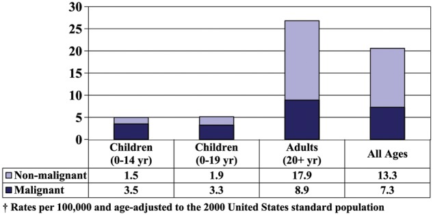
The overall average annual age-adjusted incidence rate for 2005–2009 for primary brain and CNS tumors was 20.59 per 100,000. The overall incidence rate was 5.13 per 100,000 for children 0–19 years of age (4.97 per 100,000 for children less than 15 years), and 26.81 per 100,000 for adults (20+ years). The overall incidence rates of tumors by behavior and age group (0–19 years and 20+ years) are shown in Figure 1.
Fig. 1.
Average Annual Age-Adjusted Incidence Rates† of Primary Brain and CNS Tumors by Age and Behavior.
Overall Incidence Rates by Year
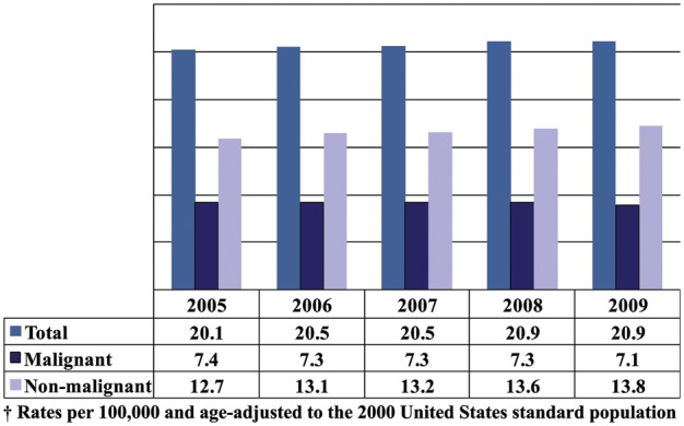
Figure 2 presents annual age-adjusted incidence rates of all primary brain and CNS tumors by behavior from 2005 through 2009. The incidence rates of all primary brain and CNS tumors during diagnostic years 2005–2009 did not differ statistically significantly from each other.
Fig. 2.
Annual Age-Adjusted Incidence Rates† of Primary Brain and CNS Tumors by Year and Behavior.
Incidence Rates by Central Cancer Registry, Age, and Behavior
The overall average annual age-adjusted incidence rates by central cancer registry, age group, and behavior are presented in Table 6. The overall average annual age-adjusted incidence rates of all primary brain and CNS tumors (malignant and non–malignant) for each individual central cancer registry ranged from 15.78 to 26.39 per 100,000. In addition, the average annual age-adjusted incidence rates of all primary malignant brain and CNS tumors ranged from 4.95 to 8.97 per 100,000, and the average annual age-adjusted incidence rates of all primary non–malignant brain and CNS tumors ranged from 8.90 to 19.02 per 100,000. Among adults 20 years of age and older, the central cancer registry–specific incidence rates ranged from 5.80 to 11.70 per 100,000 for malignant tumors and from 11.94 to 25.94 per 100,000 for non-malignant tumors. For several central cancer registries, the numbers of non-malignant tumors in those less than 20 years of age were too small to report; the highest reported incidence rate was 4.09 per 100,000 for malignant tumors and 3.61 per 100,000 for non-malignant tumors among the age group.
Table 6.
Brain and Central Nervous System Tumor Average Annual Age-Adjusted Incidence Rates† by Age, Behavior, and Central Cancer Registry, CBTRUS Statistical Report: NPCR and SEER, 2005–2009
| 0-19 Years |
20+ Years |
All Ages |
||||||||||||
|---|---|---|---|---|---|---|---|---|---|---|---|---|---|---|
| State | Malignant |
Non-Malignant |
Malignant |
Non-Malignant |
Malignant |
Non-Malignant |
All Tumors |
|||||||
| Rate | 95% CI | Rate | 95% CI | Rate | 95% CI | Rate | 95% CI | Rate | 95% CI | Rate | 95% CI | Rate | 95% CI | |
| Alabama | 2.99 | (2.57-3.45) | 0.99 | (0.76–1.26) | 8.95 | (8.51–9.40) | 13.24 | (12.70–13.79) | 7.24 | (6.90–7.58) | 9.72 | (9.34–10.13) | 16.96 | (16.45–17.49) |
| Alaska | 2.77 | (1.84–4.00) | 3.61 | (2.56–4.96) | 8.98 | (7.63–10.50) | 19.96 | (17.97–22.12) | 7.20 | (6.19–8.32) | 15.27 | (13.82–16.84) | 22.47 | (20.69–24.37) |
| Arizona | 3.23 | (2.87–3.62) | 1.59 | (1.34–1.87) | 8.67 | (8.29–9.06) | 17.10 | (16.57–17.65) | 7.11 | (6.82–7.41) | 12.65 | (12.26–13.05) | 19.76 | (19.28–20.26) |
| Arkansas | 3.42 | (2.87–4.06) | 3.51 | (2.95–4.14) | 8.63 | (8.08–9.20) | 16.18 | (15.42–16.97) | 7.13 | (6.71–7.58) | 12.55 | (11.98–13.13) | 19.68 | (18.97–20.41) |
| California | 2.95 | (2.80–3.10) | 1.74 | (1.63–1.86) | 8.29 | (8.13–8.46) | 16.79 | (16.56–17.02) | 6.76 | (6.64–6.88) | 12.47 | (12.30–12.64) | 19.23 | (19.02–19.44) |
| Colorado | 2.78 | (2.40–3.21) | 1.83 | (1.51–2.18) | 9.21 | (8.75–9.69) | 25.94 | (25.15–26.74) | 7.37 | (7.02–7.73) | 19.02 | (18.45–19.60) | 26.39 | (25.72–27.07) |
| Connecticut | 3.33 | (2.82–3.90) | 1.54 | (1.21–1.94) | 9.68 | (9.16–10.23) | 14.41 | (13.77–15.07) | 7.86 | (7.46–8.28) | 10.72 | (10.25–11.20) | 18.58 | (17.96–19.21) |
| Delaware | 3.25 | (2.29–4.48) | 2.09 | (1.34–3.11) | 9.72 | (8.68–10.85) | 15.28 | (13.98–16.68) | 7.86 | (7.07–8.73) | 11.50 | (10.54–12.52) | 19.36 | (18.10–20.68) |
| District of Columbia | 2.73 | (1.61–4.32) | – | – | 8.82 | (7.60–10.18) | 12.01 | (10.58–13.57) | 7.08 | (6.13–8.12) | 9.18 | (8.11–10.34) | 16.25 | (14.82–17.79) |
| Florida | 3.40 | (3.16–3.65) | 1.94 | (1.76–2.13) | 8.77 | (8.56–8.99) | 20.29 | (19.97–20.62) | 7.23 | (7.06–7.40) | 15.03 | (14.79–15.27) | 22.26 | (21.97–22.55) |
| Georgia | 3.04 | (2.75–3.34) | 1.59 | (1.39–1.81) | 8.44 | (8.12–8.77) | 17.41 | (16.95–17.89) | 6.89 | (6.64–7.14) | 12.87 | (12.54–13.22) | 19.76 | (19.34–20.18) |
| Hawaii | 2.85 | (2.08–3.80) | 1.48 | (0.95–2.20) | 5.80 | (5.14–6.52) | 18.27 | (17.08–19.51) | 4.95 | (4.43–5.52) | 13.45 | (12.59–14.36) | 18.40 | (17.39–19.46) |
| Idaho | 2.86 | (2.21–3.64) | 1.42 | (0.97–2.01) | 10.26 | (9.40–11.18) | 15.56 | (14.49–16.68) | 8.14 | (7.49–8.82) | 11.50 | (10.73–12.32) | 19.64 | (18.63–20.69) |
| Illinois | 2.98 | (2.73–3.24) | 1.83 | (1.64–2.04) | 8.80 | (8.53–9.08) | 18.87 | (18.47–19.27) | 7.13 | (6.93–7.34) | 13.98 | (13.69–14.27) | 21.11 | (20.76–21.47) |
| Indiana | 3.58 | (3.19–3.99) | 1.85 | (1.57–2.15) | 9.09 | (8.70–9.48) | 16.62 | (16.10–17.15) | 7.51 | (7.21–7.81) | 12.38 | (12.00–12.77) | 19.89 | (19.40–20.38) |
| Iowa | 3.30 | (2.76–3.91) | 1.57 | (1.21–2.00) | 10.11 | (9.53–10.71) | 16.90 | (16.15–17.67) | 8.16 | (7.72–8.62) | 12.50 | (11.95–13.06) | 20.66 | (19.95–21.38) |
| Kentucky | 3.43 | (2.96–3.95) | 2.33 | (1.95–2.77) | 9.46 | (8.99–9.96) | 23.68 | (22.93–24.46) | 7.73 | (7.37–8.11) | 17.56 | (17.01–18.12) | 25.29 | (24.63–25.97) |
| Louisiana | 2.97 | (2.56–3.44) | 1.68 | (1.37–2.03) | 7.78 | (7.35–8.23) | 17.32 | (16.67–17.98) | 6.40 | (6.07–6.75) | 12.83 | (12.36–13.32) | 19.23 | (18.65–19.82) |
| Maine | 3.80 | (2.89–4.91) | 1.39 | (0.87–2.12) | 9.85 | (9.03–10.73) | 12.31 | (11.37–13.31) | 8.11 | (7.47–8.80) | 9.18 | (8.49–9.91) | 17.29 | (16.34–18.28) |
| Massachusetts | 3.42 | (3.03–3.85) | 1.61 | (1.35–1.90) | 9.51 | (9.13–9.91) | 14.18 | (13.72–14.66) | 7.77 | (7.47–8.07) | 10.57 | (10.24–10.92) | 18.34 | (17.89–18.80) |
| Michigan | 3.48 | (3.17–3.81) | 1.90 | (1.68–2.15) | 9.34 | (9.03–9.66) | 18.17 | (17.74–18.61) | 7.66 | (7.42–7.90) | 13.50 | (13.19–13.82) | 21.16 | (20.76–21.56) |
| Minnesota | 2.98 | (2.59–3.42) | 1.34 | (1.09–1.64) | 8.45 | (8.03–8.87) | 11.94 | (11.45–12.44) | 6.88 | (6.56–7.21) | 8.90 | (8.54–9.27) | 15.78 | (15.30–16.27) |
| Mississippi | 2.80 | (2.32–3.35) | 1.58 | (1.23–2.00) | 8.16 | (7.62–8.73) | 16.28 | (15.51–17.08) | 6.62 | (6.21–7.05) | 12.06 | (11.51–12.64) | 18.68 | (17.99–19.40) |
| Missouri | 3.20 | (2.82–3.62) | 1.50 | (1.25–1.80) | 9.19 | (8.80–9.60) | 19.05 | (18.48–19.64) | 7.47 | (7.17–7.78) | 14.02 | (13.60–14.44) | 21.49 | (20.97–22.01) |
| Montana | 2.36 | (1.58–3.40) | 1.74 | (1.10–2.62) | 9.28 | (8.32–10.32) | 16.44 | (15.13–17.83) | 7.30 | (6.57–8.08) | 12.22 | (11.27–13.23) | 19.52 | (18.31–20.78) |
| Nebraska | 3.58 | (2.88–4.39) | 2.58 | (1.99–3.29) | 9.49 | (8.75–10.28) | 14.58 | (13.66–15.55) | 7.80 | (7.23–8.40) | 11.14 | (10.46–11.85) | 18.94 | (18.05–19.86) |
| Nevada | 2.40 | (1.91–2.96) | 0.63 | (0.39–0.95) | 8.17 | (7.58–8.78) | 14.27 | (13.49–15.10) | 6.51 | (6.07–6.98) | 10.36 | (9.79–10.95) | 16.87 | (16.15–17.61) |
| New Hampshire | 3.71 | (2.84–4.77) | 2.26 | (1.61–3.08) | 9.70 | (8.85–10.61) | 15.30 | (14.24–16.42) | 7.98 | (7.32–8.68) | 11.56 | (10.78–12.39) | 19.54 | (18.51–20.61) |
| New Jersey | 3.63 | (3.29–4.00) | 1.88 | (1.63–2.15) | 9.40 | (9.07–9.74) | 15.85 | (15.42–16.29) | 7.75 | (7.49–8.01) | 11.84 | (11.53–12.16) | 19.59 | (19.18–20.00) |
| New Mexico | 2.17 | (1.66–2.78) | 1.70 | (1.25–2.26) | 7.99 | (7.34–8.68) | 13.57 | (12.72–14.46) | 6.32 | (5.83–6.83) | 10.17 | (9.55–10.81) | 16.48 | (15.69–17.30) |
| New York | 3.57 | (3.34–3.81) | 2.37 | (2.19–2.57) | 8.93 | (8.71–9.15) | 21.07 | (20.74–21.41) | 7.39 | (7.23–7.56) | 15.71 | (15.46–15.95) | 23.10 | (22.80–23.40) |
| North Carolina | 3.20 | (2.89–3.53) | 1.91 | (1.68–2.17) | 8.78 | (8.46–9.10) | 18.03 | (17.57–18.50) | 7.18 | (6.93–7.42) | 13.41 | (13.07–13.75) | 20.58 | (20.17–21.00) |
| North Dakota | 2.96 | (1.89–4.40) | – | – | 8.90 | (7.74–10.18) | 12.05 | (10.69–13.54) | 7.19 | (6.30–8.18) | 8.95 | (7.95–10.03) | 16.14 | (14.79–17.58) |
| Ohio | 3.41 | (3.12–3.72) | 2.01 | (1.79–2.24) | 9.32 | (9.03–9.61) | 12.84 | (12.51–13.18) | 7.62 | (7.40–7.85) | 9.73 | (9.49–9.99) | 17.36 | (17.03–17.69) |
| Oklahoma | 2.92 | (2.47–3.44) | 1.42 | (1.11–1.79) | 9.44 | (8.92–9.98) | 13.32 | (12.70–13.95) | 7.57 | (7.18–7.98) | 9.90 | (9.45–10.37) | 17.47 | (16.87–18.09) |
| Oregon | 3.77 | (3.24–4.36) | 2.39 | (1.98–2.87) | 9.75 | (9.24–10.28) | 16.15 | (15.49–16.83) | 8.03 | (7.64–8.44) | 12.20 | (11.72–12.71) | 20.24 | (19.61–20.88) |
| Pennsylvania | 3.52 | (3.23–3.83) | 1.98 | (1.77–2.21) | 9.46 | (9.19–9.73) | 19.73 | (19.34–20.12) | 7.76 | (7.55–7.97) | 14.63 | (14.35–14.92) | 22.39 | (22.04–22.75) |
| Rhode Island | 3.11 | (2.23–4.23) | 1.87 | (1.22–2.75) | 8.73 | (7.85–9.69) | 17.23 | (15.98–18.56) | 7.12 | (6.44–7.86) | 12.83 | (11.91–13.80) | 19.95 | (18.80–21.15) |
| South Carolina | 3.11 | (2.67–3.59) | 1.63 | (1.32–1.98) | 8.71 | (8.27–9.17) | 16.71 | (16.08–17.35) | 7.10 | (6.76–7.46) | 12.38 | (11.93–12.85) | 19.48 | (18.91–20.07) |
| South Dakota | 2.60 | (1.74–3.74) | – | – | 8.28 | (7.27–9.39) | 13.89 | (12.58–15.30) | 6.65 | (5.88–7.49) | 10.22 | (9.26–11.24) | 16.87 | (15.64–18.17) |
| Tennessee | 3.52 | (3.13–3.95) | 2.24 | (1.93–2.59) | 8.95 | (8.57–9.34) | 19.18 | (18.62–19.76) | 7.39 | (7.10–7.70) | 14.32 | (13.91–14.74) | 21.71 | (21.20–22.23) |
| Texas | 3.41 | (3.22–3.60) | 2.28 | (2.13–2.44) | 8.66 | (8.46–8.87) | 21.80 | (21.47–22.14) | 7.16 | (7.00–7.32) | 16.20 | (15.96–16.45) | 23.36 | (23.07–23.65) |
| Utah | 4.09 | (3.53–4.72) | 1.76 | (1.39–2.19) | 8.45 | (7.81–9.13) | 20.48 | (19.47–21.53) | 7.20 | (6.71–7.71) | 15.11 | (14.38–15.86) | 22.31 | (21.43–23.21) |
| Vermont | 2.20 | (1.29–3.51) | 2.54 | (1.56–3.92) | 11.70 | (10.38–13.14) | 22.99 | (21.10–25.01) | 8.97 | (7.99–10.05) | 17.13 | (15.74–18.60) | 26.10 | (24.39–27.90) |
| Virginia | 2.97 | (2.65–3.33) | 1.19 | (0.99–1.42) | 8.13 | (7.80–8.48) | 15.41 | (14.95–15.88) | 6.65 | (6.40–6.92) | 11.33 | (10.99–11.67) | 17.98 | (17.56–18.41) |
| Washington | 3.65 | (3.26–4.08) | 2.76 | (2.42–3.14) | 9.44 | (9.05–9.85) | 24.18 | (23.55–24.82) | 7.78 | (7.48–8.09) | 18.04 | (17.58–18.51) | 25.82 | (25.27–26.38) |
| West Virginia | 3.06 | (2.36–3.89) | 1.45 | (0.99–2.05) | 9.67 | (8.98–10.41) | 16.18 | (15.27–17.13) | 7.78 | (7.24–8.35) | 11.95 | (11.29–12.65) | 19.73 | (18.87–20.62) |
| Wisconsin | 3.67 | (3.24–4.13) | 1.64 | (1.36–1.95) | 10.02 | (9.60–10.46) | 16.99 | (16.43–17.56) | 8.20 | (7.87–8.54) | 12.59 | (12.18–13.00) | 20.78 | (20.26–21.32) |
| Wyoming | 2.39 | (1.39–3.82) | – | – | 8.79 | (7.49–10.25) | 15.23 | (13.52–17.09) | 6.95 | (5.97–8.05) | 11.37 | (10.12–12.72) | 18.32 | (16.72–20.03) |
† Rates are per 100,000 and are age–adjusted to the 2000 United States standard population.
Abbreviations: CBTRUS, Central Brain Tumor Registry of the United States; NPCR, CDC's National Program of Cancer Registries; SEER, NCI's Surveillance, Epidemiology and End Results program; CI, confidence interval; NOS, not otherwise specified.
It is apparent that there is less variation by state in malignant tumor incidence rates as compared to incidence rates for tumors of non-malignant behavior, suggesting greater consistency in reporting of the malignant tumors. The central cancer registry and regional variations apparent in Table 6, especially in reported incidence rates for the non–malignant tumors, likely reflects differences in reporting practices including case ascertainment. Improvements in standardization of brain and CNS tumor collection and reporting with time will allow observation of the true variation in the incidence of brain and CNS tumors among states. Many non–malignant brain and CNS tumors are not histologically confirmed, ie the percent of diagnostically confirmed non-malignant tumors is lower than the percent of diagnostically confirmed malignant tumors. A statistically significant negative correlation exists between the proportion of tumors with non–malignant behavior and the proportion of tumors diagnostically confirmed by central cancer registry for the data presented in Table 5. Conversely, a statistically significant positive correlation is evident for the proportion of non-malignant tumors with the proportion of radiographic diagnostic confirmations.
Primary Brain and CNS Tumors: Incidence by Site, Histology, Age, Gender, Race, and Hispanic Ethnicity
Distribution of Tumors by Site and Histology
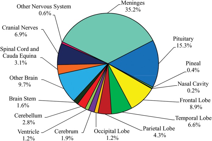
The distribution of brain and CNS tumors by site is shown in Figure 3. The most common tumor site is the meninges (35%). Twenty-one percent of tumors are located within the frontal, temporal, parietal, and occipital lobes of the brain. Cerebrum, ventricle, cerebellum, and brain stem tumors account for 7.5% of all tumors. The cranial nerves and the spinal cord/cauda equina account for 10% of all tumors. Together, the pituitary and pineal glands account for about 16% of tumors. Olfactory tumors of the nasal cavity account for less than 1% of tumors.
Fig. 3.
Distribution of Primary Brain and CNS Tumors by Site (N = 311,202).
The distribution by brain and CNS histology is shown in Figure 4. The most frequently reported histology is the predominately non–malignant meningioma, which accounts for more than one-third of all tumors, followed by glioblastoma, a malignant brain tumor. Tumors of the pituitary and nerve sheath tumors combined account for about one-fourth of all tumors, the majority of which are non-malignant. Acoustic neuromas (defined by ICD-O-3 site code C72.4 and histology code 9560) account for 65% of all nerve sheath tumors (data not shown).
Fig. 4.
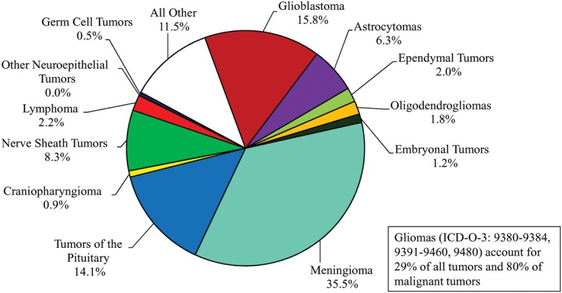
Distribution of Primary Brain and CNS Tumors by Histology (N = 311,202).
The broad category glioma represents approximately 30% of all tumors (Figure 4). The distribution of tumors by site for glioma is shown in Figure 5. About 60% of gliomas occur in the four lobes of the brain.
Fig. 5.
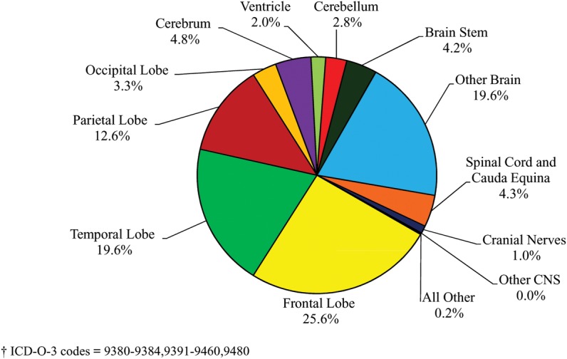
Distribution of Primary Brain and CNS Gliomas† by Site (N = 90,828).
The distribution by specific histology for glioma is illustrated in Figure 6. Glioblastoma accounts for the majority of gliomas, while astrocytoma and glioblastoma combined account for about three–fourths of gliomas.
Fig. 6.
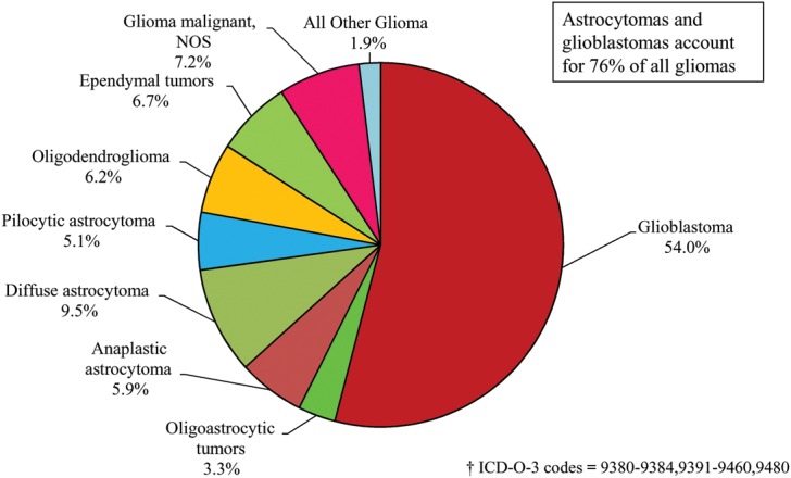
Distribution of Primary Brain and CNS Gliomas† by Histology Subtypes (N = 90,828).
Incidence of Spinal Cord Tumors
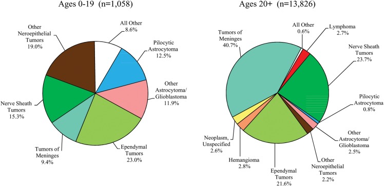
Spinal Cord Tumors are a special group of CNS tumors located in the spinal cord, spinal meninges, and cauda equina. Although these tumors account for a relatively small percentage of all brain and CNS tumors, they result in significant morbidity and are highlighted in this report. The most prevalent histologies found in the spinal cord, spinal meninges, and cauda equina are presented in Figure 7 for both children (0-19 years) and adults (20+ years). For the age group 0-19 years, the predominant histology is ependymal tumors followed by other neuroepithelial tumors, whereas tumors of the meninges account for the largest proportion of histologies among those ages 20 years and older.
Fig. 7.
Distribution of Spinal Cord, Spinal Meninges and Cauda Equina Tumors by Age Group and Histology.
Distribution of Tumors by Site and Histology in Young Adults (Ages 20-34 Years)
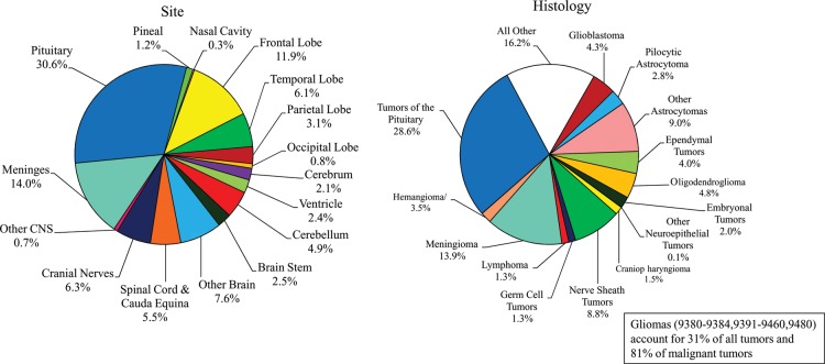
Almost 9% of all brain and CNS tumors occurred in young adults, ages 20-34 years and the distribution of these tumors by site is shown in Figure 8. Approximately 22% of tumors diagnosed in young adults are located within the frontal, temporal, parietal and occipital lobes of the brain. Cerebrum, ventricle, cerebellum and brain stem tumors combined account for about 12% of all young adult tumors. Tumors of the meninges represent 14%, while the cranial nerves and the spinal cord and cauda equina combined account for about 12%. Tumors located in the pituitary and pineal glands together account for about 32% of young adult tumors. The distribution by histology for young adults is also shown in Figure 8. Over half of reported histologies for tumors diagnosed in those 20-34 years of age are the predominately non–malignant tumors of the pituitary (29%), meningioma (14%), and nerve sheath (9%). The broad category glioma accounts for 31% of all brain and CNS tumors and about 81% of malignant tumors in young adults.
Fig. 8.
Distribution of Primary Brain and CNS Tumors by Site and Histology in Young Adults (Ages 20-34 years) (N = 26,616)
Incidence Rates by Site and Gender
Incidence counts and average annual age-adjusted rates for brain and CNS tumors by site and gender are provided in Table 7. Incidence rates were highest for tumors located in the meninges (7.18 per 100,000), followed by tumors located in the four lobes of the brain, pituitary, other areas of the brain, cranial nerves, spinal cord/cauda equina, cerebellum, cerebrum, brain stem, ventricle, other nervous system and pineal gland. Incidence rates were lowest for olfactory tumors of the nasal cavity (0.04 per 100,000). By gender, incidence rates were statistically significantly higher in females than in males for tumors located in the meninges, pituitary, and cranial nerves. Males had statistically significantly higher incidence rates of tumors located in the four lobes of the brain, cerebrum, ventricle, cerebellum, brain stem, other brain, spinal cord and cauda equina, other nervous system, pineal and olfactory tumors of the nasal cavity compared to females.
Table 7.
Brain and Central Nervous System Tumor Average Annual Age-Adjusted Incidence Rates† by Site‡ and Gender, CBTRUS Statistical Report: NPCR and SEER, 2005–2009
| ICD-O-3 Code | Site | Total |
Male |
Female |
||||||
|---|---|---|---|---|---|---|---|---|---|---|
| N | Adjusted Rate | 95% CI | N | Adjusted Rate | 95% CI | N | Adjusted Rate | 95% CI | ||
| C71.1-C71.4 | Frontal, temporal, parietal, & occipital lobes of the brain | 65,532 | 4.33 | (4.29–4.36) | 36,171 | 5.13 | (5.08–5.18) | 29,361 | 3.65 | (3.60–3.69) |
| C71.0 | Cerebrum | 6,007 | 0.40 | (0.39–0.41) | 3,148 | 0.45 | (0.43–0.46) | 2,859 | 0.37 | (0.35–0.38) |
| C71.5 | Ventricle | 3,639 | 0.25 | (0.24–0.26) | 1,993 | 0.28 | (0.26–0.29) | 1,646 | 0.22 | (0.21–0.24) |
| C71.6 | Cerebellum | 8,601 | 0.59 | (0.58–0.60) | 4,655 | 0.65 | (0.63–0.67) | 3,946 | 0.53 | (0.52–0.55) |
| C71.7 | Brain stem | 4,924 | 0.34 | (0.33–0.35) | 2,606 | 0.36 | (0.35–0.38) | 2,318 | 0.32 | (0.31–0.33) |
| C71.8–C71.9 | Other brain | 30,334 | 2.00 | (1.98–2.03) | 15,763 | 2.28 | (2.25–2.32) | 14,571 | 1.77 | (1.74–1.80) |
| C72.0–C72.1 | Spinal cord and cauda equina | 9,538 | 0.64 | (0.63–0.65) | 4,867 | 0.67 | (0.65–0.69) | 4,671 | 0.61 | (0.59–0.62) |
| C72.2–C72.5 | Cranial nerves | 21,489 | 1.41 | (1.39–1.43) | 10,055 | 1.38 | (1.35–1.41) | 11,434 | 1.44 | (1.42–1.47) |
| C72.8–C72.9 | Other nervous system | 1,902 | 0.13 | (0.12–0.13) | 1,013 | 0.14 | (0.13–0.15) | 889 | 0.11 | (0.11–0.12) |
| C70.0–C70.9 | Meninges (cerebral & spinal) | 109,660 | 7.18 | (7.14–7.22) | 28,856 | 4.27 | (4.22–4.32) | 80,804 | 9.68 | (9.61–9.75) |
| C75.1–C75.2 | Pituitary | 47,614 | 3.19 | (3.16–3.22) | 21,473 | 3.02 | (2.98–3.06) | 26,141 | 3.45 | (3.40–3.49) |
| C75.3 | Pineal | 1,391 | 0.10 | (0.09–0.10) | 831 | 0.11 | (0.11–0.12) | 560 | 0.08 | (0.07–0.08) |
| C30.0 (9522–9523) | Olfactory tumors of the nasal cavity | 571 | 0.04 | (0.03–0.04) | 339 | 0.05 | (0.04–0.05) | 232 | 0.03 | (0.03–0.03) |
| Total | 311,202 | 20.59 | (20.52–20.66) | 131,770 | 18.80 | (18.69–18.90) | 179,432 | 22.25 | (22.15–22.35) | |
† Rates are per 100,000 and are age adjusted to the 2000 US standard population.
‡ The sites referred to in this table are loosely based on the categories and site codes defined in the SEER site/histology validation list.
Abbreviations: CBTRUS, Central Brain Tumor Registry of the United States; NPCR, CDC's National Program of Cancer Registries; SEER, NCI's Surveillance, Epidemiology and End Results program; CI, confidence interval.
Incidence Rates by Major Histology Groupings and Specific Histologies
Tables 8 through 16 present incidence rates by major histology groupings and specific histologies. Among major histology groupings, incidence rates were highest for tumors of the meninges (7.49 per 100,000), followed by tumors of the neuroepithelial tissue (6.60 per 100,000 person–years), tumors of the sellar region (3.12 per 100,000) and tumors of the cranial and spinal nerves (1.70 per 100,000) (Table 8).
Table 8.
Distribution and Average Annual Age-Adjusted Incidence Rates† of Brain and Central Nervous System Tumors by Major Histology Groupings and Histology, CBTRUS Statistical Report: NPCR and SEER, 2005–2009
| Total |
Malignant |
Non-malignant |
|||||||||
|---|---|---|---|---|---|---|---|---|---|---|---|
| Histology | N | % of All Tumors | Median Age | Rate | (95% CI) | N | Rate | (95% CI) | N | Rate | (95% CI) |
| Tumors of Neuroepithelial Tissue | 99,063 | 31.8 | 55 | 6.60 | (6.56–6.64) | 92,418 | 6.14 | (6.10–6.18) | 6,645 | 0.46 | (0.45–0.47) |
| Pilocytic astrocytoma | 4,636 | 1.5 | 13 | 0.33 | (0.32–0.34) | 4,636 | 0.33 | (0.32–0.34) | – | – | – |
| Diffuse astrocytoma | 8,616 | 2.8 | 48 | 0.58 | (0.57–0.60) | 8,616 | 0.58 | (0.57–0.60) | – | – | – |
| Anaplastic astrocytoma | 5,374 | 1.7 | 54 | 0.36 | (0.35–0.37) | 5,374 | 0.36 | (0.35–0.37) | – | – | – |
| Unique astrocytoma variants | 938 | 0.3 | 22 | 0.07 | (0.06–0.07) | 613 | 0.04 | (0.04–0.05) | 325 | 0.02 | (0.02–0.03) |
| Glioblastoma | 49,088 | 15.8 | 64 | 3.19 | (3.16–3.22) | 49,088 | 3.19 | (3.16–3.22) | – | – | – |
| Oligodendroglioma | 3,973 | 1.3 | 43 | 0.27 | (0.26–0.28) | 3,973 | 0.27 | (0.26–0.28) | – | – | – |
| Anaplastic oligodendroglioma | 1,687 | 0.5 | 49 | 0.11 | (0.11–0.12) | 1,685 | 0.11 | (0.11–0.12) | – | – | – |
| Oligoastrocytic tumors | 3,020 | 1.0 | 42 | 0.21 | (0.20–0.21) | 3,019 | 0.21 | (0.20–0.21) | – | – | – |
| Ependymal tumors | 6,117 | 2.0 | 43 | 0.41 | (0.40–0.42) | 3,906 | 0.26 | (0.26–0.27) | 2,211 | 0.15 | (0.14–0.16) |
| Glioma malignant, NOS | 6,574 | 2.1 | 40 | 0.45 | (0.44–0.46) | 6,574 | 0.45 | (0.44–0.46) | – | – | – |
| Choroid plexus tumors | 772 | 0.2 | 19 | 0.05 | (0.05–0.06) | 133 | 0.01 | (0.01–0.01) | 639 | 0.04 | (0.04–0.05) |
| Other neuroepithelial tumors | 95 | 0.0 | 38 | 0.01 | (0.01–0.01) | 59 | 0.00 | (0.00–0.01) | 36 | 0.00 | (0.00–0.00) |
| Neuronal and mixed neuronal–glial tumors | 3,887 | 1.2 | 27 | 0.27 | (0.26–0.28) | 806 | 0.05 | (0.05–0.06) | 3,081 | 0.21 | (0.21–0.22) |
| Tumors of the pineal region | 579 | 0.2 | 34 | 0.04 | (0.04–0.04) | 324 | 0.02 | (0.02–0.03) | 255 | 0.02 | (0.02–0.02) |
| Embryonal tumors | 3,707 | 1.2 | 8 | 0.26 | (0.25–0.27) | 3,612 | 0.25 | (0.24–0.26) | 95 | 0.01 | (0.01–0.01) |
| Tumors of Cranial and Spinal Nerves | 25,942 | 8.3 | 54 | 1.70 | (1.68–1.73) | 241 | 0.02 | (0.01–0.02) | 25,701 | 1.69 | (1.67–1.71) |
| Nerve sheath tumors | 25,926 | 8.3 | 54 | 1.70 | (1.68–1.72) | 241 | 0.02 | (0.01–0.02) | 25,685 | 1.69 | (1.67–1.71) |
| Other tumors of cranial and spinal nerves | 16 | 0.0 | 58 | 0.00 | (0.00–0.00) | – | – | – | 16 | 0.00 | (0.00–0.00) |
| Tumors of Meninges | 114,363 | 36.7 | 65 | 7.49 | (7.45–7.54) | 2,587 | 0.17 | (0.16–0.18) | 111,776 | 7.32 | (7.28–7.37) |
| Meningioma | 110,359 | 35.5 | 65 | 7.22 | (7.18–7.27) | 1,878 | 0.12 | (0.12–0.13) | 108,481 | 7.10 | (7.06–7.14) |
| Mesenchymal tumors | 1,192 | 0.4 | 47 | 0.08 | (0.08–0.09) | 374 | 0.03 | (0.02–0.03) | 818 | 0.06 | (0.05–0.06) |
| Primary melanocytic lesions | 106 | 0.0 | 50 | 0.01 | (0.01–0.01) | 71 | 0.01 | (0.00–0.01) | 35 | 0.00 | (0.00–0.00) |
| Other neoplasms related to the meninges | 2,706 | 0.9 | 48 | 0.18 | (0.17–0.19) | 264 | 0.02 | (0.02–0.02) | 2,442 | 0.16 | (0.16–0.17) |
| Lymphomas and Hemopoietic Neoplasms | 6,956 | 2.2 | 64 | 0.46 | (0.45–0.47) | 6,920 | 0.46 | (0.45–0.47) | 36 | 0.00 | (0.00–0.00) |
| Lymphoma | 6,774 | 2.2 | 65 | 0.45 | (0.44–0.46) | 6,773 | 0.45 | (0.44–0.46) | – | – | – |
| Other hemopoietic neoplasms | 182 | 0.1 | 51 | 0.01 | (0.01–0.01) | 147 | 0.01 | (0.01–0.01) | 35 | 0.00 | (0.00–0.00) |
| Germ Cell Tumors and Cysts | 1,418 | 0.5 | 17 | 0.10 | (0.09–0.10) | 947 | 0.07 | (0.06–0.07) | 471 | 0.03 | (0.03–0.04) |
| Germ cell tumors, cysts and heterotopias | 1,418 | 0.5 | 17 | 0.10 | (0.09–0.10) | 947 | 0.07 | (0.06–0.07) | 471 | 0.03 | (0.03–0.04) |
| Tumors of Sellar Region | 46,562 | 15.0 | 50 | 3.12 | (3.09–3.15) | 140 | 0.01 | (0.01–0.01) | 46,422 | 3.11 | (3.08–3.14) |
| Tumors of the pituitary | 43,882 | 14.1 | 51 | 2.94 | (2.91–2.97) | 133 | 0.01 | (0.01–0.01) | 43,749 | 2.93 | (2.90–2.96) |
| Craniopharyngioma | 2,680 | 0.9 | 41 | 0.18 | (0.18–0.19) | – | – | – | 2,673 | 0.18 | (0.18–0.19) |
| Unclassified Tumors | 16,898 | 5.4 | 65 | 1.12 | (1.10–1.13) | 6,442 | 0.42 | (0.41–0.43) | 10,456 | 0.70 | (0.68–0.71) |
| Hemangioma | 3,240 | 1.0 | 49 | 0.22 | (0.21–0.23) | 20 | 0.00 | (0.00–0.00) | 3,220 | 0.22 | (0.21–0.22) |
| Neoplasm, unspecified | 13,566 | 4.4 | 71 | 0.89 | (0.88–0.91) | 6,402 | 0.42 | (0.41–0.43) | 7,164 | 0.47 | (0.46–0.49) |
| All other | 92 | 0.0 | 61 | 0.01 | (0.01–0.01) | 20 | 0.00 | (0.00–0.00) | 72 | 0.01 | (0.00–0.01) |
| Total‡ | 311,202 | 100.0 | 59 | 20.59 | (20.52–20.66) | 109,695 | 7.28 | (7.24–7.33) | 201,507 | 13.31 | (13.25–13.37) |
† Rates are per 100,000 and are age–adjusted to the 2000 US standard population.
‡ Refers to all brain tumors including histologies not presented in this table.
– Counts are not presented when fewer than 16 cases were reported for the specific histology category. The suppressed cases are included in the counts for totals.
Abbreviations: CBTRUS, Central Brain Tumor Registry of the United States; NPCR, CDC's National Program of Cancer Registries; SEER, NCI's Surveillance, Epidemiology and End Results program; CI, confidence interval; NOS, not otherwise specified.
Incidence rates also varied by specific brain and CNS histology (Table 8). Incidence rates were highest for meningiomas (7.22 per 100,000), glioblastomas (3.19 per 100,000), tumors of the pituitary (2.94 per 100,000), and nerve sheath tumors (1.70 per 100,000). The incidence rate for glioma was 6.03 per 100,000, a major contributor to the magnitude of the neuroepithelial tissue rate (data not shown). Acoustic neuromas, included under tumors of cranial and spinal nerves, comprise the majority (65%; 1.10 per 100,000) of nerve sheath tumors (1.70 per 100,000) and account for 5% of all primary brain and CNS tumors (data not shown).
Incidence Rates by Behavior and Histology
Brain and CNS tumor incidence rates by behavior (malignant and non-malignant) are presented in Table 8. For those with malignant behavior, the incidence rate was highest for glioblastoma (3.19 per 100,000) followed by diffuse astrocytoma (0.58 per 100,000), and lymphoma (0.45 per 100,000. Meningioma (7.10 per 100,000), tumors of the pituitary (2.93 per 100,000), and nerve sheath (1.69 per 100,000) tumors were the non-malignant histologies with the highest incidence rates.
Median Age at Diagnosis
The median age at diagnosis for all primary brain and CNS tumors is 59 years (Table 8). The histology-specific median ages range from 8 to 71 years. Pilocytic astrocytoma, choroid plexus tumors, neuronal and mixed neuronal-glial tumors, tumors of the pineal region, embryonal tumors, and germ cell tumors and cysts are histologies with younger median age at diagnosis onset. Meningioma and glioblastoma are primarily diagnosed at older ages. Unclassified tumors have a median age of 65 years suggesting that younger individuals may receive more specific tumor identification and classification.
Incidence Rates by Gender and Histology
Incidence rates by histology and gender are presented in Table 9. Incidence rates for all primary brain and CNS tumors combined are higher among females (22.25 per 100,000 person–years) than males (18.80 per 100,000 person–years). The difference between these incidence rates is statistically significant. Incidence rates for tumors of the neuroepithelial tissue are 1.4 times greater in males as compared to females, while tumors of the meninges are 2.2 times greater in females as compared to males. Incidence rates for tumors of the neuroepithelial and tumors of the meninges were statistically significantly different between males and females. The incidence rate of gliomas is higher in males (7.16 per 100,000 person–years) than in females (5.06 per 100,000 person–years). Similar patterns were found for individual histologies with incidence rates higher in males, especially for germ cell tumors, most glial tumors, lymphomas, and embryonal tumors, or comparable between males and females, with the notable exception of meningiomas and tumors of the pituitary, which are more common in women. Incidence rate ratios (male: female) for selected histologies are shown in Figure 9.
Table 9.
Brain and Central Nervous System Tumor Average Annual Age–Adjusted Incidence Rates† by Major Histology Groupings, Histology and Gender, CBTRUS Statistical Report: NPCR and SEER, 2005–2009
| Histology | Total |
Male |
Female |
|||
|---|---|---|---|---|---|---|
| Rate | 95% CI | Rate | 95% CI | Rate | 95% CI | |
| Tumors of Neuroepithelial Tissue | 6.60 | (6.56–6.64) | 7.77 | (7.70–7.84) | 5.59 | (5.54–5.64) |
| Pilocytic astrocytoma | 0.33 | (0.32–0.34) | 0.33 | (0.32–0.34) | 0.32 | (0.31–0.34) |
| Diffuse astrocytoma | 0.58 | (0.57–0.60) | 0.68 | (0.66–0.70) | 0.50 | (0.48–0.52) |
| Anaplastic astrocytoma | 0.36 | (0.35–0.37) | 0.43 | (0.42–0.45) | 0.30 | (0.28–0.31) |
| Unique astrocytoma variants | 0.07 | (0.06–0.07) | 0.07 | (0.06–0.08) | 0.06 | (0.06–0.07) |
| Glioblastoma | 3.19 | (3.16–3.22) | 3.98 | (3.94–4.03) | 2.53 | (2.49–2.56) |
| Oligodendroglioma | 0.27 | (0.26–0.28) | 0.30 | (0.29–0.31) | 0.24 | (0.23–0.25) |
| Anaplastic oligodendroglioma | 0.11 | (0.11–0.12) | 0.13 | (0.12–0.14) | 0.10 | (0.09–0.11) |
| Oligoastrocytic tumors | 0.21 | (0.20–0.21) | 0.24 | (0.23–0.25) | 0.18 | (0.17–0.19) |
| Ependymal tumors | 0.41 | (0.40–0.42) | 0.46 | (0.45–0.48) | 0.37 | (0.35–0.38) |
| Glioma malignant, NOS | 0.45 | (0.44–0.46) | 0.48 | (0.46–0.49) | 0.43 | (0.41–0.44) |
| Choroid plexus tumors | 0.05 | (0.05–0.06) | 0.05 | (0.05–0.06) | 0.05 | (0.05–0.06) |
| Other neuroepithelial tumors | 0.01 | (0.01–0.01) | 0.01 | (0.00–0.01) | 0.01 | (0.01–0.01) |
| Neuronal and mixed neuronal–glial tumors | 0.27 | (0.26–0.28) | 0.29 | (0.28–0.30) | 0.25 | (0.24–0.26) |
| Tumors of the pineal region | 0.04 | (0.04–0.04) | 0.03 | (0.03–0.04) | 0.05 | (0.04–0.05) |
| Embryonal tumors | 0.26 | (0.25–0.27) | 0.29 | (0.28–0.31) | 0.22 | (0.21–0.24) |
| Tumors of Cranial and Spinal Nerves | 1.70 | (1.68–1.73) | 1.70 | (1.67–1.73) | 1.71 | (1.69–1.74) |
| Nerve sheath tumors | 1.70 | (1.68–1.72) | 1.70 | (1.67–1.73) | 1.71 | (1.68–1.74) |
| Other tumors of cranial and spinal nerves | 0.00 | (0.00–0.00) | – | – | – | – |
| Tumors of Meninges | 7.49 | (7.45–7.54) | 4.58 | (4.53–4.63) | 10.00 | (9.93–10.07) |
| Meningioma | 7.22 | (7.18–7.27) | 4.28 | (4.23–4.33) | 9.76 | (9.69–9.83) |
| Mesenchymal tumors | 0.08 | (0.08–0.09) | 0.08 | (0.08–0.09) | 0.08 | (0.07–0.09) |
| Primary melanocytic lesions | 0.01 | (0.01–0.01) | 0.01 | (0.01–0.01) | 0.01 | (0.00–0.01) |
| Other neoplasms related to the meninges | 0.18 | (0.17–0.19) | 0.21 | (0.20–0.22) | 0.16 | (0.15–0.17) |
| Lymphomas and Hemopoietic Neoplasms | 0.46 | (0.45–0.47) | 0.54 | (0.52–0.55) | 0.40 | (0.38–0.41) |
| Lymphoma | 0.45 | (0.44–0.46) | 0.52 | (0.51–0.54) | 0.39 | (0.37–0.40) |
| Other hemopoietic neoplasms | 0.01 | (0.01–0.01) | 0.01 | (0.01–0.02) | 0.01 | (0.01–0.01) |
| Germ Cell Tumors and Cysts | 0.10 | (0.09–0.10) | 0.13 | (0.12–0.14) | 0.06 | (0.06–0.07) |
| Germ cell tumors, cysts and heterotopias | 0.10 | (0.09–0.10) | 0.13 | (0.12–0.14) | 0.06 | (0.06–0.07) |
| Tumors of Sellar Region | 3.12 | (3.09–3.15) | 2.96 | (2.92–3.00) | 3.36 | (3.32–3.41) |
| Tumors of the pituitary | 2.94 | (2.91–2.97) | 2.78 | (2.74–2.81) | 3.18 | (3.14–3.22) |
| Craniopharyngioma | 0.18 | (0.18–0.19) | 0.18 | (0.17–0.19) | 0.18 | (0.17–0.19) |
| Unclassified Tumors | 1.12 | (1.10–1.13) | 1.12 | (1.10–1.15) | 1.12 | (1.10–1.14) |
| Hemangioma | 0.22 | (0.21–0.23) | 0.20 | (0.19–0.21) | 0.23 | (0.22–0.25) |
| Neoplasm, unspecified | 0.89 | (0.88–0.91) | 0.92 | (0.89–0.94) | 0.88 | (0.86–0.90) |
| All other | 0.01 | (0.01–0.01) | 0.01 | (0.01–0.01) | 0.01 | (0.00–0.01) |
| Total‡ | 20.59 | (20.52–20.66) | 18.80 | (18.69–18.90) | 22.25 | (22.15–22.35) |
† Rates are per 100,000 and are age–adjusted to the 2000 US standard population.
‡ Refers to all brain tumors including histologies not presented in this table.
– Counts are not presented when fewer than 16 cases were reported for the specific histology category. Suppressed cases are included in the total count.
Abbreviations: CBTRUS, Central Brain Tumor Registry of the United States; NPCR, CDC's National Program of Cancer Registries; SEER, NCI's Surveillance, Epidemiology and End Results program; CI, confidence interval; NOS, not otherwise specified.
Fig. 9.
Patterns by Gender for Selected Histologies.
Incidence Rates by Race and Histology
Incidence rates by histology and race are shown in Table 10. Incidence rates for all primary brain and CNS tumors combined are substantially and statistically significantly lower for race groups AIAN (13.15 per 100,000) and API (12.98 per 100,000) compared with whites (20.61 per 100,000) and blacks (20.12 per 100,000). Incidence rates for most histologies are statistically significantly higher for whites than black, AIAN, and API race groups. An exception is observed for meningioma, tumors of the pituitary, and craniopharyngioma where the rates for blacks significantly exceed those observed for white, AIAN, and API races. It should also be noted that the average annual incidence rate for tumors of the cranial and spinal nerves in the API group is statistically significantly higher than those rates observed for black or AIAN races.
Table 10.
Brain and Central Nervous System Tumor Average Annual Age–Adjusted Incidence Rates† by Major Histology Groupings, Histology and Race, CBTRUS Statistical Report: NPCR and SEER, 2005–2009
| Histology | White |
Black |
AIAN |
API |
||||
|---|---|---|---|---|---|---|---|---|
| Rate | 95% CI | Rate | 95% CI | Rate | 95% CI | Rate | 95% CI | |
| Tumors of Neuroepithelial Tissue | 7.07 | (7.02–7.12) | 3.80 | (3.71–3.90) | 3.56 | (3.24–3.90) | 2.71 | (2.59–2.84) |
| Pilocytic astrocytoma | 0.35 | (0.34–0.36) | 0.23 | (0.21–0.25) | 0.22 | (0.15–0.31) | 0.14 | (0.12–0.18) |
| Diffuse astrocytoma | 0.63 | (0.61–0.64) | 0.33 | (0.31–0.36) | 0.46 | (0.36–0.60) | 0.25 | (0.21–0.29) |
| Anaplastic astrocytoma | 0.39 | (0.38–0.40) | 0.17 | (0.15–0.19) | 0.21 | (0.14–0.30) | 0.16 | (0.13–0.20) |
| Unique astrocytoma variants | 0.07 | (0.06–0.07) | 0.06 | (0.05–0.07) | – | – | – | – |
| Glioblastoma | 3.44 | (3.40–3.47) | 1.67 | (1.61–1.74) | 1.50 | (1.28–1.74) | 1.07 | (0.99–1.16) |
| Oligodendroglioma | 0.30 | (0.29–0.31) | 0.13 | (0.11–0.15) | 0.13 | (0.08–0.20) | 0.12 | (0.09–0.15) |
| Anaplastic oligodendroglioma | 0.12 | (0.12–0.13) | 0.05 | (0.04–0.06) | – | – | 0.06 | (0.04–0.08) |
| Oligoastrocytic tumors | 0.23 | (0.22–0.24) | 0.09 | (0.08–0.11) | 0.13 | (0.08–0.21) | 0.11 | (0.09–0.14) |
| Ependymal tumors | 0.44 | (0.43–0.45) | 0.25 | (0.23–0.28) | 0.24 | (0.17–0.33) | 0.20 | (0.17–0.24) |
| Glioma malignant, NOS | 0.46 | (0.45–0.48) | 0.35 | (0.32–0.38) | 0.22 | (0.15–0.31) | 0.25 | (0.21–0.29) |
| Choroid plexus tumors | 0.06 | (0.05–0.06) | 0.03 | (0.02–0.04) | – | – | 0.03 | (0.02–0.04) |
| Other neuroepithelial tumors | 0.01 | (0.01–0.01) | – | – | – | – | – | – |
| Neuronal and mixed neuronal–glial tumors | 0.28 | (0.27–0.29) | 0.20 | (0.18–0.22) | 0.14 | (0.09–0.21) | 0.15 | (0.12–0.18) |
| Tumors of the pineal region | 0.04 | (0.03–0.04) | 0.05 | (0.04–0.06) | – | – | – | – |
| Embryonal tumors | 0.28 | (0.27–0.29) | 0.18 | (0.16–0.20) | 0.13 | (0.08–0.20) | 0.15 | (0.12–0.18) |
| Tumors of Cranial and Spinal Nerves | 1.78 | (1.76–1.81) | 0.78 | (0.74–0.83) | 0.75 | (0.61–0.92) | 1.25 | (1.16–1.33) |
| Nerve sheath tumors | 1.78 | (1.76–1.81) | 0.78 | (0.74–0.83) | 0.75 | (0.61–0.92) | 1.24 | (1.16–1.33) |
| Other tumors of cranial and spinal nerves | – | – | – | – | – | – | – | – |
| Tumors of Meninges | 7.28 | (7.23–7.33) | 8.75 | (8.60–8.91) | 5.05 | (4.63–5.50) | 5.61 | (5.42–5.81) |
| Meningioma | 7.00 | (6.96–7.05) | 8.55 | (8.40–8.70) | 4.86 | (4.44–5.30) | 5.41 | (5.22–5.61) |
| Mesenchymal tumors | 0.08 | (0.08–0.09) | 0.06 | (0.05–0.08) | – | – | 0.06 | (0.04–0.08) |
| Primary melanocytic lesions | 0.01 | (0.01–0.01) | – | – | – | – | – | – |
| Other neoplasms related to the meninges | 0.19 | (0.18–0.19) | 0.14 | (0.12–0.16) | 0.12 | (0.07–0.19) | 0.14 | (0.11–0.17) |
| Lymphomas and Hemopoietic Neoplasms | 0.46 | (0.44–0.47) | 0.41 | (0.38–0.45) | 0.31 | (0.22–0.43) | 0.41 | (0.36–0.47) |
| Lymphoma | 0.44 | (0.43–0.46) | 0.40 | (0.37–0.43) | 0.29 | (0.20–0.41) | 0.40 | (0.35–0.46) |
| Other hemopoietic neoplasms | 0.01 | (0.01–0.01) | – | – | – | – | – | – |
| Germ Cell Tumors and Cysts | 0.10 | (0.09–0.11) | 0.07 | (0.06–0.08) | – | – | 0.13 | (0.11–0.16) |
| Germ cell tumors, cysts and heterotopias | 0.10 | (0.09–0.11) | 0.07 | (0.06–0.08) | – | – | 0.13 | (0.11–0.16) |
| Tumors of Sellar Region | 2.81 | (2.78–2.84) | 5.17 | (5.06–5.28) | 2.53 | (2.27–2.82) | 2.31 | (2.19–2.43) |
| Tumors of the pituitary | 2.64 | (2.61–2.67) | 4.90 | (4.80–5.01) | 2.39 | (2.13–2.66) | 2.17 | (2.06–2.29) |
| Craniopharyngioma | 0.17 | (0.16–0.18) | 0.26 | (0.24–0.29) | 0.14 | (0.09–0.23) | 0.14 | (0.11–0.17) |
| Unclassified Tumors | 1.11 | (1.09–1.13) | 1.13 | (1.08–1.19) | 0.90 | (0.73–1.10) | 0.56 | (0.50–0.63) |
| Hemangioma | 0.23 | (0.22–0.23) | 0.15 | (0.13–0.17) | 0.11 | (0.07–0.18) | 0.16 | (0.13–0.19) |
| Neoplasm, unspecified | 0.88 | (0.86–0.90) | 0.98 | (0.93–1.04) | 0.76 | (0.60–0.95) | 0.40 | (0.35–0.46) |
| All other | 0.01 | (0.01–0.01) | – | – | – | – | – | – |
| Total‡ | 20.61 | (20.53–20.69) | 20.12 | (19.90–20.35) | 13.15 | (12.50–13.82) | 12.98 | (12.70–13.27) |
† Rates are per 100,000 and are age–adjusted to the 2000 US standard population.
‡ Refers to all brain tumors including histologies not presented in this table.
– Counts and rates are not presented when fewer than 16 cases were reported for the specific histology category. The suppressed cases are included in the counts and rates for totals.
Abbreviations: CBTRUS, Central Brain Tumor Registry of the United States; NPCR, CDC's National Program of Cancer Registries; SEER, NCI's Surveillance, Epidemiology and End Results program; CI, confidence interval; NOS, not otherwise specified; AIAN, American Indian/Alaskan Native; API, Asian Pacific Islander.
Incidence rate ratios (white: black) for selected histologies are shown in Figure 10. Incidence rates for anaplastic astrocytoma, glioblastoma, oligodendroglioma, oligoastrocytic tumors, and nerve sheath tumors are two or more times greater in whites than in blacks. Incidence rates for pilocytic astrocytoma, ependymal tumors, embryonal tumors, lymphoma and germ cell tumors also are significantly higher among whites than blacks. In contrast, incidence rates for meningioma and tumors of the pituitary are statistically significantly higher among blacks than whites.
Fig. 10.
Patterns by Race for Selected Histologies.
Incidence Rates by Hispanic Ethnicity and Histology
Incidence rates by Hispanic ethnicity and histology are shown in Table 11. The overall incidence rate for primary brain and CNS tumors among Hispanics is 19.36 per 100,000 and among non–Hispanics is 20.81 per 100,000. The difference between these two incidence rates is statistically significant, with rates among non-Hispanics exceeding those observed for Hispanics overall and for most histologies. Only the incidence rate for tumors of the pituitary is statistically significantly higher in Hispanics than non-Hispanics.
Table 11.
Brain and Central Nervous System Tumor Average Annual Age-Adjusted Incidence Rates† By Major Histology Groupings, Histology, and Hispanic Ethnicity‡, CBTRUS Statistical Report: NPCR and SEER, 2005–2009
| Histology | Hispanic |
Non-Hispanic |
||
|---|---|---|---|---|
| Rate | 95% CI | Rate | 95% CI | |
| Tumors of Neuroepithelial Tissue | 5.08 | (4.96-5.19) | 6.81 | (6.77-6.86) |
| Pilocytic astrocytoma | 0.23 | (0.21-0.25) | 0.35 | (0.34-0.36) |
| Diffuse astrocytoma | 0.43 | (0.39-0.46) | 0.61 | (0.59-0.62) |
| Anaplastic astrocytoma | 0.26 | (0.24-0.29) | 0.37 | (0.36-0.38) |
| Unique astrocytoma variants | 0.05 | (0.05-0.07) | 0.07 | (0.06-0.07) |
| Glioblastoma | 2.40 | (2.31-2.49) | 3.26 | (3.23-3.29) |
| Oligodendroglioma | 0.21 | (0.19-0.24) | 0.28 | (0.27-0.29) |
| Anaplastic oligodendroglioma | 0.09 | (0.07-0.10) | 0.12 | (0.11-0.12) |
| Oligoastrocytic tumors | 0.15 | (0.14-0.17) | 0.22 | (0.21-0.22) |
| Ependymal tumors | 0.34 | (0.31-0.37) | 0.43 | (0.42-0.44) |
| Glioma malignant, NOS | 0.37 | (0.34-0.40) | 0.47 | (0.45-0.48) |
| Choroid plexus tumors | 0.05 | (0.04-0.06) | 0.05 | (0.05-0.06) |
| Other neuroepithelial tumors | – | – | 0.01 | (0.01-0.01) |
| Neuronal and mixed neuronal-glial tumors | 0.19 | (0.17-0.21) | 0.28 | (0.28-0.29) |
| Tumors of the pineal region | 0.03 | (0.03-0.04) | 0.04 | (0.04-0.04) |
| Embryonal tumors | 0.27 | (0.25-0.29) | 0.26 | (0.25-0.27) |
| Tumors of Cranial and Spinal Nerves | 1.26 | (1.20-1.32) | 1.77 | (1.74-1.79) |
| Nerve sheath tumors | 1.26 | (1.20-1.32) | 1.76 | (1.74-1.79) |
| Other tumors of cranial and spinal nerves | – | – | – | – |
| Tumors of Meninges | 7.40 | (7.24-7.55) | 7.53 | (7.48-7.57) |
| Meningioma | 7.14 | (6.99-7.30) | 7.26 | (7.21-7.30) |
| Mesenchymal tumors | 0.07 | (0.05-0.08) | 0.08 | (0.08-0.09) |
| Primary melanocytic lesions | – | – | 0.01 | (0.01-0.01) |
| Other neoplasms related to the meninges | 0.18 | (0.16-0.21) | 0.18 | (0.17-0.19) |
| Lymphomas and Hemopoietic Neoplasms | 0.49 | (0.45-0.53) | 0.45 | (0.44-0.47) |
| Lymphoma | 0.48 | (0.44-0.52) | 0.44 | (0.43-0.45) |
| Other hemopoietic neoplasms | 0.01 | (0.01-0.02) | 0.01 | (0.01-0.01) |
| Germ Cell Tumors and Cysts | 0.11 | (0.10-0.13) | 0.10 | (0.09-0.10) |
| Germ cell tumors, cysts and heterotopias | 0.11 | (0.10-0.13) | 0.10 | (0.09-0.10) |
| Tumors of Sellar Region | 3.82 | (3.72-3.92) | 3.04 | (3.01-3.07) |
| Tumors of the pituitary | 3.63 | (3.53-3.73) | 2.86 | (2.83-2.89) |
| Craniopharyngioma | 0.19 | (0.17-0.21) | 0.18 | (0.17-0.19) |
| Unclassified Tumors | 1.21 | (1.15-1.27) | 1.11 | (1.09-1.12) |
| Hemangioma | 0.20 | (0.18-0.22) | 0.22 | (0.21-0.23) |
| Neoplasm, unspecified | 1.00 | (0.95-1.06) | 0.88 | (0.87-0.90) |
| All other | – | – | 0.01 | (0.01-0.01) |
| Total§ | 19.36 | (19.13-19.60) | 20.81 | (20.73-20.88) |
† Rates are per 100,000 and age-adjusted to the 2000 US standard population.
‡ Hispanic ethnicity is not mutually exclusive of race; Classified using the North American Association of Central Cancer Registries Hispanic Identification Algorithm, version 2 (NHIA v2).
§ Refers to all brain tumors including histologies not presented in this table.
- Counts and rates are not presented when fewer than 16 cases were reported for the specific histology category. The suppressed cases are included in the counts and rates for totals.
Abbreviations: CBTRUS, Central Brain Tumor Registry of the United States; NPCR, CDC's National Program of Cancer Registries; SEER, NCI's Surveillance, Epidemiology and End Results program; CI, confidence interval; NOS, not otherwise specified.
Incidence Rates by Age and Histology
The age–specific incidence rates by histology are presented in Table 12. The incidence for all brain and CNS tumors is highest among the 85+ year olds (75.27 per 100,000) and lowest among children ages 0-19 years (5.13 per 100,000). However, the distribution patterns of histologies within age groups differ substantially as is apparent in Table 12. For example, the incidence rates of pilocytic astrocytoma, germ cell tumors and embryonal tumors are higher in the younger age groups and decrease with advancing age. This is in contrast to the incidence rate of meningioma, which increases progressively with age. Age–specific incidence rates for selected histologies are graphically displayed in Figure 11. Figure 12 shows the most common and second most common brain and CNS tumor histologies by age at occurrence.
Table 12.
Brain and Central Nervous System Tumor Average Annual Age-Adjusted and Age-Specific Incidence Rates† by Major Histology Groupings, Histology and Age at Diagnosis, CBTRUS Statistical Report: NPCR and SEER, 2005–2009
| Histology | Age at Diagnosis |
Age at Diagnosis |
||||||||||||||||
|---|---|---|---|---|---|---|---|---|---|---|---|---|---|---|---|---|---|---|
| 0-14 |
0-19 |
20-34 |
35-44 |
45-54 |
55-64 |
65-74 |
75-84 |
85 + |
||||||||||
| Rate | 95% CI | Rate | 95% CI | Rate | 95% CI | Rate | 95% CI | Rate | 95% CI | Rate | 95% CI | Rate | 95% CI | Rate | 95% CI | Rate | 95% CI | |
| Tumors of Neuroepithelial Tissue | 3.77 | (3.70-3.84) | 3.51 | (3.45-3.57) | 3.26 | (3.20-3.33) | 4.49 | (4.40-4.59) | 6.98 | (6.87-7.10) | 11.92 | (11.75-12.09) | 17.57 | (17.30-17.84) | 19.37 | (19.03-19.71) | 12.14 | (11.72-12.57) |
| Pilocytic astrocytoma | 0.87 | (0.84-0.91) | 0.80 | (0.78-0.83) | 0.24 | (0.23-0.26) | 0.12 | (0.10-0.13) | 0.09 | (0.08-0.10) | 0.08 | (0.07-0.10) | 0.07 | (0.06-0.09) | 0.07 | (0.05-0.09) | – | – |
| Diffuse astrocytoma | 0.27 | (0.25-0.29) | 0.27 | (0.25-0.28) | 0.49 | (0.46-0.51) | 0.64 | (0.61-0.67) | 0.64 | (0.61-0.68) | 0.84 | (0.80-0.89) | 1.14 | (1.07-1.21) | 1.28 | (1.19-1.37) | 0.71 | (0.61-0.82) |
| Anaplastic astrocytoma | 0.08 | (0.07-0.09) | 0.08 | (0.07-0.09) | 0.26 | (0.24-0.28) | 0.35 | (0.33-0.38) | 0.43 | (0.41-0.46) | 0.67 | (0.63-0.71) | 0.92 | (0.86-0.98) | 0.96 | (0.88-1.04) | 0.40 | (0.33-0.48) |
| Unique astrocytoma variants | 0.10 | (0.09-0.11) | 0.11 | (0.10-0.12) | 0.06 | (0.05-0.07) | 0.04 | (0.03-0.05) | 0.04 | (0.03-0.05) | 0.04 | (0.03-0.05) | 0.04 | (0.03-0.06) | 0.06 | (0.05-0.09) | – | – |
| Glioblastoma | 0.13 | (0.12-0.15) | 0.14 | (0.13-0.15) | 0.39 | (0.37-0.41) | 1.21 | (1.16-1.25) | 3.66 | (3.58-3.74) | 8.16 | (8.02-8.30) | 13.21 | (12.98-13.45) | 14.64 | (14.34-14.94) | 8.96 | (8.59-9.33) |
| Oligodendroglioma | 0.04 | (0.04-0.05) | 0.06 | (0.05-0.07) | 0.32 | (0.30-0.34) | 0.49 | (0.46-0.52) | 0.42 | (0.39-0.45) | 0.33 | (0.30-0.36) | 0.25 | (0.22-0.28) | 0.18 | (0.15-0.22) | 0.07 | (0.05-0.12) |
| Anaplastic oligodendroglioma | 0.01 | (0.01-0.01) | 0.01 | (0.01-0.02) | 0.09 | (0.08-0.10) | 0.17 | (0.15-0.19) | 0.18 | (0.17-0.20) | 0.22 | (0.20-0.25) | 0.19 | (0.17-0.22) | 0.14 | (0.11-0.17) | – | – |
| Oligoastrocytic tumors | 0.03 | (0.02-0.04) | 0.03 | (0.03-0.04) | 0.28 | (0.26-0.30) | 0.35 | (0.32-0.37) | 0.28 | (0.26-0.31) | 0.26 | (0.24-0.29) | 0.23 | (0.20-0.26) | 0.16 | (0.13-0.19) | – | – |
| Ependymal tumors | 0.28 | (0.26-0.30) | 0.27 | (0.26-0.29) | 0.36 | (0.33-0.38) | 0.49 | (0.46-0.52) | 0.58 | (0.55-0.62) | 0.59 | (0.55-0.62) | 0.55 | (0.50-0.59) | 0.37 | (0.32-0.42) | 0.11 | (0.07-0.16) |
| Glioma malignant, NOS | 0.70 | (0.67-0.73) | 0.58 | (0.56-0.61) | 0.23 | (0.22-0.25) | 0.24 | (0.22-0.26) | 0.28 | (0.25-0.30) | 0.39 | (0.36-0.42) | 0.68 | (0.63-0.73) | 1.22 | (1.14-1.31) | 1.55 | (1.40-1.71) |
| Choroid plexus tumors | 0.11 | (0.10-0.12) | 0.10 | (0.09-0.11) | 0.04 | (0.03-0.05) | 0.04 | (0.03-0.05) | 0.03 | (0.03-0.04) | 0.04 | (0.03-0.05) | 0.03 | (0.02-0.04) | 0.04 | (0.03-0.06) | – | – |
| Other neuroepithelial tumors | 0.01 | (0.01-0.01) | 0.01 | (0.01-0.01) | 0.01 | (0.00-0.01) | – | – | 0.01 | (0.01-0.01) | – | – | – | – | – | – | – | – |
| Neuronal and mixed neuronal-glial tumors | 0.33 | (0.31-0.35) | 0.36 | (0.34-0.38) | 0.29 | (0.27-0.30) | 0.23 | (0.21-0.25) | 0.21 | (0.19-0.23) | 0.22 | (0.19-0.24) | 0.19 | (0.16-0.22) | 0.18 | (0.15-0.22) | 0.08 | (0.05-0.12) |
| Tumors of the pineal region | 0.04 | (0.03-0.05) | 0.04 | (0.04-0.05) | 0.04 | (0.04-0.05) | 0.04 | (0.03-0.05) | 0.05 | (0.04-0.06) | 0.04 | (0.03-0.05) | 0.04 | (0.02-0.05) | – | – | – | – |
| Embryonal tumors | 0.78 | (0.75-0.81) | 0.65 | (0.62-0.67) | 0.18 | (0.17-0.20) | 0.10 | (0.09-0.12) | 0.08 | (0.07-0.09) | 0.05 | (0.04-0.07) | 0.04 | (0.03-0.06) | 0.05 | (0.03-0.07) | – | – |
| Tumors of Cranial and Spinal Nerves | 0.24 | (0.23-0.26) | 0.27 | (0.26-0.29) | 0.80 | (0.76-0.83) | 1.75 | (1.69-1.81) | 2.90 | (2.83-2.97) | 3.90 | (3.81-4.00) | 4.21 | (4.08-4.34) | 3.26 | (3.12-3.40) | 1.71 | (1.55-1.87) |
| Nerve sheath tumors | 0.24 | (0.23-0.26) | 0.27 | (0.26-0.29) | 0.79 | (0.76-0.83) | 1.75 | (1.69-1.81) | 2.90 | (2.83-2.97) | 3.90 | (3.81-4.00) | 4.20 | (4.07-4.34) | 3.26 | (3.12-3.40) | 1.70 | (1.55-1.87) |
| Other tumors of cranial and spinal nerves | – | – | – | – | – | – | – | – | – | – | – | – | – | – | – | – | – | – |
| Tumors of Meninges | 0.15 | (0.14-0.17) | 0.21 | (0.20-0.23) | 1.51 | (1.47-1.56) | 4.64 | (4.55-4.74) | 8.79 | (8.66-8.92) | 14.47 | (14.28-14.66) | 24.74 | (24.42-25.06) | 35.47 | (35.01-35.93) | 45.11 | (44.29-45.94) |
| Meningioma | 0.09 | (0.08-0.10) | 0.14 | (0.12-0.15) | 1.27 | (1.23-1.31) | 4.31 | (4.22-4.40) | 8.41 | (8.29-8.53) | 14.02 | (13.83-14.20) | 24.26 | (23.95-24.58) | 35.06 | (34.60-35.52) | 44.90 | (44.08-45.73) |
| Mesenchymal tumors | 0.04 | (0.03-0.05) | 0.04 | (0.03-0.04) | 0.06 | (0.06-0.07) | 0.10 | (0.08-0.11) | 0.10 | (0.09-0.11) | 0.14 | (0.12-0.15) | 0.14 | (0.12-0.17) | 0.12 | (0.09-0.15) | 0.08 | (0.05-0.13) |
| Primary melanocytic lesions | – | – | 0.01 | (0.00-0.01) | – | – | – | – | 0.01 | (0.01-0.02) | 0.01 | (0.01-0.02) | 0.02 | (0.01-0.03) | – | – | – | – |
| Other neoplasms related to the meninges | 0.02 | (0.01-0.02) | 0.03 | (0.03-0.04) | 0.17 | (0.16-0.19) | 0.23 | (0.21-0.25) | 0.27 | (0.25-0.29) | 0.31 | (0.28-0.34) | 0.32 | (0.29-0.36) | 0.27 | (0.23-0.32) | 0.13 | (0.09-0.18) |
| Lymphomas and Hemopoietic Neoplasms | 0.02 | (0.01-0.02) | 0.02 | (0.02-0.03) | 0.12 | (0.11-0.14) | 0.28 | (0.26-0.30) | 0.48 | (0.45-0.51) | 0.92 | (0.88-0.97) | 1.86 | (1.77-1.95) | 2.23 | (2.11-2.35) | 1.06 | (0.94-1.20) |
| Lymphoma | 0.01 | (0.01-0.02) | 0.02 | (0.01-0.02) | 0.12 | (0.11-0.13) | 0.27 | (0.25-0.29) | 0.47 | (0.44-0.50) | 0.90 | (0.85-0.95) | 1.83 | (1.74-1.92) | 2.21 | (2.09-2.33) | 1.05 | (0.93-1.18) |
| Other hemopoietic neoplasms | 0.01 | (0.00-0.01) | 0.01 | (0.01-0.01) | 0.01 | (0.00-0.01) | 0.01 | (0.01-0.02) | 0.01 | (0.01-0.02) | 0.02 | (0.02-0.03) | 0.03 | (0.02-0.04) | – | – | – | – |
| Germ Cell Tumors and Cysts | 0.18 | (0.16-0.19) | 0.20 | (0.19-0.22) | 0.11 | (0.10-0.12) | 0.05 | (0.04-0.06) | 0.03 | (0.02-0.04) | 0.03 | (0.02-0.03) | 0.03 | (0.02-0.05) | 0.03 | (0.02-0.04) | – | – |
| Germ cell tumors, cysts and heterotopias | 0.18 | (0.16-0.19) | 0.20 | (0.19-0.22) | 0.11 | (0.10-0.12) | 0.05 | (0.04-0.06) | 0.03 | (0.02-0.04) | 0.03 | (0.02-0.03) | 0.03 | (0.02-0.05) | 0.03 | (0.02-0.04) | – | – |
| Tumors of Sellar Region | 0.39 | (0.37-0.41) | 0.66 | (0.63-0.68) | 2.68 | (2.62-2.74) | 3.65 | (3.56-3.73) | 4.17 | (4.08-4.25) | 5.04 | (4.93-5.15) | 6.78 | (6.61-6.95) | 6.36 | (6.16-6.56) | 4.14 | (3.90-4.40) |
| Tumors of the pituitary | 0.18 | (0.17-0.20) | 0.47 | (0.45-0.49) | 2.56 | (2.50-2.62) | 3.48 | (3.40-3.56) | 3.96 | (3.88-4.05) | 4.80 | (4.69-4.91) | 6.53 | (6.37-6.70) | 6.17 | (5.98-6.37) | 4.07 | (3.82-4.32) |
| Craniopharyngioma | 0.21 | (0.19-0.22) | 0.19 | (0.18-0.21) | 0.13 | (0.11-0.14) | 0.17 | (0.15-0.19) | 0.21 | (0.19-0.23) | 0.24 | (0.22-0.26) | 0.25 | (0.22-0.28) | 0.18 | (0.15-0.22) | 0.08 | (0.05-0.12) |
| Unclassified Tumors | 0.22 | (0.20-0.24) | 0.25 | (0.24-0.27) | 0.50 | (0.47-0.52) | 0.72 | (0.69-0.76) | 0.96 | (0.92-1.00) | 1.44 | (1.38-1.50) | 2.54 | (2.44-2.64) | 5.16 | (4.99-5.34) | 11.09 | (10.69-11.51) |
| Hemangioma | 0.05 | (0.05-0.06) | 0.07 | (0.06-0.08) | 0.18 | (0.16-0.19) | 0.26 | (0.24-0.28) | 0.31 | (0.29-0.34) | 0.35 | (0.32-0.38) | 0.39 | (0.36-0.44) | 0.36 | (0.31-0.41) | 0.25 | (0.20-0.32) |
| Neoplasm, unspecified | 0.16 | (0.15-0.18) | 0.18 | (0.17-0.19) | 0.32 | (0.30-0.34) | 0.46 | (0.43-0.49) | 0.64 | (0.61-0.68) | 1.07 | (1.02-1.12) | 2.13 | (2.04-2.22) | 4.78 | (4.61-4.95) | 10.81 | (10.41-11.22) |
| All other | – | – | – | – | – | – | – | – | – | – | – | – | – | – | – | – | – | – |
| Total‡ | 4.97 | (4.89-5.05) | 5.13 | (5.06-5.20) | 8.98 | (8.88-9.09) | 15.58 | (15.41-15.75) | 24.31 | (24.10-24.52) | 37.72 | (37.41-38.02) | 57.72 | (57.24-58.21) | 71.86 | (71.21-72.52) | 75.27 | (74.21-76.34) |
† Rates are per 100,000 and age-adjusted to the 2000 US. standard population.
‡ Refers to all brain tumors including histologies not presented in this table.
- Counts and rates are not presented when fewer than 16 cases were reported for the specific histology category. The suppressed cases are included in the counts and rates for totals.
Abbreviations: CBTRUS, Central Brain Tumor Registry of the United States; NPCR, CDC's National Program of Cancer Registries; SEER, NCI's Surveillance, Epidemiology and End Results program; CI, confidence interval; NOS, not otherwise specified.
Fig. 11.
Age-Specific Incidence Rates of Primary Brain and CNS Tumors by Selected Histologies.
Fig. 12.
Most Common Primary Brain and CNS Tumors by Age.
Childhood Primary Brain and CNS Tumors: Incidence by Site, Histology, Gender, and Age
Childhood Brain Tumors
Brain and CNS tumors are the second most common malignancy among children; leukemias as a group are the most common.21,22 However, brain and CNS tumors are the most common form of solid tumors in children.21 About 7% of the reported brain and CNS tumors during 2005–2009 occurred in children ages 0-19 years.
Distribution of Tumors by Site and Histology
The distribution of brain and CNS tumors for children ages 0-19 years by site is shown in Figure 13. The largest percentage of childhood tumors (17%) are located within the frontal, temporal, parietal and occipital lobes of the brain. Cerebrum, ventricle, cerebellum, and brain stem tumors account for 6%, 6%, 16%, and 10% of all childhood tumors, respectively. The listing, Other Brain, account for 14% and tumors of the meninges represent 3% of all childhood tumors. The cranial nerves and the spinal cord and cauda equina account for 6% and 5%, respectively. Tumors located in the pituitary and pineal glands together account for about 16% of all childhood brain and CNS tumors.
Fig. 13.
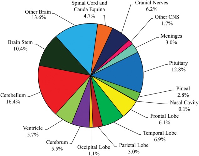
Distribution of Childhood (Ages 0-19 years) Primary Brain and CNS Tumors by Site (N = 20,709).
Figure 14 presents the most common brain and CNS histologies in children ages 0–14 years and adolescents ages 15-19 years. For children ages 0-14 years, pilocytic astrocytomas, embryonal tumors, and malignant glioma, NOS, account for 18%, 15%, and 14%, respectively. The most common histologies in adolescents ages 15–19 years include tumors of the pituitary (23%) and pilocytic astrocytoma (11%) (Figure 14). The broad category glioma accounts for 53% of tumors in children ages 0-14 years and 37% in adolescents ages 15–19 years.
Fig. 14.
Distribution of Childhood Primary Brain and CNS Tumors by Histology.
Childhood Incidence Rates by Histology and Gender
The incidence rates of the most common childhood tumors by gender are shown in Table 13. The overall incidence rate for childhood brain and CNS tumors (ages 0–19 years) is 5.13 per 100,000. Among major histology groupings, incidence rates were highest for tumors of the neuroepithelial tissue (3.51 per 100,000). Pilocytic astrocytoma (0.80 per 100,000), embryonal tumors (0.65 per 100,000), and glioma malignant, NOS (0.58 per 100,000) have the highest rates among individual histologies. Germ cell tumors are more than twice as common in males compared to females. Conversely, the incidence rate of tumors of the pituitary for females is more than two and one-half times the rate observed for males. Differences in incidence rates between males and females for ependymal tumors, embryonal tumors, germ cell tumors, and tumors of the pituitary are statistically significant. Due to small numbers for some tumors caution when interpreting and comparing incidence rates is required.
Table 13.
Selected Childhood (Ages 0–19) Brain and Central Nervous System Tumor Average Annual Age–Adjusted Incidence Rates† by Major Histology Groupings, Histology and Gender, CBTRUS Statistical Report: NPCR and SEER, 2005–2009
| Histology | Total |
Male |
Female |
|||
|---|---|---|---|---|---|---|
| Rate | 95% CI | Rate | 95% CI | Rate | 95% CI | |
| Tumors of Neuroepithelial Tissue | 3.51 | (3.45–3.57) | 3.70 | (3.62–3.78) | 3.31 | (3.23–3.40) |
| Pilocytic astrocytoma | 0.80 | (0.78–0.83) | 0.81 | (0.77–0.85) | 0.79 | (0.75–0.83) |
| Diffuse astrocytoma | 0.27 | (0.25–0.28) | 0.28 | (0.26–0.30) | 0.26 | (0.23–0.28) |
| Anaplastic astrocytoma | 0.08 | (0.07–0.09) | 0.09 | (0.08–0.11) | 0.07 | (0.06–0.08) |
| Unique astrocytoma variants | 0.11 | (0.10–0.12) | 0.11 | (0.10–0.12) | 0.11 | (0.09–0.12) |
| Glioblastoma | 0.14 | (0.13–0.15) | 0.16 | (0.14–0.18) | 0.12 | (0.11–0.14) |
| Oligodendroglioma | 0.06 | (0.05–0.07) | 0.06 | (0.05–0.08) | 0.05 | (0.04–0.06) |
| Anaplastic oligodendroglioma | 0.01 | (0.01–0.02) | 0.01 | (0.01–0.02) | 0.01 | (0.01–0.02) |
| Oligoastrocytic tumors | 0.03 | (0.03–0.04) | 0.03 | (0.02–0.04) | 0.04 | (0.03–0.05) |
| Ependymal tumors | 0.27 | (0.26–0.29) | 0.31 | (0.29–0.33) | 0.24 | (0.22–0.26) |
| Glioma malignant, NOS | 0.58 | (0.56–0.61) | 0.56 | (0.53–0.59) | 0.61 | (0.58–0.65) |
| Choroid plexus tumors | 0.10 | (0.09–0.11) | 0.10 | (0.09–0.12) | 0.09 | (0.07–0.10) |
| Other neuroepithelial tumors | 0.01 | (0.01–0.01) | – | – | 0.01 | (0.01–0.02) |
| Neuronal and mixed neuronal–glial tumors | 0.36 | (0.34–0.38) | 0.39 | (0.36–0.42) | 0.33 | (0.31–0.36) |
| Tumors of the pineal region | 0.04 | (0.04–0.05) | 0.04 | (0.03–0.05) | 0.04 | (0.03–0.05) |
| Embryonal tumors | 0.65 | (0.62–0.67) | 0.74 | (0.70–0.78) | 0.55 | (0.52–0.58) |
| Tumors of Cranial and Spinal Nerves | 0.27 | (0.26–0.29) | 0.27 | (0.25–0.29) | 0.28 | (0.25–0.30) |
| Nerve sheath tumors | 0.27 | (0.26–0.29) | 0.27 | (0.25–0.29) | 0.28 | (0.25–0.30) |
| Other tumors of cranial and spinal nerves | – | – | – | – | – | – |
| Tumors of Meninges | 0.21 | (0.20–0.23) | 0.20 | (0.18–0.22) | 0.23 | (0.21–0.25) |
| Meningioma | 0.14 | (0.12–0.15) | 0.13 | (0.11–0.14) | 0.14 | (0.13–0.16) |
| Mesenchymal tumors | 0.04 | (0.03–0.04) | 0.03 | (0.02–0.04) | 0.05 | (0.04–0.06) |
| Primary melanocytic lesions | 0.01 | (0.00–0.01) | 0.01 | (0.00–0.01) | 0.01 | (0.00–0.01) |
| Other neoplasms related to the meninges | 0.03 | (0.03–0.04) | 0.04 | (0.03–0.05) | 0.03 | (0.03–0.04) |
| Lymphomas and Hemopoietic Neoplasms | 0.02 | (0.02–0.03) | 0.03 | (0.02–0.04) | 0.02 | (0.01–0.02) |
| Lymphoma | 0.02 | (0.01–0.02) | 0.02 | (0.01–0.03) | 0.01 | (0.01–0.02) |
| Other hemopoietic neoplasms | 0.01 | (0.01–0.01) | 0.01 | (0.01–0.01) | 0.01 | (0.00–0.01) |
| Germ Cell Tumors and Cysts | 0.20 | (0.19–0.22) | 0.28 | (0.26–0.31) | 0.12 | (0.11–0.14) |
| Germ cell tumors, cysts and heterotopias | 0.20 | (0.19–0.22) | 0.28 | (0.26–0.31) | 0.12 | (0.11–0.14) |
| Tumors of Sellar Region | 0.66 | (0.63–0.68) | 0.45 | (0.42–0.48) | 0.87 | (0.83–0.92) |
| Tumors of the pituitary | 0.47 | (0.45–0.49) | 0.26 | (0.24–0.28) | 0.68 | (0.64–0.72) |
| Craniopharyngioma | 0.19 | (0.18–0.21) | 0.19 | (0.17–0.21) | 0.19 | (0.18–0.21) |
| Unclassified Tumors | 0.25 | (0.24–0.27) | 0.26 | (0.24–0.29) | 0.25 | (0.22–0.27) |
| Hemangioma | 0.07 | (0.06–0.08) | 0.08 | (0.07–0.09) | 0.07 | (0.06–0.08) |
| Neoplasm, unspecified | 0.18 | (0.17–0.19) | 0.18 | (0.17–0.20) | 0.18 | (0.16–0.20) |
| All other | – | – | – | – | – | – |
| Total‡ | 5.13 | (5.06–5.20) | 5.19 | (5.09–5.29) | 5.08 | (4.98–5.18) |
† Rates are per 100,000 and are age–adjusted to the 2000 US standard population.
‡ Refers to all brain tumors including histologies not presented in this table.
– Counts and rates are not presented when fewer than 16 cases were reported for the specific histology category. Suppressed cases are included in the total counts and rates.
Abbreviations: CBTRUS, Central Brain Tumor Registry of the United States; NPCR, CDC's National Program of Cancer Registries; SEER, NCI's Surveillance, Epidemiology and End Results program; CI, confidence interval; NOS, not otherwise specified.
Childhood Incidence Rates by Histology and Race
Table 14 shows incidence rates by histology and race for children ages 0-19 years. Incidence rates were highest among whites (5.31 per 100,000) compared with black (3.96 per 100,000), AIAN (3.40 per 100,000) or API (3.07 per 100,000) race groups. The observed overall incidence rate differences between whites and each of the three other race groups are statistically significant. Total brain and CNS tumor incidence rates between black and AIAN races are not significantly different. However, the total average annual incidence rate for the API race group is statistically significantly lower than the rate observed for black children ages 0-19 years. Children ages 0-19 years of API races have statistically significantly higher rates of germ cell tumors and cysts than either white or black races. Conversely, API incidence rate for tumors of the sellar region is statistically significantly lower than those observed for white, black, or AIAN race groups.
Table 14.
Childhood (Ages 0–19) Brain and Central Nervous System Tumor Average Annual Age–Adjusted Incidence Rates† by Major Histology Groupings and Race, CBTRUS Statistical Report: NPCR and SEER, 2005–2009
| White |
Black |
AIAN |
API |
|||||
|---|---|---|---|---|---|---|---|---|
| Rate | 95% CI | Rate | 95% CI | Rate | 95% CI | Rate | 95% CI | |
| Tumors of Neuroepithelial Tissue | 3.67 | (3.60–3.74) | 2.69 | (2.56–2.82) | 1.96 | (1.62–2.36) | 1.96 | (1.77–2.16) |
| Pilocytic astrocytoma | 0.85 | (0.82–0.89) | 0.58 | (0.52–0.64) | – | – | 0.37 | (0.29–0.46) |
| Diffuse astrocytoma | 0.28 | (0.26–0.30) | 0.21 | (0.18–0.25) | – | – | 0.16 | (0.11–0.22) |
| Anaplastic astrocytoma | 0.08 | (0.07–0.09) | 0.06 | (0.05–0.09) | – | – | – | – |
| Unique astrocytoma variants | 0.11 | (0.09–0.12) | 0.12 | (0.09–0.15) | – | – | – | – |
| Glioblastoma | 0.14 | (0.12–0.15) | 0.16 | (0.13–0.19) | – | – | 0.12 | (0.08–0.18) |
| Oligodendroglioma | 0.06 | (0.05–0.07) | 0.06 | (0.04–0.08) | – | – | – | – |
| Anaplastic oligodendroglioma | 0.01 | (0.01–0.02) | – | – | – | – | – | – |
| Oligoastrocytic tumors | 0.04 | (0.03–0.05) | – | – | – | – | – | – |
| Ependymal tumors | 0.29 | (0.27–0.31) | 0.20 | (0.16–0.23) | 0.27 | (0.15–0.43) | 0.24 | (0.17–0.31) |
| Glioma malignant, NOS | 0.60 | (0.58–0.63) | 0.43 | (0.38–0.49) | – | – | 0.37 | (0.29–0.46) |
| Choroid plexus tumors | 0.11 | (0.10–0.12) | 0.04 | (0.03–0.06) | – | – | – | – |
| Other neuroepithelial tumors | 0.01 | (0.01–0.01) | – | – | – | – | – | – |
| Neuronal and mixed neuronal–glial tumors | 0.39 | (0.36–0.41) | 0.25 | (0.22–0.30) | – | – | 0.14 | (0.09–0.20) |
| Tumors of the pineal region | 0.03 | (0.03–0.04) | 0.08 | (0.06–0.11) | – | – | – | – |
| Embryonal tumors | 0.69 | (0.66–0.72) | 0.46 | (0.41–0.51) | – | – | 0.40 | (0.32–0.50) |
| Tumors of Cranial and Spinal Nerves | 0.28 | (0.26–0.30) | 0.21 | (0.18–0.25) | 0.20 | (0.10–0.34) | 0.14 | (0.09–0.20) |
| Nerve sheath tumors | 0.28 | (0.26–0.30) | 0.21 | (0.18–0.25) | – | – | 0.14 | (0.09–0.20) |
| Other tumors of cranial and spinal nerves | – | – | – | – | – | – | – | – |
| Tumors of Meninges | 0.22 | (0.20–0.24) | 0.16 | (0.13–0.20) | – | – | 0.12 | (0.08–0.18) |
| Meningioma | 0.14 | (0.12–0.15) | 0.11 | (0.09–0.14) | – | – | 0.09 | (0.05–0.14) |
| Mesenchymal tumors | 0.04 | (0.03–0.05) | – | – | – | – | – | – |
| Primary melanocytic lesions | 0.01 | (0.00–0.01) | – | – | – | – | – | – |
| Other neoplasms related to the meninges | 0.04 | (0.03–0.04) | 0.03 | (0.02–0.04) | – | – | – | – |
| Lymphomas and Hemopoietic Neoplasms | 0.02 | (0.02–0.03) | – | – | – | – | – | – |
| Lymphoma | 0.01 | (0.01–0.02) | – | – | – | – | – | – |
| Other hemopoietic neoplasms | 0.01 | (0.01–0.01) | – | – | – | – | – | – |
| Germ Cell Tumors and Cysts | 0.21 | (0.19–0.22) | 0.14 | (0.11–0.17) | – | – | 0.33 | (0.26–0.43) |
| Germ cell tumors, cysts and heterotopias | 0.21 | (0.19–0.22) | 0.14 | (0.11–0.17) | – | – | 0.33 | (0.26–0.43) |
| Tumors of Sellar Region | 0.65 | (0.62–0.67) | 0.58 | (0.52–0.64) | 0.77 | (0.56–1.03) | 0.38 | (0.30–0.48) |
| Tumors of the pituitary | 0.46 | (0.43–0.48) | 0.40 | (0.35–0.45) | 0.60 | (0.42–0.83) | 0.28 | (0.21–0.37) |
| Craniopharyngioma | 0.19 | (0.17–0.21) | 0.18 | (0.15–0.22) | – | – | 0.10 | (0.06–0.15) |
| Unclassified Tumors | 0.27 | (0.25–0.28) | 0.17 | (0.14–0.20) | – | – | 0.11 | (0.07–0.17) |
| Hemangioma | 0.08 | (0.07–0.09) | 0.04 | (0.02–0.05) | – | – | – | – |
| Neoplasm, unspecified | 0.19 | (0.17–0.20) | 0.13 | (0.10–0.16) | – | – | – | – |
| All other | – | – | – | – | – | – | – | – |
| Total‡ | 5.31 | (5.23–5.39) | 3.96 | (3.81–4.12) | 3.40 | (2.95–3.91) | 3.07 | (2.83–3.32) |
† Rates are per 100,000 and are age–adjusted to the 2000 US standard population.
‡ Refers to all brain tumors including histologies not presented in this table.
– Counts and rates are not presented when fewer than 16 cases were reported for the specific histology category. Suppressed cases are included in the total counts and rates.
Abbreviations: CBTRUS, Central Brain Tumor Registry of the United States; NPCR, CDC's National Program of Cancer Registries; SEER, NCI's Surveillance, Epidemiology and End Results program; CI, confidence interval; NOS, not otherwise specified; AIAN, American Indian/Alaskan Native; API, Asian Pacific Islander.
Childhood Incidence rates by Histology and Hispanic Ethnicity
Incidence rates for children ages 0-19 years by Hispanic ethnicity are shown in Table 15. The non-Hispanic rate (5.30 per 100,000) is statistically significantly higher than the observed rate for Hispanics (4.55 per 100,000). This difference is apparent for incidence rates of tumors of neuroepithelial tissue and tumors of cranial and spinal nerves. Conversely, incidence rates for tumors of the pituitary are statistically significantly higher among Hispanic children ages 0-19 years than their non-Hispanic counterparts.
Table 15.
Selected Childhood (Ages 0-19) Brain and Central Nervous System Tumor Average Annual Age-Adjusted Incidence Rates† By Major Histology Groupings, Histology, and Hispanic Ethnicity‡, CBTRUS Statistical Report: NPCR and SEER, 2005–2009
| Histology | Hispanic |
Non-Hispanic |
||
|---|---|---|---|---|
| Rate | 95% CI | Rate | 95% CI | |
| Tumors of Neuroepithelial Tissue | 2.80 | (2.69-2.91) | 3.71 | (3.64-3.77) |
| Pilocytic astrocytoma | 0.58 | (0.53-0.64) | 0.86 | (0.83-0.90) |
| Diffuse astrocytoma | 0.21 | (0.18-0.24) | 0.28 | (0.27-0.30) |
| Anaplastic astrocytoma | 0.07 | (0.06-0.10) | 0.08 | (0.07-0.09) |
| Unique astrocytoma variants | 0.09 | (0.07-0.11) | 0.11 | (0.10-0.12) |
| Glioblastoma | 0.13 | (0.10-0.15) | 0.14 | (0.13-0.16) |
| Oligodendroglioma | 0.04 | (0.03-0.06) | 0.06 | (0.05-0.07) |
| Anaplastic oligodendroglioma | – | – | 0.01 | (0.01-0.02) |
| Oligoastrocytic tumors | 0.02 | (0.01-0.03) | 0.04 | (0.03-0.04) |
| Ependymal tumors | 0.26 | (0.23-0.30) | 0.28 | (0.26-0.30) |
| Glioma malignant, NOS | 0.44 | (0.40-0.49) | 0.62 | (0.60-0.65) |
| Choroid plexus tumors | 0.09 | (0.07-0.11) | 0.10 | (0.09-0.11) |
| Other neuroepithelial tumors | – | – | 0.01 | (0.01-0.01) |
| Neuronal and mixed neuronal-glial tumors | 0.25 | (0.21-0.28) | 0.39 | (0.37-0.42) |
| Tumors of the pineal region | 0.04 | (0.03-0.05) | 0.04 | (0.04-0.05) |
| Embryonal tumors | 0.57 | (0.52-0.62) | 0.67 | (0.64-0.70) |
| Tumors of Cranial and Spinal Nerves | 0.23 | (0.20-0.26) | 0.28 | (0.27-0.30) |
| Nerve sheath tumors | 0.23 | (0.20-0.26) | 0.28 | (0.27-0.30) |
| Other tumors of cranial and spinal nerves | – | – | – | – |
| Tumors of Meninges | 0.18 | (0.15-0.21) | 0.22 | (0.20-0.24) |
| Meningioma | 0.09 | (0.07-0.11) | 0.15 | (0.13-0.16) |
| Mesenchymal tumors | 0.03 | (0.02-0.05) | 0.04 | (0.03-0.05) |
| Primary melanocytic lesions | – | – | – | – |
| Other neoplasms related to the meninges | 0.05 | (0.04-0.07) | 0.03 | (0.03-0.04) |
| Lymphomas and Hemopoietic Neoplasms | 0.03 | (0.02-0.05) | 0.02 | (0.02-0.03) |
| Lymphoma | 0.02 | (0.01-0.04) | 0.01 | (0.01-0.02) |
| Other hemopoietic neoplasms | 0.01 | (0.00-0.02) | 0.01 | (0.00-0.01) |
| Germ Cell Tumors and Cysts | 0.23 | (0.20-0.26) | 0.20 | (0.18-0.22) |
| Germ cell tumors, cysts and heterotopias | 0.23 | (0.20-0.26) | 0.20 | (0.18-0.22) |
| Tumors of Sellar Region | 0.79 | (0.73-0.85) | 0.63 | (0.60-0.66) |
| Tumors of the pituitary | 0.58 | (0.52-0.63) | 0.44 | (0.42-0.46) |
| Craniopharyngioma | 0.21 | (0.18-0.25) | 0.19 | (0.17-0.20) |
| Unclassified Tumors | 0.29 | (0.26-0.33) | 0.24 | (0.23-0.26) |
| Hemangioma | 0.08 | (0.07-0.11) | 0.07 | (0.06-0.08) |
| Neoplasm, unspecified | 0.21 | (0.18-0.24) | 0.17 | (0.16-0.19) |
| All other | – | – | – | – |
| Total§ | 4.55 | (4.40-4.69) | 5.30 | (5.22-5.38) |
† Rates are per 100,000 and are age-adjusted to the 2000 US standard population.
‡ Hispanic ethnicity is not mutually exclusive of race; Classified using the North American Association of Central Cancer Registries Hispanic Identification Algorithm, version 2 (NHIA v2).
§ Refers to all brain tumors including histologies not presented in this table.
- Counts and rates are not presented when fewer than 16 cases were reported for the specific histology category. The suppressed cases are included in the counts and rates for totals.
Abbreviations: CBTRUS, Central Brain Tumor Registry of the United States; NPCR, CDC's National Program of Cancer Registries; SEER, NCI's Surveillance, Epidemiology and End Results program; CI, confidence interval; NOS, not otherwise specified.
Childhood Incidence Rates by Age and Histology
The detailed age–specific incidence rates by histology for children age groups 0-4 years, 5-9 years, 10-14 years, 15-19 years, 0-19 years, and 0-14 years are shown in Table 16. The overall incidence rates for age groups 0-4 years and 15-19 years statistically significantly exceeded those observed in age groups 5-9 years and 10-14 years. It should also be noted that individual histology distributions vary substantially within these childhood age groups. The incidence rates of pilocytic astrocytoma, malignant glioma NOS, ependymal tumors, choroid plexus tumors and embryonal tumors decrease with increasing age groups.
Table 16.
Selected Childhood Brain and Central Nervous System Tumor, Average Annual Age-Adjusted and Age-Specific Incidence Rates† by Major Histology Groupings, Histology and Age at Diagnosis, CBTRUS Statistical Report: NPCR and SEER, 2005–2009
| Age At Diagnosis |
Age at Diagnosis |
|||||||||||
|---|---|---|---|---|---|---|---|---|---|---|---|---|
| 0-4 |
5-9 |
10-14 |
15-19 |
0-19‡ |
0-14‡ |
|||||||
| Rate | 95% CI | Rate | 95% CI | Rate | 95% CI | Rate | 95% CI | Rate | 95% CI | Rate | 95% CI | |
| Tumors of Neuroepithelial Tissue | 4.50 | (4.37-4.63) | 3.68 | (3.56-3.81) | 3.17 | (3.06-3.28) | 2.74 | (2.64-2.84) | 3.51 | (3.45-3.57) | 3.77 | (3.70-3.84) |
| Pilocytic astrocytoma | 0.87 | (0.82-0.93) | 0.89 | (0.83-0.95) | 0.86 | (0.80-0.92) | 0.59 | (0.55-0.64) | 0.80 | (0.78-0.83) | 0.87 | (0.84-0.91) |
| Diffuse astrocytoma | 0.33 | (0.29-0.37) | 0.22 | (0.19-0.25) | 0.26 | (0.23-0.30) | 0.26 | (0.23-0.29) | 0.27 | (0.25-0.28) | 0.27 | (0.25-0.29) |
| Anaplastic astrocytoma | 0.05 | (0.04-0.07) | 0.08 | (0.07-0.10) | 0.09 | (0.07-0.11) | 0.09 | (0.08-0.11) | 0.08 | (0.07-0.09) | 0.08 | (0.07-0.09) |
| Unique astrocytoma variants | 0.06 | (0.05-0.08) | 0.10 | (0.08-0.12) | 0.14 | (0.11-0.16) | 0.13 | (0.11-0.16) | 0.11 | (0.10-0.12) | 0.10 | (0.09-0.11) |
| Glioblastoma | 0.09 | (0.07-0.11) | 0.14 | (0.12-0.17) | 0.16 | (0.14-0.19) | 0.17 | (0.14-0.19) | 0.14 | (0.13-0.15) | 0.13 | (0.12-0.15) |
| Oligodendroglioma | 0.03 | (0.02-0.04) | 0.04 | (0.03-0.05) | 0.06 | (0.04-0.08) | 0.11 | (0.09-0.13) | 0.06 | (0.05-0.07) | 0.04 | (0.04-0.05) |
| Anaplastic oligodendroglioma | – | – | – | – | – | – | 0.02 | (0.02-0.04) | 0.01 | (0.01-0.02) | 0.01 | (0.01-0.01) |
| Oligoastrocytic tumors | 0.03 | (0.02-0.04) | 0.03 | (0.02-0.04) | 0.03 | (0.02-0.05) | 0.05 | (0.04-0.07) | 0.03 | (0.03-0.04) | 0.03 | (0.02-0.04) |
| Ependymal tumors | 0.40 | (0.36-0.44) | 0.23 | (0.20-0.26) | 0.21 | (0.19-0.24) | 0.26 | (0.23-0.30) | 0.27 | (0.26-0.29) | 0.28 | (0.26-0.30) |
| Glioma malignant, NOS | 0.86 | (0.81-0.92) | 0.81 | (0.75-0.87) | 0.43 | (0.39-0.47) | 0.24 | (0.22-0.28) | 0.58 | (0.56-0.61) | 0.70 | (0.67-0.73) |
| Choroid plexus tumors | 0.26 | (0.23-0.29) | 0.04 | (0.03-0.06) | 0.04 | (0.03-0.05) | 0.05 | (0.04-0.06) | 0.10 | (0.09-0.11) | 0.11 | (0.10-0.12) |
| Other neuroepithelial tumors | – | – | – | – | – | – | – | – | 0.01 | (0.01-0.01) | 0.01 | (0.01-0.01) |
| Neuronal and mixed neuronal-glial tumors | 0.24 | (0.21-0.28) | 0.30 | (0.27-0.34) | 0.43 | (0.39-0.48) | 0.46 | (0.42-0.51) | 0.36 | (0.34-0.38) | 0.33 | (0.31-0.35) |
| Tumors of the pineal region | 0.05 | (0.04-0.07) | 0.04 | (0.03-0.05) | 0.03 | (0.02-0.05) | 0.04 | (0.03-0.05) | 0.04 | (0.04-0.05) | 0.04 | (0.03-0.05) |
| Embryonal tumors | 1.21 | (1.15-1.28) | 0.76 | (0.70-0.81) | 0.39 | (0.35-0.43) | 0.25 | (0.22-0.28) | 0.65 | (0.62-0.67) | 0.78 | (0.75-0.81) |
| Tumors of Cranial and Spinal Nerves | 0.27 | (0.24-0.30) | 0.21 | (0.18-0.24) | 0.26 | (0.22-0.29) | 0.35 | (0.32-0.39) | 0.27 | (0.26-0.29) | 0.24 | (0.23-0.26) |
| Nerve sheath tumors | 0.27 | (0.24-0.30) | 0.21 | (0.18-0.24) | 0.26 | (0.22-0.29) | 0.35 | (0.32-0.39) | 0.27 | (0.26-0.29) | 0.24 | (0.23-0.26) |
| Other tumors of cranial and spinal nerves | – | – | – | – | – | – | – | – | – | – | – | – |
| Tumors of Meninges | 0.14 | (0.12-0.16) | 0.10 | (0.08-0.12) | 0.21 | (0.18-0.24) | 0.39 | (0.36-0.43) | 0.21 | (0.20-0.23) | 0.15 | (0.14-0.17) |
| Meningioma | 0.07 | (0.05-0.09) | 0.06 | (0.05-0.08) | 0.14 | (0.12-0.17) | 0.26 | (0.23-0.30) | 0.14 | (0.12-0.15) | 0.09 | (0.08-0.10) |
| Mesenchymal tumors | 0.06 | (0.04-0.07) | 0.03 | (0.02-0.04) | 0.03 | (0.02-0.04) | 0.03 | (0.02-0.05) | 0.04 | (0.03-0.04) | 0.04 | (0.03-0.05) |
| Primary melanocytic lesions | – | – | – | – | – | – | – | – | 0.01 | (0.00-0.01) | – | – |
| Other neoplasms related to the meninges | – | – | – | – | 0.03 | (0.02-0.05) | 0.09 | (0.07-0.11) | 0.03 | (0.03-0.04) | 0.02 | (0.01-0.02) |
| Lymphomas and Hemopoietic Neoplasms | – | – | 0.02 | (0.01-0.03) | 0.02 | (0.01-0.03) | 0.03 | (0.02-0.05) | 0.02 | (0.02-0.03) | 0.02 | (0.01-0.02) |
| Lymphoma | – | – | – | – | – | – | 0.02 | (0.02-0.04) | 0.02 | (0.01-0.02) | 0.01 | (0.01-0.02) |
| Other hemopoietic neoplasms | – | – | – | – | – | – | – | – | 0.01 | (0.01-0.01) | 0.01 | (0.00-0.01) |
| Germ Cell Tumors and Cysts | 0.13 | (0.11-0.15) | 0.14 | (0.11-0.16) | 0.27 | (0.23-0.30) | 0.29 | (0.26-0.32) | 0.20 | (0.19-0.22) | 0.18 | (0.16-0.19) |
| Germ cell tumors, cysts and heterotopias | 0.13 | (0.11-0.15) | 0.14 | (0.11-0.16) | 0.27 | (0.23-0.30) | 0.29 | (0.26-0.32) | 0.20 | (0.19-0.22) | 0.18 | (0.16-0.19) |
| Tumors of Sellar Region | 0.17 | (0.15-0.20) | 0.37 | (0.33-0.41) | 0.61 | (0.57-0.67) | 1.46 | (1.39-1.53) | 0.66 | (0.63-0.68) | 0.39 | (0.37-0.41) |
| Tumors of the pituitary | 0.03 | (0.02-0.05) | 0.11 | (0.09-0.13) | 0.40 | (0.36-0.44) | 1.31 | (1.24-1.38) | 0.47 | (0.45-0.49) | 0.18 | (0.17-0.20) |
| Craniopharyngioma | 0.14 | (0.12-0.16) | 0.26 | (0.23-0.29) | 0.22 | (0.19-0.25) | 0.15 | (0.13-0.18) | 0.19 | (0.18-0.21) | 0.21 | (0.19-0.22) |
| Unclassified Tumors | 0.22 | (0.19-0.25) | 0.18 | (0.15-0.21) | 0.26 | (0.22-0.29) | 0.36 | (0.33-0.40) | 0.25 | (0.24-0.27) | 0.22 | (0.20-0.24) |
| Hemangioma | 0.05 | (0.04-0.06) | 0.04 | (0.03-0.05) | 0.08 | (0.06-0.09) | 0.13 | (0.11-0.15) | 0.07 | (0.06-0.08) | 0.05 | (0.05-0.06) |
| Neoplasm, unspecified | 0.17 | (0.15-0.20) | 0.14 | (0.12-0.17) | 0.18 | (0.15-0.21) | 0.23 | (0.20-0.26) | 0.18 | (0.17-0.19) | 0.16 | (0.15-0.18) |
| All other | – | – | – | – | – | – | – | – | – | – | – | – |
| Total§ | 5.43 | (5.29-5.58) | 4.70 | (4.56-4.84) | 4.79 | (4.65-4.93) | 5.63 | (5.49-5.77) | 5.13 | (5.06-5.20) | 4.97 | (4.89-5.05) |
† Rates are per 100,000 and are age-adjusted to the 2000 US standard population.
§ Refers to all brain tumors including histologies not presented in this table.
- Counts and rates are not presented when fewer than 16 cases were reported for the specific histology category. The suppressed cases are included in the counts and rates for totals.
Abbreviations: CBTRUS, Central Brain Tumor Registry of the United States; NPCR, CDC's National Program of Cancer Registries; SEER, NCI's Surveillance, Epidemiology and End Results program; CI, confidence interval; NOS, not otherwise specified.
Age–specific incidence rates for selected histologies are graphically shown in Figure 15. The U-shaped overall incidence rate pattern across the four age group categories is apparent on the graph. Sharp declines in incidence rates between age groups for the broad gliomas and embryonal tumor histology groups are evident. The incidence decline rate for pilocytic astrocytoma is substantial from the 10-14 years to the 15-19 years age group. In contrast, ependymal tumor incidence rates are highest in the 0-4 years age group then decline and remain relatively stable across the 5-9 years, 10-14 years, and 15-19 years age groups.
Fig. 15.
Age-Specific Incidence of Childhood Brain and CNS Tumors by Selected Histologies.
Childhood Incidence Rates by Histology Defined by ICCC
Table 17 presents the CBTRUS childhood brain and CNS tumor data used for this report according to the International Classification of Childhood Cancer (ICCC) grouping system for pediatric cancers.23 As shown, the Table 17 age group category total 0-19 age group count and age-specific and adjusted rates are equivalent to those presented throughout this report. However, the histology grouping scheme differences are apparent and reflect different approaches to the description of childhood brain and CNS tumors.
Table 17.
Age-Adjusted and Age-Specific Incidence Rates† for Childhood Brain and Central Nervous System Tumors: Malignant and Non-Malignant by International Classification of Childhood Cancer (ICCC), CBTRUS Statistical Report: NPCR and SEER, 2005–2009
| ICCC Category | 0-19‡ years |
0-14‡ years |
< 1 year |
1-4 years |
5-9 years |
10-14 years |
15-19 years |
||||||||
|---|---|---|---|---|---|---|---|---|---|---|---|---|---|---|---|
| Count | Rate | 95% CI | Rate | 95% CI | Rate | 95% CI | Rate | 95% CI | Rate | 95% CI | Rate | 95% CI | Rate | 95% CI | |
| II Lymphomas and reticuloendothelial neoplasms | 69 | 0.02 | (0.01-0.02) | 0.01 | (0.01-0.02) | – | – | – | – | 0.02 | (0.01-0.03) | 0.02 | (0.01-0.03) | 0.03 | (0.02-0.04) |
| III CNS and misc intracranial and intraspinal neoplasms | 17,654 | 4.38 | (4.31-4.44) | 4.32 | (4.25-4.40) | 4.53 | (4.24-4.83) | 4.82 | (4.67-4.97) | 4.21 | (4.08-4.34) | 4.03 | (3.91-4.16) | 4.53 | (4.41-4.67) |
| III(a) Ependymomas and choroid plexus tumor | 1,506 | 0.37 | (0.35-0.39) | 0.39 | (0.37-0.41) | 0.93 | (0.80-1.07) | 0.59 | (0.54-0.64) | 0.27 | (0.24-0.31) | 0.25 | (0.22-0.29) | 0.31 | (0.28-0.35) |
| III(b) Astrocytomas | 6,277 | 1.56 | (1.52-1.60) | 1.66 | (1.61-1.70) | 1.25 | (1.10-1.41) | 1.92 | (1.83-2.02) | 1.61 | (1.53-1.69) | 1.58 | (1.50-1.66) | 1.27 | (1.21-1.34) |
| III(c) Intracranial and intraspinal embryonal tumors | 2,408 | 0.60 | (0.57-0.62) | 0.72 | (0.69-0.75) | 1.19 | (1.04-1.35) | 1.04 | (0.97-1.11) | 0.73 | (0.67-0.78) | 0.37 | (0.34-0.41) | 0.23 | (0.21-0.27) |
| III(d) Other gliomas | 2,135 | 0.53 | (0.51-0.56) | 0.58 | (0.55-0.61) | 0.26 | (0.20-0.34) | 0.61 | (0.56-0.67) | 0.70 | (0.65-0.76) | 0.49 | (0.44-0.53) | 0.40 | (0.37-0.44) |
| III(e) Other specified intracranial and intraspinal neoplasms | 4,602 | 1.14 | (1.11-1.17) | 0.82 | (0.79-0.86) | 0.61 | (0.51-0.73) | 0.51 | (0.47-0.57) | 0.76 | (0.70-0.81) | 1.17 | (1.10-1.24) | 2.08 | (2.00-2.17) |
| III(f) Unspecified intracranial and intraspinal neoplasms | 726 | 0.18 | (0.17-0.19) | 0.16 | (0.15-0.18) | 0.29 | (0.22-0.38) | 0.14 | (0.12-0.17) | 0.14 | (0.12-0.16) | 0.18 | (0.15-0.21) | 0.23 | (0.20-0.26) |
| IV Neuroblastoma and other peripheral nervous cell tumors | 206 | 0.05 | (0.04-0.06) | 0.06 | (0.05-0.07) | 0.28 | (0.21-0.37) | 0.09 | (0.07-0.11) | 0.03 | (0.02-0.04) | 0.02 | (0.02-0.04) | 0.03 | (0.02-0.04) |
| IX Soft tissue and other extraosseous sarcomas | 1,667 | 0.41 | (0.39-0.43) | 0.35 | (0.33-0.37) | 0.56 | (0.46-0.67) | 0.33 | (0.29-0.37) | 0.28 | (0.25-0.32) | 0.39 | (0.35-0.43) | 0.60 | (0.55-0.65) |
| X(a) Intracranial & intraspinal germ cell tumors | 823 | 0.20 | (0.19-0.22) | 0.18 | (0.16-0.19) | 0.38 | (0.30-0.48) | 0.06 | (0.05-0.08) | 0.14 | (0.11-0.16) | 0.27 | (0.23-0.30) | 0.29 | (0.26-0.32) |
| All other categories | 290 | 0.07 | (0.06-0.08) | 0.04 | (0.04-0.05) | – | – | 0.04 | (0.02-0.05) | 0.03 | (0.02-0.05) | 0.06 | (0.05-0.08) | 0.15 | (0.13-0.18) |
| Total§ | 20,709 | 5.13 | (5.06-5.20) | 4.97 | (4.89-5.05) | 5.82 | (5.49-6.16) | 5.33 | (5.18-5.50) | 4.70 | (4.56-4.84) | 4.79 | (4.65-4.93) | 5.63 | (5.49-5.77) |
† Rates are per 100,000 and are ‡age adjusted to the 2000 U.S. standard population.
§ Refers to all brain tumors including histologies not presented in this table.
- Counts and rates are not presented when fewer than 16 cases were reported for the specific ICCC category. The suppressed cases are included in the counts and rates for totals.
Abbreviations: ICCC, International Classification of Childhood Cancer; CBTRUS, Central Brain Tumor Registry of the United States; NPCR, CDC's National Program of Cancer Registries; SEER, NCI's Surveillance, Epidemiology and End Results program; CI, confidence interval.
Mortality Rates, Expected Incidence, and Survival
Estimated Mortality Rates from Brain and CNS Malignant Tumors by State and Gender
Table 18 shows average annual age-adjusted mortality rates from primary malignant brain and CNS tumors in the United States during 2005–2009 by state and gender. The aggregate total rate is 4.28 deaths per 100,000. However, there is considerable variation by individual state, which ranges from a low of 2.30 deaths per 100,000 to a high of 5.53 deaths per 100,000. Males have statistically significantly higher mortality rates from brain and CNS tumors than females in the U.S.
Table 18.
Average Annual Age–Adjusted Mortality Rates† for Malignant Brain and Central Nervous System Cancer Overall and by State and Gender, United States, 2005–2009‡
| State | Total |
Males |
Females |
||||||
|---|---|---|---|---|---|---|---|---|---|
| N | Rate | 95% CI | N | Rate | 95% CI | N | Rate | 95% CI | |
| Alabama | 1,148 | 4.58 | (4.31–4.85) | 626 | 5.55 | (5.12–6.01) | 522 | 3.76 | (3.45–4.11) |
| Alaska | 111 | 4.20 | (3.39–5.14) | 65 | 4.96 | (3.67–6.52) | 46 | 3.56 | (2.55–4.81) |
| Arizona | 1,336 | 4.11 | (3.90–4.34) | 743 | 4.87 | (4.52–5.23) | 593 | 3.46 | (3.19–3.76) |
| Arkansas | 775 | 4.95 | (4.60–5.31) | 417 | 5.90 | (5.34–6.50) | 358 | 4.15 | (3.73–4.62) |
| California | 7,457 | 4.31 | (4.21–4.41) | 4,225 | 5.32 | (5.16–5.49) | 3,232 | 3.46 | (3.34–3.58) |
| Colorado | 1,016 | 4.40 | (4.13–4.68) | 586 | 5.45 | (5.00–5.92) | 430 | 3.51 | (3.18–3.86) |
| Connecticut | 809 | 4.22 | (3.93–4.52) | 436 | 5.07 | (4.60–5.58) | 373 | 3.52 | (3.16–3.90) |
| Delaware | 212 | 4.54 | (3.95–5.20) | 119 | 5.75 | (4.75–6.89) | 93 | 3.60 | (2.90–4.43) |
| Washington DC | 101 | 3.47 | (2.82–4.23) | 57 | 4.44 | (3.33–5.78) | 44 | 2.67 | (1.93–3.60) |
| Florida | 4,385 | 3.95 | (3.83–4.07) | 2,455 | 4.81 | (4.62–5.01) | 1,930 | 3.19 | (3.05–3.34) |
| Georgia | 1,671 | 3.87 | (3.68–4.06) | 913 | 4.67 | (4.36–5.00) | 758 | 3.22 | (2.99–3.46) |
| Hawaii | 171 | 2.39 | (2.04–2.78) | 98 | 2.92 | (2.36–3.57) | 73 | 1.95 | (1.52–2.47) |
| Idaho | 379 | 5.14 | (4.63–5.69) | 202 | 5.63 | (4.87–6.48) | 177 | 4.62 | (3.96–5.36) |
| Illinois | 2,523 | 3.89 | (3.74–4.05) | 1,387 | 4.74 | (4.49–5.00) | 1,136 | 3.19 | (3.01–3.39) |
| Indiana | 1,526 | 4.62 | (4.39–4.86) | 819 | 5.46 | (5.08–5.85) | 707 | 3.93 | (3.64–4.23) |
| Iowa | 918 | 5.53 | (5.17–5.90) | 526 | 6.95 | (6.36–7.57) | 392 | 4.33 | (3.90–4.80) |
| Kansas | 735 | 5.07 | (4.70–5.45) | 422 | 6.39 | (5.79–7.03) | 313 | 3.93 | (3.50–4.40) |
| Kentucky | 1,021 | 4.49 | (4.21–4.77) | 565 | 5.46 | (5.01–5.94) | 456 | 3.66 | (3.33–4.02) |
| Louisiana | 1,007 | 4.53 | (4.25–4.82) | 565 | 5.63 | (5.17–6.13) | 442 | 3.61 | (3.28–3.96) |
| Maine | 403 | 5.12 | (4.63–5.66) | 233 | 6.47 | (5.65–7.38) | 170 | 4.00 | (3.41–4.67) |
| Maryland | 1,094 | 3.78 | (3.56–4.02) | 590 | 4.57 | (4.20–4.97) | 504 | 3.15 | (2.88–3.44) |
| Massachusetts | 1,517 | 4.25 | (4.03–4.47) | 847 | 5.38 | (5.02–5.76) | 670 | 3.35 | (3.10–3.62) |
| Michigan | 2,602 | 4.86 | (4.67–5.05) | 1,458 | 5.99 | (5.68–6.31) | 1,144 | 3.95 | (3.72–4.19) |
| Minnesota | 1,165 | 4.33 | (4.09–4.59) | 644 | 5.18 | (4.78–5.60) | 521 | 3.60 | (3.30–3.93) |
| Mississippi | 693 | 4.67 | (4.32–5.03) | 362 | 5.50 | (4.93–6.10) | 331 | 3.99 | (3.57–4.45) |
| Missouri | 1,468 | 4.58 | (4.35–4.82) | 793 | 5.45 | (5.07–5.84) | 675 | 3.86 | (3.57–4.17) |
| Montana | 248 | 4.53 | (3.97–5.14) | 147 | 5.53 | (4.66–6.52) | 101 | 3.63 | (2.94–4.43) |
| Nebraska | 475 | 5.13 | (4.67–5.61) | 256 | 6.01 | (5.29–6.80) | 219 | 4.30 | (3.74–4.93) |
| Nevada | 517 | 4.10 | (3.75–4.48) | 318 | 5.21 | (4.64–5.84) | 199 | 3.09 | (2.67–3.55) |
| New Hampshire | 338 | 4.76 | (4.26–5.30) | 202 | 6.11 | (5.28–7.03) | 136 | 3.62 | (3.03–4.30) |
| New Jersey | 1,720 | 3.69 | (3.51–3.87) | 970 | 4.65 | (4.36–4.96) | 750 | 2.90 | (2.70–3.12) |
| New Mexico | 375 | 3.66 | (3.30–4.05) | 216 | 4.48 | (3.89–5.13) | 159 | 2.95 | (2.50–3.45) |
| New York | 3,861 | 3.70 | (3.58–3.82) | 2,193 | 4.74 | (4.54–4.95) | 1,668 | 2.87 | (2.74–3.02) |
| North Carolina | 1,893 | 4.05 | (3.87–4.24) | 1,044 | 5.01 | (4.71–5.33) | 849 | 3.28 | (3.06–3.51) |
| North Dakota | 174 | 5.02 | (4.29–5.83) | 89 | 5.63 | (4.51–6.95) | 85 | 4.42 | (3.51–5.50) |
| Ohio | 2,794 | 4.44 | (4.27–4.61) | 1,527 | 5.36 | (5.09–5.64) | 1,267 | 3.66 | (3.46–3.88) |
| Oklahoma | 897 | 4.68 | (4.38–5.00) | 491 | 5.60 | (5.11–6.13) | 406 | 3.85 | (3.47–4.25) |
| Oregon | 1,056 | 5.19 | (4.88–5.52) | 624 | 6.56 | (6.05–7.10) | 432 | 4.05 | (3.67–4.46) |
| Pennsylvania | 3,008 | 4.10 | (3.95–4.25) | 1,623 | 4.95 | (4.71–5.20) | 1,385 | 3.41 | (3.23–3.60) |
| Rhode Island | 252 | 4.24 | (3.73–4.80) | 139 | 5.24 | (4.39–6.20) | 113 | 3.43 | (2.81–4.15) |
| South Carolina | 1,027 | 4.33 | (4.07–4.61) | 571 | 5.42 | (4.97–5.89) | 456 | 3.47 | (3.16–3.81) |
| South Dakota | 210 | 4.93 | (4.28–5.66) | 113 | 5.66 | (4.65–6.82) | 97 | 4.32 | (3.48–5.30) |
| Tennessee | 1,644 | 4.99 | (4.75–5.24) | 897 | 6.03 | (5.64–6.45) | 747 | 4.14 | (3.85–4.45) |
| Texas | 4,490 | 4.18 | (4.06–4.31) | 2,409 | 4.89 | (4.70–5.10) | 2,081 | 3.60 | (3.45–3.76) |
| Utah | 480 | 4.46 | (4.07–4.89) | 276 | 5.32 | (4.70–6.01) | 204 | 3.65 | (3.16–4.19) |
| Vermont | 158 | 4.47 | (3.79–5.25) | 82 | 4.93 | (3.90–6.16) | 76 | 3.99 | (3.13–5.03) |
| Virginia | 1,517 | 3.87 | (3.68–4.08) | 854 | 4.87 | (4.54–5.22) | 663 | 3.07 | (2.84–3.31) |
| Washington | 1,707 | 5.12 | (4.88–5.38) | 1,010 | 6.48 | (6.08–6.90) | 697 | 3.94 | (3.65–4.25) |
| West Virginia | 498 | 4.57 | (4.18–5.00) | 266 | 5.31 | (4.68–6.00) | 232 | 3.99 | (3.48–4.55) |
| Wisconsin | 1,469 | 4.90 | (4.65–5.16) | 838 | 6.06 | (5.65–6.49) | 631 | 3.91 | (3.60–4.23) |
| Wyoming | 121 | 4.40 | (3.64–5.28) | 66 | 4.94 | (3.79–6.32) | 55 | 3.91 | (2.93–5.11) |
| United States | 67,172 | 4.28 | (4.25–4.31) | 37,374 | 5.23 | (5.18–5.29) | 29,798 | 3.48 | (3.44–3.52) |
† Rates are per 100,000 and are age–adjusted to the 2000 US standard population.
‡ Estimated by CBTRUS using Surveillance, Epidemiology, and End Results (SEER) Program (www.seer.cancer.gov) SEER*Stat Database: Mortality – All COD, Aggregated With State, Total U.S. (1969–2009) <Katrina/Rita Population Adjustment>, National Cancer Institute, DCCPS, Surveillance Research Program, Surveillance Systems Branch, released April 2012. Underlying mortality data provided by NCHS (www.cdc.gov/nchs). Database: Mortality – All COD, Aggregated With State, Total U.S. (1969–2009) <Katrina/Rita Population Adjustment>
Abbreviations: SEER, Survival, Epidemiology and End Results; NCHS, National Center for Health Statistics; CI, confidence interval.
Estimated Numbers of Expected Cases of All Primary Brain and CNS Tumors by State
The estimated numbers of cases of all primary brain and CNS tumors for 2012 and 2013 by state are shown in Table 19. The estimated numbers of cases of malignant and non–malignant tumors by state were calculated using the CBTRUS age–specific incidence rates (2005–2009) for 18 age groups, race (white, black, and other), and gender and applying them to the 2012 and 2013 population projections for each state and the District of Columbia. The total number of new cases of primary brain and CNS tumors for all 50 states and the District of Columbia in 2012 is estimated to be 68,530 with 24,170 being malignant and 44,360 being non-malignant. For 2013, the estimates are 69,720 primary brain and CNS cases of which 24,560 and 45,160 would be expected to be malignant and non-malignant, respectively.
Table 19.
Brain and Central Nervous System Tumors, Estimated Number of Cases†‡ and by Behavior by State, 2012, 2013
| STATE | 2012 Estimated New Cases |
2013 Estimated New Cases |
||||
|---|---|---|---|---|---|---|
| Total | Malignant | Non-Malignant | Total | Malignant | Non-Malignant | |
| Alabama | 1,070 | 360 | 710 | 1,090 | 370 | 720 |
| Alaska | 130 | 50 | 90 | 140 | 50 | 90 |
| Arizona | 1,530 | 560 | 970 | 1,590 | 580 | 1,010 |
| Arkansas | 670 | 240 | 430 | 680 | 240 | 440 |
| California | 7,560 | 2,670 | 4,890 | 7,700 | 2,710 | 4,980 |
| Colorado | 1,090 | 400 | 690 | 1,120 | 410 | 710 |
| Connecticut | 810 | 290 | 520 | 820 | 290 | 530 |
| Delaware | 210 | 70 | 140 | 220 | 80 | 140 |
| District of Columbia | 130 | – | 90 | 130 | – | 90 |
| Florida | 4,780 | 1,680 | 3,100 | 4,890 | 1,710 | 3,180 |
| Georgia | 2,090 | 700 | 1,380 | 2,150 | 720 | 1,420 |
| Hawaii | 260 | 80 | 180 | 260 | 80 | 180 |
| Idaho | 350 | 130 | 220 | 360 | 130 | 220 |
| Illinois | 2,770 | 970 | 1,800 | 2,800 | 980 | 1,820 |
| Indiana | 1,430 | 520 | 910 | 1,450 | 520 | 930 |
| Iowa | 700 | 260 | 440 | 710 | 260 | 450 |
| Kansas | 620 | 230 | 390 | 630 | 230 | 400 |
| Kentucky | 990 | 360 | 630 | 1,000 | 360 | 640 |
| Louisiana | 940 | 320 | 630 | 950 | 320 | 630 |
| Maine | 340 | 120 | 210 | 340 | 130 | 220 |
| Maryland | 1,260 | 410 | 850 | 1,280 | 420 | 860 |
| Massachusetts | 1,510 | 540 | 970 | 1,520 | 540 | 980 |
| Michigan | 2,280 | 800 | 1,480 | 2,310 | 810 | 1,490 |
| Minnesota | 1,180 | 430 | 750 | 1,200 | 440 | 760 |
| Mississippi | 640 | 210 | 430 | 640 | 210 | 430 |
| Missouri | 1,370 | 490 | 880 | 1,390 | 500 | 900 |
| Montana | 240 | 90 | 150 | 240 | 90 | 150 |
| Nebraska | 400 | 150 | 250 | 400 | 150 | 260 |
| Nevada | 600 | 220 | 390 | 630 | 230 | 400 |
| New Hampshire | 320 | 120 | 200 | 330 | 120 | 210 |
| New Jersey | 1,950 | 680 | 1,280 | 1,970 | 680 | 1,290 |
| New Mexico | 450 | 160 | 290 | 460 | 170 | 290 |
| New York | 4,380 | 1,500 | 2,890 | 4,430 | 1,510 | 2,920 |
| North Carolina | 2,130 | 730 | 1,400 | 2,190 | 750 | 1,430 |
| North Dakota | 150 | 50 | 90 | 150 | 50 | 90 |
| Ohio | 2,660 | 940 | 1,710 | 2,690 | 950 | 1,730 |
| Oklahoma | 800 | 290 | 520 | 810 | 290 | 520 |
| Oregon | 900 | 330 | 570 | 920 | 340 | 590 |
| Pennsylvania | 3,020 | 1,070 | 1,950 | 3,050 | 1,080 | 1,970 |
| Rhode Island | 250 | 90 | 160 | 250 | 90 | 160 |
| South Carolina | 1,070 | 360 | 710 | 1,100 | 370 | 730 |
| South Dakota | 190 | 70 | 120 | 190 | 70 | 120 |
| Tennessee | 1,450 | 510 | 940 | 1,480 | 520 | 960 |
| Texas | 5,070 | 1,830 | 3,240 | 5,200 | 1,880 | 3,330 |
| Utah | 540 | 210 | 330 | 550 | 210 | 340 |
| Vermont | 150 | 60 | 100 | 160 | 60 | 100 |
| Virginia | 1,750 | 600 | 1,150 | 1,780 | 610 | 1,170 |
| Washington | 1,490 | 540 | 950 | 1,530 | 560 | 980 |
| West Virginia | 440 | 160 | 280 | 450 | 160 | 280 |
| Wisconsin | 1,300 | 470 | 830 | 1,320 | 480 | 840 |
| Wyoming | 120 | 50 | 80 | 120 | 50 | 80 |
| United States | 68,530 | 24,170 | 44,360 | 69,720 | 24,560 | 45,160 |
† Source: Estimation based on CBTRUS NPCR and SEER 2005–2009 data.
‡ Rounded to the nearest 10.
- Estimated number is less than 50.
Relative Survival Rates for Malignant Brain and CNS Tumors by Tumor Location (Site)
Relative survival estimates by brain and CNS tumor location (site) are presented in Table 20. Individuals diagnosed from 1995 through 2009 with tumors located in the cerebrum, the frontal, temporal, parietal and occipital lobes of the brain, other brain, and other nervous system have poor short- and long-term survival rates. In contrast, those with tumor locations in the cerebellum, spinal cord and cauda equina, cranial nerves, pituitary and pineal glands and nasal cavity are observed to have better survival outcomes with ten–year survival rates ranging from 60% to as high as more than 90% (cranial nerves).
Table 20.
One-, Two-, Five-, and Ten-Year Relative Survival Rates† for Malignant Brain and Central Nervous System Tumors by Site‡, SEER 18 Registries, 1995-2009§
| 1-Yr |
2-Yr |
5-Yr |
10-Yr |
|||||||
|---|---|---|---|---|---|---|---|---|---|---|
| ICD-O-3 CODE | SITE‡ | N | % | 95% CI | % | 95% CI | % | 95% CI | % | 95% CI |
| C71.1 | Frontal lobe of the brain | 13,584 | 60.1 | (59.3-61.0) | 45.4 | (44.5-46.3) | 34.4 | (33.5-35.3) | 26.1 | (25.1-27.2) |
| C71.2 | Temporal lobe of the brain | 9,759 | 55.0 | (54.0-56.0) | 33.9 | (32.9-34.9) | 22.3 | (21.4-23.3) | 17.1 | (16.0-18.2) |
| C71.3 | Parietal lobe of the brain | 6,655 | 47.5 | (46.3-48.8) | 28.7 | (27.6-29.9) | 18.8 | (17.7-19.9) | 12.9 | (11.7-14.2) |
| C71.4 | Occipital lobe of the brain | 1,657 | 50.0 | (47.5-52.5) | 30.6 | (28.3-33.0) | 20.9 | (18.7-23.2) | 16.8 | (14.3-19.4) |
| C71.0 | Cerebrum | 2,979 | 48.2 | (46.4-50.1) | 34.0 | (32.2-35.8) | 25.8 | (24.1-27.6) | 22.5 | (20.6-24.4) |
| C71.5 | Ventricle | 1,069 | 75.4 | (72.6-77.9) | 67.6 | (64.6-70.5) | 61.9 | (58.6-65.0) | 57.1 | (53.2-60.8) |
| C71.6 | Cerebellum | 3,380 | 84.6 | (83.3-85.8) | 78.4 | (76.9-79.8) | 70.8 | (69.0-72.5) | 65.9 | (63.8-67.9) |
| C71.7 | Brain stem | 2,706 | 69.3 | (67.5-71.1) | 56.3 | (54.3-58.2) | 47.8 | (45.7-49.9) | 42.7 | (40.3-45.1) |
| C71.8-C71.9 | Other brain | 13,529 | 42.4 | (41.5-43.2) | 29.8 | (29.0-30.6) | 21.2 | (20.4-21.9) | 16.5 | (15.7-17.4) |
| C72.0-C72.1 | Spinal cord and cauda equina | 2,025 | 89.0 | (87.5-90.4) | 84.6 | (82.8-86.2) | 79.6 | (77.4-81.6) | 75.4 | (72.3-78.2) |
| C72.2-C72.5 | Cranial nerves | 609 | 95.8 | (93.8-97.2) | 94.3 | (91.9-96.0) | 91.4 | (88.4-93.6) | 90.7 | (87.2-93.4) |
| C72.8-C72.9 | Other nervous system | 581 | 60.0 | (55.8-64.0) | 50.1 | (45.7-54.4) | 42.5 | (37.8-47.1) | 36.7 | (31.0-42.5) |
| C70.0-C70.9 | Meninges (cerebral and spinal) | 1,164 | 82.0 | (79.5-84.2) | 74.9 | (72.0-77.6) | 64.2 | (60.7-67.5) | 55.3 | (50.5-59.9) |
| C75.1-C75.2 | Pituitary and craniopharyngeal duct | 242 | 84.1 | (78.5-88.3) | 80.6 | (74.5-85.4) | 71.5 | (64.3-77.5) | 62.9 | (53.8-70.7) |
| C75.3 | Pineal | 615 | 87.8 | (84.8-90.2) | 80.4 | (76.8-83.5) | 73.3 | (69.1-77.0) | 67.1 | (61.5-72.0) |
| C30.0 (9522-9523) | Olfactory tumors of the nasal cavity | 430 | 90.2 | (86.7-92.8) | 82.7 | (78.4-86.3) | 75.3 | (70.0-79.8) | 60.8 | (53.4-67.4) |
| All Codes | All Sites | 60,984 | 57.5 | (57.1-57.9) | 43.2 | (42.8-43.6) | 33.8 | (33.3-34.2) | 28.0 | (27.5-28.5) |
† The cohort analysis of survival rates was utilized for calculating the survival estimates presented in this table. Long-term cohort-based survival estimates reflect the survival experience of individuals diagnosed over the time period, and they may not necessarily reflect the long-term survival outlook of newly diagnosed cases.
‡ The sites referred to in this table are based on the categories and site codes defined in the SEER Site/Histology Validation List.
§ Estimated by CBTRUS using Surveillance, Epidemiology, and End Results (SEER) Program (www.seer.cancer.gov) SEER*Stat Database: Incidence - SEER 18 Regs Research Data + Hurricane Katrina Impacted Louisiana Cases, Nov 2011 Sub (1973-2009 varying) - Linked To County Attributes - Total U.S., 1969-2010 Counties, National Cancer Institute, DCCPS, Surveillance Research Program, Cancer Statistics Branch, released April 2012, based on the November 2011 submission.
Abbreviation: SEER, Survival, Epidemiology and End Results; CI, confidence interval.
Survival Rates for Malignant Brain and CNS Tumors by Histology and Age
Survival estimates for malignant brain tumors by histology and age at diagnosis are presented in Tables 21 and 22. The one– through ten–year relative survival rates by histology are shown in Table 21. The estimated five– and ten–year relative survival rates for malignant brain and CNS tumors are 33.8% and 28.0%, respectively. However, there is a large variation in survival estimates depending upon tumor histologies (Table 21). For example, five–year survival rates are 94% for pilocytic astrocytomas but are less than 5% for glioblastomas. Survival generally decreases with older age at diagnosis (Table 22). Children and young adults generally have better survival outcomes for most histologies.
Table 21.
One-, Two-, Three-, Four-, Five-, and Ten-Year Relative Survival Rates†‡ for Selected Malignant Brain and Central Nervous System Tumors by Histology, SEER 18 Registries, 1995-2009§
| 1-Yr |
2-Yr |
3-Yr |
4-Yr |
5-Yr |
10-Yr |
||||||||
|---|---|---|---|---|---|---|---|---|---|---|---|---|---|
| Histology | N | % | 95% CI | % | 95% CI | % | 95% CI | % | 95% CI | % | 95% CI | % | 95% CI |
| Pilocytic astrocytoma | 2,967 | 97.8 | (97.2-98.3) | 96.6 | (95.9-97.3) | 95.6 | (94.7-96.4) | 94.8 | (93.8-95.6) | 94.1 | (93.1-95.1) | 91.4 | (89.8-92.8) |
| Diffuse astrocytoma | 5,430 | 71.6 | (70.3-72.8) | 60.7 | (59.4-62.1) | 54.5 | (53.1-56.0) | 50.1 | (48.6-51.5) | 47.1 | (45.6-48.6) | 36.0 | (34.2-37.8) |
| Anaplastic astrocytoma | 3,173 | 60.1 | (58.3-61.8) | 41.5 | (39.6-43.3) | 33.3 | (31.5-35.1) | 29.1 | (27.4-30.9) | 25.9 | (24.2-27.7) | 17.6 | (15.7-19.5) |
| Glioblastoma | 25,628 | 35.7 | (35.1-36.3) | 13.6 | (13.2-14.1) | 7.8 | (7.4-8.1) | 5.7 | (5.4-6.1) | 4.7 | (4.4-5.0) | 2.3 | (2.0-2.7) |
| Oligodendroglioma | 2,978 | 94.2 | (93.2-95.0) | 89.9 | (88.7-91.0) | 85.7 | (84.3-87.0) | 82.1 | (80.5-83.6) | 79.1 | (77.3-80.7) | 63.2 | (60.5-65.7) |
| Anaplastic oligodendroglioma | 1,165 | 81.0 | (78.6-83.3) | 66.9 | (64.0-69.7) | 59.8 | (56.7-62.8) | 54.3 | (51.1-57.4) | 49.4 | (46.1-52.6) | 34.2 | (29.8-38.5) |
| Ependymal tumors | 2,264 | 93.7 | (92.6-94.7) | 89.4 | (87.9-90.7) | 86.2 | (84.5-87.7) | 83.9 | (82.0-85.6) | 82.5 | (80.5-84.3) | 77.6 | (74.9-80.1) |
| Oligoastrocytic tumors | 1,639 | 87.9 | (86.2-89.5) | 76.6 | (74.4-78.8) | 70.2 | (67.7-72.5) | 64.5 | (61.8-67.1) | 60.2 | (57.3-63.0) | 47.2 | (43.5-50.9) |
| Glioma malignant, NOS | 3,644 | 61.9 | (60.2-63.5) | 50.4 | (48.7-52.1) | 47.0 | (45.2-48.7) | 44.7 | (42. 9-46.4) | 43.3 | (41.5-45.1) | 38.3 | (36.2-40.4) |
| Neuronal and mixed neuronal-glial tumors | 523 | 90.6 | (87.5-93.0) | 82.6 | (78.7-85.9) | 79.1 | (74.9-82.8) | 75.4 | (70.8-79.4) | 74.6 | (69.8-78.7) | 60.1 | (53.1-66.3) |
| Embryonal tumors | 2,464 | 82.2 | (80.6-83.7) | 72.0 | (70.1-73.8) | 67.1 | (65.0-69.0) | 63.8 | (61.7-65.8) | 61.2 | (59.0-63.3) | 54.2 | (51.7-56.6) |
| Lymphoma | 4,142 | 47.8 | (46.2-49.3) | 38.7 | (37.1-40.3) | 34.0 | (32.5-35.6) | 30.9 | (29.3-32.4) | 28.4 | (26.8-30.0) | 21.1 | (19.2-23.1) |
| Total: All Brain and Other Nervous System | 60,984 | 57.5 | (57.1-57.9) | 43.2 | (42.8-43.6) | 38.2 | (37.8-38.6) | 35.5 | (35.1-35.9) | 33.8 | (33.3-34.2) | 28.0 | (27.5-28.5) |
† The cohort analysis of survival rates was utilized for calculating the survival estimates presented in this table. Long-term cohort-based survival estimates reflect the survival experience of individuals diagnosed over the time period, and they may not necessarily reflect the long-term survival outlook of newly diagnosed cases.
‡ Rates are an estimate of the percentage of patients alive at one, two, three, four, five, and ten year, respectively.
§ Estimated by CBTRUS using Surveillance, Epidemiology, and End Results (SEER) Program (www.seer.cancer.gov) SEER*Stat Database: Incidence - SEER 18 Regs Research Data + Hurricane Katrina Impacted Louisiana Cases, Nov 2011 Sub (1973-2009 varying) - Linked To County Attributes - Total U.S., 1969-2010 Counties, National Cancer Institute, DCCPS, Surveillance Research Program, Cancer Statistics Branch, released April 2012, based on the November 2011 submission.
Includes histologies not listed in this table.
Abbreviation: SEER, Survival, Epidemiology and End Results; CI, confidence interval; NOS, not otherwise specified.
Table 22.
One-, Two-, Five-, and Ten-Year Relative Survival Rates†‡ for Selected Malignant Brain and Central Nervous System Tumor Histologys by Age Groups, SEER 18 Registries, 1995-2009§
| Histology | Age Group | 1-Yr |
2-Yr |
5-Yr |
10-Yr |
|||||
|---|---|---|---|---|---|---|---|---|---|---|
| N | % | 95% CI | % | 95% CI | % | 95% CI | % | 95% CI | ||
| Pilocytic astrocytoma | 0-14 yr | 1,782 | 98.6 | (97.9-99.1) | 98.3 | (97.6-98.9) | 97.1 | (96.1-97.9) | 95.8 | (94.2-96.9) |
| 0-19 yr | 2,145 | 98.4 | (97.8-98.9) | 98.2 | (97.4-98.7) | 96.8 | (95.8-97.5) | 95.6 | (94.2-96.7) | |
| 20-44 yr | 619 | 96.6 | (94.8-97.8) | 94.8 | (92.6-96.3) | 89.8 | (86.7-92.2) | 84.9 | (80.3-88.4) | |
| 45-54 yr | 113 | 93.7 | (86.7-97.1) | 86.0 | (77.3-91.5) | 77.2 | (66.6-84.8) | 70.8 | (57.2-80.8) | |
| 55-64 yr | 53 | 96.6 | (84.0-99.0) | 88.6 | (73.7-95.3) | 86.6 | (71.1-94.1) | 64.6 | (40.3-81.0) | |
| 65-74 yr | – | – | – | – | – | – | – | – | – | |
| 75+ yr | – | – | – | – | – | – | – | – | – | |
| Diffuse astrocytoma | 0-14 yr | 641 | 91.7 | (89.3-93.7) | 87.5 | (84.5-89.9) | 83.0 | (79.6-86.0) | 81.2 | (77.1-84.6) |
| 0-19 yr | 801 | 92.6 | (90.5-94.3) | 87.8 | (85.3-90.0) | 83.2 | (80.1-85.8) | 80.7 | (77.1-83.8) | |
| 20-44 yr | 1,941 | 92.6 | (91.3-93.7) | 84.5 | (82.7-86.1) | 64.7 | (62.2-67.1) | 44.8 | (41.4-48.1) | |
| 45-54 yr | 859 | 73.1 | (69.9-76.0) | 58.7 | (55.1-62.1) | 42.2 | (38.4-45.9) | 30.2 | (25.5-35.1) | |
| 55-64 yr | 731 | 53.4 | (49.6-57.1) | 33.2 | (29.5-36.8) | 20.2 | (16.9-23.7) | 11.3 | (7.9-15.4) | |
| 65-74 yr | 582 | 36.3 | (32.2-40.4) | 22.7 | (19.1-26.5) | 12.6 | (9.5-16.2) | 6.6 | (3.2-11.7) | |
| 75+ yr | 516 | 20.5 | (16.9-24.3) | 10.4 | (7.7-13.6) | 4.4 | (2.3-7.4) | – | – | |
| Anaplastic astrocytoma | 0-14 yr | 176 | 58.2 | (50.4-65.3) | 40.0 | (32.5-47.4) | 29.8 | (22.9-37.1) | 25.0 | (18.1-32.3) |
| 0-19 yr | 240 | 64.3 | (57.8-70.1) | 42.1 | (35.6-48.5) | 31.2 | (25.1-37.6) | 26.2 | (19.9-32.9) | |
| 20-44 yr | 1,030 | 86.9 | (84.6-88.9) | 71.6 | (68.5-74.4) | 48.8 | (45.2-52.3) | 33.6 | (29.4-37.9) | |
| 45-54 yr | 558 | 69.8 | (65.7-73.5) | 47.2 | (42.8-51.6) | 28.0 | (23.9-32.3) | 15.6 | (11.2-20.7) | |
| 55-64 yr | 544 | 50.0 | (45.6-54.3) | 24.8 | (21.0-28.8) | 10.2 | (7.3-13.7) | 6.7 | (3.8-10.7) | |
| 65-74 yr | 448 | 30.9 | (26.5-35.3) | 12.7 | (9.6-16.2) | 4.2 | (2.2-7.1) | – | – | |
| 75+ yr | 353 | 15.2 | (11.5-19.5) | 4.8 | (2.7-8.0) | – | – | – | – | |
| Glioblastoma | 0-14 yr | 222 | 50.1 | (43.1-56.7) | 28.5 | (22.2-35.1) | 21.3 | (15.6-27.7) | 13.7 | (8.3-20.5) |
| 0-19 yr | 322 | 57.1 | (51.3-62.5) | 31.7 | (26.3-37.3) | 18.2 | (13.6-23.3) | 11.9 | (7.5-17.3) | |
| 20-44 yr | 2,543 | 67.1 | (65.1-68.9) | 35.4 | (33.5-37.4) | 16.6 | (15.0-18.3) | 9.8 | (8.1-11.6) | |
| 45-54 yr | 4,559 | 53.7 | (52.2-55.2) | 20.6 | (19.4-21.9) | 5.8 | (5.0-6.7) | 2.9 | (2.0-3.9) | |
| 55-64 yr | 6,584 | 41.6 | (40.4-42.8) | 14.2 | (13.3-15.1) | 3.9 | (3.4-4.6) | 0.9 | (0.4-1.9) | |
| 65-74 yr | 6,279 | 24.0 | (22.9-25.1) | 6.9 | (6.2-7.6) | 1.6 | (1.2-2.1) | 0.5 | (0.2-1.2) | |
| 75+ yr | 5,341 | 9.7 | (8.8-10.5) | 2.6 | (2.1-3.1) | 0.8 | (0.5-1.2) | – | – | |
| Oligodendroglioma | 0-14 yr | 136 | 97.0 | (92.2-98.9) | 96.2 | (91.1-98.4) | 93.6 | (87.5-96.8) | 90.4 | (81.5-95.1) |
| 0-19 yr | 232 | 97.4 | (94.3-98.9) | 95.6 | (91.8-97.6) | 92.5 | (87.9-95.4) | 90.0 | (84.0-93.9) | |
| 20-44 yr | 1,548 | 98.2 | (97.3-98.8) | 95.7 | (94.5-96.7) | 85.2 | (83.0-87.1) | 67.8 | (64.2-71.1) | |
| 45-54 yr | 648 | 94.4 | (92.2-96.0) | 89.4 | (86.5-91.7) | 77.8 | (73.8-81.3) | 58.8 | (52.4-64.7) | |
| 55-64 yr | 330 | 87.5 | (83.1-90.8) | 78.3 | (73.0-82.8) | 64.3 | (57.6-70.2) | 47.2 | (38.2-55.7) | |
| 65-74 yr | 139 | 77.1 | (68.7-83.5) | 69.1 | (59.9-76.6) | 47.1 | (36.7-56.8) | 34.2 | (19.5-49.4) | |
| 75+ yr | 81 | 61.3 | (48.9-71.5) | 45.7 | (33.0-57.5) | 36.6 | (22.0-51.4) | – | – | |
| Anaplastic oligodendroglioma | 0-14 yr | – | – | – | – | – | – | – | – | – |
| 0-19 yr | – | – | – | – | – | – | – | – | – | |
| 20-44 yr | 465 | 93.6 | (90.9-95.5) | 82.2 | (78.2-85.5) | 65.2 | (60.1-69.8) | 45.3 | (38.0-52.3) | |
| 45-54 yr | 281 | 83.5 | (78.4-87.4) | 70.1 | (64.0-75.3) | 53.8 | (46.9-60.1) | 40.1 | (32.2-47.9) | |
| 55-64 yr | 225 | 74.8 | (68.3-80.2) | 58.3 | (51.0-65.0) | 37.5 | (30.0-45.0) | 22.6 | (13.3-33.3) | |
| 65-74 yr | 116 | 52.6 | (42.7-61.5) | 33.9 | (24.9-43.2) | 15.0 | (8.4-23.5) | – | – | |
| 75+ yr | – | – | – | – | – | – | – | – | – | |
| Ependymal tumors | 0-14 yr | 540 | 93.2 | (90.7-95.1) | 85.6 | (82.2-88.5) | 71.2 | (66.7-75.3) | 62.5 | (56.8-67.6) |
| 0-19 yr | 645 | 93.7 | (91.5-95.4) | 86.7 | (83.6-89.2) | 73.8 | (69.7-77.4) | 64.4 | (59.2-69.2) | |
| 20-44 yr | 736 | 96.1 | (94.3-97.3) | 94.9 | (93.0-96.4) | 91.3 | (88.6-93.4) | 88.2 | (84.4-91.1) | |
| 45-54 yr | 404 | 94.3 | (91.3-96.2) | 90.6 | (87.0-93.3) | 84.3 | (79.4-88.1) | 82.7 | (77.4-86.9) | |
| 55-64 yr | 280 | 92.2 | (88.0-95.0) | 87.8 | (82.7-91.5) | 83.8 | (77.2-88.6) | 83.7 | (76.2-89.0) | |
| 65-74 yr | 132 | 89.6 | (82.0-94.1) | 79.5 | (70.0-86.3) | 75.1 | (64.6-82.9) | 66.4 | (50.4-78.3) | |
| 75+ yr | 67 | 77.3 | (64.2-86.1) | 72.2 | (57.0-82.7) | 65.3 | (47.7-78.2) | 30.2 | (9.0-55.1) | |
| Oligoastrocytic tumors | 0-14 yr | 69 | 93.8 | (84.2-97.7) | 84.5 | (72.0-91.7) | 79.9 | (66.3-88.5) | 72.3 | (54.8-84.0) |
| 0-19 yr | 114 | 92.7 | (85.8-96.3) | 85.2 | (76.6-90.9) | 82.8 | (73.6-89.0) | 76.2 | (63.7-84.9) | |
| 20-44 yr | 892 | 96.2 | (94.6-97.3) | 89.1 | (86.7-91.1) | 70.3 | (66.5-73.8) | 55.3 | (50.1-60.2) | |
| 45-54 yr | 322 | 88.2 | (83.9-91.3) | 75.0 | (69.4-79.6) | 58.3 | (51.6-64.4) | 41.2 | (31.3-50.7) | |
| 55-64 yr | 173 | 71.4 | (63.6-77.8) | 47.4 | (39.0-55.2) | 28.3 | (20.4-36.8) | 21.8 | (13.1-31.9) | |
| 65-74 yr | 94 | 63.3 | (52.2-72.5) | 36.7 | (26.4-47.0) | 23.6 | (14.5-34.0) | – | – | |
| 75+ yr | – | – | – | – | – | – | – | – | – | |
| Glioma malignant, NOS | 0-14 yr | 1,191 | 75.1 | (72.5-77.6) | 61.6 | (58.6-64.4) | 57.6 | (54.5-60.5) | 56.3 | (53.1-59.3) |
| 0-19 yr | 1,319 | 76.6 | (74.1-78.8) | 63.5 | (60.6-66.1) | 59.0 | (56.1-61.8) | 57.5 | (54.5-60.5) | |
| 20-44 yr | 700 | 87.6 | (84.9-89.9) | 77.8 | (74.3-80.9) | 64.6 | (60.3-68.5) | 50.0 | (44.4-55.3) | |
| 45-54 yr | 371 | 71.4 | (66.4-75.8) | 56.9 | (51.4-62.0) | 46.1 | (40.3-51.8) | 39.1 | (32.0-46.1) | |
| 55-64 yr | 311 | 50.5 | (44.7-56.1) | 33.8 | (28.2-39.4) | 24.2 | (18.9-30.0) | 21.3 | (14.8-28.7) | |
| 65-74 yr | 341 | 33.2 | (28.1-38.4) | 21.6 | (17.0-26.5) | 15.8 | (11.5-20.7) | 11.8 | (7.0-17.9) | |
| 75+ yr | 602 | 14.9 | (12.0-18.0) | 9.8 | (7.4-12.7) | 6.2 | (3.8-9.3) | 4.1 | (1.5-8.8) | |
| Neuronal and mixed neuronal-glial tumors | 0-14 yr | – | – | – | – | – | – | – | – | – |
| 0-19 yr | 57 | 92.5 | (81.0-97.1) | 81.9 | (67.9-90.2) | 81.9 | (67.9-90.2) | 68.5 | (35.5-87.2) | |
| 20-44 yr | 138 | 96.4 | (91.3-98.5) | 91.8 | (85.4-95.4) | 80.6 | (71.7-87.0) | 68.3 | (56.2-77.7) | |
| 45-54 yr | 131 | 93.4 | (87.2-96.6) | 90.3 | (83.2-94.5) | 77.9 | (68.1-85.1) | 63.4 | (49.7-74.3) | |
| 55-64 yr | 103 | 90.4 | (82.1-94.9) | 75.2 | (64.4-83.1) | 64.4 | (52.3-74.2) | 53.1 | (38.2-66.0) | |
| 65-74 yr | 51 | 75.7 | (60.5-85.7) | 68.4 | (52.2-80.1) | 67.0 | (50.2-79.3) | 38.5 | (18.3-58.5) | |
| 75+ yr | – | – | – | – | – | – | – | – | – | |
| Embryonal tumors | 0-14 yr | 1,644 | 80.8 | (78.8-82.7) | 70.3 | (68.0-72.6) | 61.9 | (59.2-64.4) | 55.5 | (52.5-58.4) |
| 0-19 yr | 1,810 | 81.8 | (79.9-83.6) | 71.1 | (68.9-73.2) | 61.9 | (59.4-64.3) | 56.0 | (53.2-58.8) | |
| 20-44 yr | 514 | 86.9 | (83.5-89.5) | 79.8 | (75.8-83.1) | 64.5 | (59.6-68.9) | 55.4 | (49.6-60.9) | |
| 45-54 yr | 68 | 78.6 | (66.2-86.8) | 65.0 | (51.4-75.6) | 52.3 | (36.6-65.8) | 29.5 | (13.2-47.9) | |
| 55-64 yr | – | – | – | – | – | – | – | – | – | |
| 65-74 yr | – | – | – | – | – | – | – | – | – | |
| 75+ yr | – | – | – | – | – | – | – | – | – | |
| Lymphoma | 0-14 yr | – | – | – | – | – | – | – | – | – |
| 0-19 yr | 60 | 83.0 | (70.6-90.5) | 77.3 | (64.0-86.2) | 73.2 | (59.2-83.0) | 61.8 | (40.7-77.3) | |
| 20-44 yr | 1,013 | 39.4 | (36.3-42.4) | 33.1 | (30.2-36.1) | 27.9 | (25.0-30.9) | 21.7 | (18.4-25.2) | |
| 45-54 yr | 678 | 56.8 | (52.9-60.5) | 48.4 | (44.4-52.2) | 37.9 | (33.8-42.0) | 28.5 | (23.5-33.7) | |
| 55-64 yr | 771 | 60.3 | (56.6-63.7) | 50.0 | (46.2-53.6) | 36.2 | (32.3-40.1) | 27.0 | (22.6-31.7) | |
| 65-74 yr | 878 | 47.6 | (44.1-51.0) | 37.4 | (33.9-40.9) | 22.3 | (19.0-25.9) | 12.4 | (8.3-17.3) | |
| 75+ yr | 742 | 34.8 | (31.1-38.4) | 23.1 | (19.7-26.6) | 13.2 | (10.0-16.8) | 9.7 | (6.5-13.6) | |
| Total: All Brain and CNS | 0-14 yr | 7,183 | 85.7 | (84.8-86.5) | 78.1 | (77.1-79.1) | 72.2 | (71.1-73.3) | 68.1 | (66.8-69.4) |
| 0-19 yr | 8,778 | 86.9 | (86.1-87.6) | 79.3 | (78.4-80.1) | 73.0 | (72.0-74.0) | 69.0 | (67.8-70.2) | |
| 20-44 yr | 13,023 | 83.4 | (82.8-84.1) | 71.9 | (71.1-72.7) | 57.7 | (56.7-58.6) | 45.7 | (44.5-46.9) | |
| 45-54 yr | 9,506 | 66.4 | (65.4-67.3) | 45.1 | (44.0-46.1) | 31.7 | (30.6-32.8) | 24.4 | (23.1-25.7) | |
| 55-64 yr | 10,709 | 50.7 | (49.7-51.7) | 28.4 | (27.5-29.3) | 17.6 | (16.8-18.5) | 12.9 | (11.9-13.9) | |
| 65-74 yr | 9,737 | 32.4 | (31.4-33.4) | 17.2 | (16.4-18.0) | 10.0 | (9.3-10.7) | 7.0 | (6.1-7.9) | |
| 75+ yr | 9,231 | 16.6 | (15.8-17.4) | 9.0 | (8.4-9.7) | 5.7 | (5.1-6.4) | 3.8 | (3.0-4.8) | |
† The cohort analysis of survival rates was utilized for calculating the survival estimates presented in this table. Long-term cohort-based survival estimates reflect the survival experience of individuals diagnosed over the time period, and they may not necessarily reflect the long-term survival outlook of newly diagnosed cases.
‡ Rates are an estimate of the percentage of patients alive at one, two, five, and ten year, respectively. Rates were not presented for categories with 50 or less cases and were suppressed for rates where less than 16 cases were surviving within a category.
§ Estimated by CBTRUS using Surveillance, Epidemiology, and End Results (SEER) Program (www.seer.cancer.gov) SEER*Stat Database: Incidence - SEER 18 Regs Research Data + Hurricane Katrina Impacted Louisiana Cases, Nov 2011 Sub (1973-2009 varying) - Linked To County Attributes - Total U.S., 1969-2010 Counties, National Cancer Institute, DCCPS, Surveillance Research Program, Cancer Statistics Branch, released April 2012, based on the November 2011 submission.
Includes histologies not listed in this table
Abbreviation: SEER, Survival, Epidemiology and End Results; CI, confidence interval; NOS, not otherwise specified.
Descriptive Summary of Meningioma and Glioblastoma
The findings in the CBTRUS Statistical Report 2005–2009 are synthesized to describe the two most common histologies, meningioma and glioblastoma. Meningiomas are the most frequently reported tumors and account for more than 35% of tumors reported to CBTRUS (Table 8; Figure 4). Ninety–eight percent of meningiomas reported to CBTRUS had a non–malignant behavior code (Table 8). Of the non-malignant meningiomas, 49% were histologically confirmed, while 49% were radiographically confirmed. Meningiomas are more common in older adults (Table 12) and are uncommon in children. The incidence of meningiomas increases with increasing age. The rates for meningiomas increase dramatically after age 65 and continue to be high even among the population aged 85 years and older (Table 12). Meningiomas are more than twice as common in females as compared to males (Table 9). The incidence of meningiomas is statistically significantly higher in blacks than whites (Table 10). As only malignant meningiomas were reported in the SEER database prior to 2004, insufficient time has passed to estimate 5-year survival for non-malignant meningiomas, and therefore, survival estimates were not generated for this report. However, information about meningioma survival estimates was obtained from a manuscript that used the National Cancer Data Base and showed the overall five–year survival rate for meningioma to be 69% (70% for benign and 55% for malignant).24 In addition, a recent study of 12,284 patients with a diagnosis of nonmalignant intracranial meningioma reported to SEER for diagnosis years 2004-2007 found that over 85% of patients survived three years after diagnosis, and resection was associated with improved survival.25
Glioblastomas are the second most frequently reported histology and the most common malignancy. They account for 16% of all primary brain tumors (Table 8; Figure 4). Glioblastomas are more common in older adults (Table 12) and are uncommon in children. Glioblastomas comprise approximately 3% of all brain and CNS tumors reported among 0–19 year olds (Table 4). The incidence of glioblastomas increases with increasing age, with rates highest in the 75 to 84 years olds (Table 12). Glioblastomas are 1.6 times more common in males (Table 9). Glioblastomas are two to three times higher among whites as compared to black, AIAN and API race groups (Table 10). The relative survival estimates for glioblastoma are quite low; less than 5% of patients survived five years post diagnosis (Table 21). Glioblastoma survival estimates are somewhat higher for the small number of patients who are diagnosed under age 20 (Table 22).
Concluding Comment
The CBTRUS Statistical Report: Primary Brain and Central Nervous System Tumors Diagnosed in the United States in 2005–2009 presents a comprehensive view of current population-based incidence and related surveillance measures on primary malignant and non-malignant brain and CNS tumors collected and reported by central cancer registries that cover approximately 97% of the United States population. This report aims to serve as a useful resource for researchers, patient families as well as clinicians. In keeping with its mission, CBTRUS continually revises its reports mindful of the broader surveillance community in which it works while balancing the input it receives from the clinical and research community, especially those comments from neuropathologists. In this way, the CBTRUS facilitates communication between the cancer surveillance and the brain tumor research and clinical communities and contributes meaningful insight into the descriptive epidemiology of all primary brain and CNS tumors in the United States.
Abbreviations
AIAN – American Indian/Alaskan Native
API – Asian Pacific Islander
CBTRUS – Central Brain Tumor Registry of the United States
CDC –Centers for Disease Control and Prevention
CSS –Cancer Surveillance System
CI – Confidence interval
CNS – central nervous system
ICD-O-3 – International Classification of Diseases for Oncology, Third Edition
ICCC – International Classification of Childhood Cancer
NAACCR – North American Association of Central Cancer Registries
NCDB – National Cancer Data Base
NCHS – National Center for Health Statistics
NCI –National Cancer Institute
NOS – Not otherwise specified
NPCR – National Program of Cancer Registries
SEER – Surveillance, Epidemiology and End Results
SPSS –Statistical Packages for the Social Sciences
UDS – Uniform Data Standards
US – United States
VHA – Veterans Health Administration
WHO – World Health Organization
Appendix
Appendix A.
2000 U.S. Standard Population
| Age Group | 2000 U.S. | Age Group | 2000 U.S. | Age Group | 2000 U.S. |
|---|---|---|---|---|---|
| 0-4 | 18,986,520 | 45-49 | 19,805,793 | Total | 274,633,642 |
| 5-9 | 19,919,840 | 50-54 | 17,224,359 | ||
| 10-14 | 20,056,779 | 55-59 | 13,307,234 | ||
| 15-19 | 19,819,518 | 60-64 | 10,654,272 | ||
| 20-24 | 18,257,225 | 65-69 | 9,409,940 | ||
| 25-29 | 17,722,067 | 70-74 | 8,725,574 | ||
| 30-34 | 19,511,370 | 75-79 | 7,414,559 | ||
| 35-39 | 22,179,956 | 80-84 | 4,900,234 | ||
| 40-44 | 22,479,229 | 85 + | 4,259,173 |
Appendix B.
Average Annual Populations† for 2005–2009, By Age, Gender and Race
| Age Group | White | Black | AIAN | API | Total |
|---|---|---|---|---|---|
| Male | |||||
| 0-4 | 8,037,698 | 1,622,527 | 159,258 | 566,834 | 10,386,317 |
| 5-9 | 7,793,666 | 1,544,900 | 139,946 | 509,083 | 9,987,595 |
| 10-14 | 7,888,319 | 1,627,045 | 139,972 | 479,511 | 10,134,847 |
| 15-19 | 8,338,842 | 1,715,900 | 153,160 | 486,529 | 10,694,432 |
| 20-24 | 8,410,996 | 1,576,574 | 151,010 | 519,412 | 10,657,993 |
| 25-29 | 8,228,350 | 1,428,057 | 137,050 | 594,488 | 10,387,945 |
| 30-34 | 7,691,310 | 1,233,338 | 117,988 | 633,887 | 9,676,522 |
| 35-39 | 8,218,599 | 1,248,410 | 113,550 | 616,955 | 10,197,514 |
| 40-44 | 8,663,105 | 1,268,913 | 113,563 | 545,955 | 10,591,536 |
| 45-49 | 9,048,219 | 1,247,998 | 110,007 | 492,833 | 10,899,056 |
| 50-54 | 8,355,171 | 1,071,350 | 94,187 | 426,708 | 9,947,415 |
| 55-59 | 7,320,322 | 847,538 | 76,093 | 355,164 | 8,599,117 |
| 60-64 | 5,772,849 | 579,643 | 54,121 | 257,295 | 6,663,908 |
| 65-69 | 4,288,055 | 419,179 | 37,341 | 191,508 | 4,936,083 |
| 70-74 | 3,336,984 | 321,809 | 25,633 | 140,338 | 3,824,763 |
| 75-79 | 2,737,962 | 230,861 | 17,366 | 98,399 | 3,084,587 |
| 80-84 | 1,963,511 | 144,799 | 10,494 | 64,648 | 2,183,452 |
| 85+ | 1,444,483 | 88,929 | 7,413 | 49,597 | 1,590,423 |
| Total | 117,538,441 | 18,217,770 | 1,658,152 | 7,029,142 | 144,443,505 |
| Female | |||||
| 0-4 | 7,662,890 | 1,567,047 | 154,968 | 546,376 | 9,931,281 |
| 5-9 | 7,416,719 | 1,496,348 | 135,425 | 494,935 | 9,543,427 |
| 10-14 | 7,484,741 | 1,576,475 | 135,679 | 461,321 | 9,658,217 |
| 15-19 | 7,865,425 | 1,667,329 | 148,312 | 460,551 | 10,141,617 |
| 20-24 | 7,830,296 | 1,533,923 | 142,567 | 503,546 | 10,010,332 |
| 25-29 | 7,725,919 | 1,475,962 | 126,944 | 621,460 | 9,950,284 |
| 30-34 | 7,280,280 | 1,350,815 | 109,913 | 664,590 | 9,405,598 |
| 35-39 | 7,905,500 | 1,393,178 | 108,591 | 649,284 | 10,056,553 |
| 40-44 | 8,501,828 | 1,436,436 | 113,580 | 583,877 | 10,635,721 |
| 45-49 | 9,052,206 | 1,435,380 | 115,242 | 539,788 | 11,142,615 |
| 50-54 | 8,501,688 | 1,260,910 | 100,959 | 484,785 | 10,348,341 |
| 55-59 | 7,595,609 | 1,029,493 | 81,966 | 415,962 | 9,123,030 |
| 60-64 | 6,170,532 | 739,907 | 59,158 | 301,314 | 7,270,911 |
| 65-69 | 4,800,958 | 568,866 | 41,640 | 223,716 | 5,635,180 |
| 70-74 | 3,985,644 | 464,588 | 30,806 | 176,326 | 4,657,364 |
| 75-79 | 3,606,677 | 375,056 | 22,573 | 138,650 | 4,142,956 |
| 80-84 | 3,039,413 | 275,902 | 15,663 | 96,536 | 3,427,514 |
| 85 + | 3,181,222 | 241,124 | 15,156 | 84,328 | 3,521,830 |
| Total | 119,607,548 | 19,888,738 | 1,659,141 | 7,447,345 | 148,602,772 |
† Population data source for 49 population-based geographic regions: Estimates from the United States. Bureau of the Census <http://seer.cancer.gov/popdata/index.html>.
Abbreviations: AIAN, American Indian Alaskan Native; API, Asian Pacific Islander.
Appendix C.
Average Annual Populations† for 2005–2009 By Age, Gender, and Hispanic Ethnicity
| Age Group | Hispanic | Non-Hispanic | Total |
|---|---|---|---|
| Male | |||
| 0-4 | 2,591,526 | 7,794,790 | 10,386,317 |
| 5-9 | 2,206,575 | 7,781,020 | 9,987,595 |
| 10-14 | 1,994,101 | 8,140,746 | 10,134,847 |
| 15-19 | 1,937,795 | 8,756,636 | 10,694,432 |
| 20-24 | 2,015,006 | 8,642,987 | 10,657,993 |
| 25-29 | 2,191,048 | 8,196,897 | 10,387,945 |
| 30-34 | 2,071,604 | 7,604,918 | 9,676,522 |
| 35-39 | 1,872,881 | 8,324,633 | 10,197,514 |
| 40-44 | 1,630,191 | 8,961,346 | 10,591,536 |
| 45-49 | 1,339,787 | 9,559,269 | 10,899,056 |
| 50-54 | 1,013,168 | 8,934,247 | 9,947,415 |
| 55-59 | 748,483 | 7,850,634 | 8,599,117 |
| 60-64 | 520,940 | 6,142,969 | 6,663,908 |
| 65-69 | 366,071 | 4,570,012 | 4,936,083 |
| 70-74 | 271,035 | 3,553,729 | 3,824,763 |
| 75-79 | 199,994 | 2,884,594 | 3,084,587 |
| 80-84 | 128,533 | 2,054,919 | 2,183,452 |
| 85 + | 92,899 | 1,497,524 | 1,590,423 |
| Total | 23,191,636 | 121,251,869 | 144,443,505 |
| Female | |||
| 0-4 | 2,485,956 | 7,445,325 | 9,931,281 |
| 5-9 | 2,106,961 | 7,436,465 | 9,543,427 |
| 10-14 | 1,900,666 | 7,757,551 | 9,658,217 |
| 15-19 | 1,799,832 | 8,341,785 | 10,141,617 |
| 20-24 | 1,732,051 | 8,278,281 | 10,010,332 |
| 25-29 | 1,778,951 | 8,171,333 | 9,950,284 |
| 30-34 | 1,724,996 | 7,680,602 | 9,405,598 |
| 35-39 | 1,621,039 | 8,435,514 | 10,056,553 |
| 40-44 | 1,464,772 | 9,170,949 | 10,635,721 |
| 45-49 | 1,265,587 | 9,877,029 | 11,142,615 |
| 50-54 | 1,007,437 | 9,340,904 | 10,348,341 |
| 55-59 | 786,589 | 8,336,441 | 9,123,030 |
| 60-64 | 581,503 | 6,689,407 | 7,270,911 |
| 65-69 | 434,780 | 5,200,400 | 5,635,180 |
| 70-74 | 344,316 | 4,313,047 | 4,657,364 |
| 75-79 | 272,247 | 3,870,710 | 4,142,956 |
| 80-84 | 191,020 | 3,236,494 | 3,427,514 |
| 85 + | 176,572 | 3,345,258 | 3,521,830 |
| Total | 21,675,276 | 126,927,496 | 148,602,772 |
† Population data source for 49 population-based geographic regions: Estimates from the U.S. Census Bureau. <http://seer.cancer.gov/popdata/index.html>.
Disclaimer
The Central Brain Tumor Registry of the United States (CBTRUS) is a not-for-profit corporation which gathers and disseminates epidemiologic data on primary brain and central nervous system tumors in order to facilitate research and establish awareness of the disease. CBTRUS makes no representations or warranties, and gives no other assurances or guarantees, express or implied, with respect to the accuracy or completeness of the data presented. The information provided in this report is not intended to assist in the evaluation, diagnosis or treatment of individual diseases. Persons with questions regarding individual diseases should contact their own physician to obtain medical assistance. The contents in this report are solely the responsibility of the authors and do not necessarily represent the official views of the Centers for Disease Control and Prevention.
References
- 1. http://www.cdc.gov/cancer/npcr/pdf/npcr_css.pdf . [Google Scholar]
- 2. http://www.cdc.gov/cancer/npcr/npcrpdfs/publaw.pdf . [Google Scholar]
- 3. http://seer.cancer.gov/about/overview.html . [Google Scholar]
- 4. http://www.soc-neuro-onc.org/about-sno/ [Google Scholar]
- 5. http://www.gpo.gov/fdsys/pkg/PLAW-107publ260/pdf/PLAW-107publ260.pdf . [Google Scholar]
- 6.Fritz A, Percy C, Jack A, Shanmugaratnam K, Sobin L, Perkin DM, Whelan S. Third edition. World Health Organization; 2000. International Classification of Diseases for Oncology. [Google Scholar]
- 7. http://www.naaccr.org/ [Google Scholar]
- 8.Louis DN, Ohgaki H, Wiestler OD, Cavanee WK. WHO Classification of Tumours of the Central Nervous System. Lyon: IARC; 2007. [DOI] [PMC free article] [PubMed] [Google Scholar]
- 9.Kleihues P, Burger PC, Scheithauer BW. The new WHO classification of brain tumors. Brain Pathology. 1993;3:255–268. doi: 10.1111/j.1750-3639.1993.tb00752.x. [DOI] [PubMed] [Google Scholar]
- 10.McCarthy BJ, Surawicz T, Bruner JM, Kruchko C, Davis F. Consensus conference on brain tumor definition for registration. Neuro–Oncology. 2002;4:1:34–145. doi: 10.1215/15228517-4-2-134. (Posted to Neuro–Oncology [serial online], Doc. 01–059, February 21, 2002. <neuro-oncoogy.mc.duke.edu. >) [DOI] [PMC free article] [PubMed] [Google Scholar]
- 11.2004. ICD–O–3 SEER Site/Histology Validation List. Surveillance, Epidemiology, and End Results (SEER) Program, National Cancer Institute, June 21 [http://seer.cancer.gov/icd-o-3/sitetype.d06212004.pdf. ]
- 12. Surveillance Research Program, National Cancer Institute SEER*Stat software (www.seer.cancer.gov/seerstat. ) version 7.0.9. [Google Scholar]
- 13.Statistical Packages for Social Sciences Statistics for Windows, version 18.0.0, Released July 30. Chicago, IL): SPSS Inc; 2009. [Google Scholar]
- 14.US population data. Surveillance, Epidemiology, and End Results (SEER) Program Populations. 1990–2009 (http://seer.cancer.gov/popdata/ ), National Cancer Institute, DCCPS, Surveillance Research Program, Cancer Statistics Branch, released November 2011. [Google Scholar]
- 15.Tiwari RC, Clegg LX, Zou Z. Efficient interval estimation for age-adjusted cancer rates. Statistical Methods in Medical Research. 2006;15(6):547–569. doi: 10.1177/0962280206070621. [DOI] [PubMed] [Google Scholar]
- 16.NAACCR Race and Ethnicity Work Group. NAACCR Guideline for Enhancing Hispanic/Latino Identification: Revised NAACCR Hispanic/Latino Identification Algorithm [NHIA v2.2.1].1. Springfield (IL): North American Association of Central Cancer Registries; September 2011. [Google Scholar]
- 17. Surveillance, Epidemiology, and End Results (SEER) Program (www.seer.cancer.gov. ) SEER*Stat Database: Mortality - All COD, Aggregated With State, Total U.S. (1969-2009) <Katrina/Rita Population Adjustment>, National Cancer Institute, DCCPS, Surveillance Research Program, Surveillance Systems Branch, released April 2012.Underlying mortality data provided by NCHS (www.cdc.gov/nchs. )
- 18. Surveillance, Epidemiology, and End Results (SEER) Program (www.seer.cancer.gov. ) SEER*Stat Database: Incidence - SEER 18 Regs Research Data + Hurricane Katrina Impacted LouisianaCases, Nov 2011 Sub, Vintage 2009 Pops (2000-2009) <Katrina/Rita Population Adjustment> - Linked To County Attributes - Total U.S., 1969-2010 Counties, National Cancer Institute, DCCPS, Surveillance Research Program, Surveillance Systems Branch, released April 2012, based on the November 2011 submission. [Google Scholar]
- 19.Clegg LX, Feuer EJ, Midthune D, Fay MP, Hankey BF. Impact of reporting delay and reporting error on cancer incidence rates and trends. J Natl Cancer Inst. 2002;94:1537–1545. doi: 10.1093/jnci/94.20.1537. [DOI] [PubMed] [Google Scholar]
- 20.Howlader H, Ries LA, Stinchcomb DG, Edwards BK. The Impact of Underreported Veterans Affairs Data on National Cancer Statistics: Analysis Using Population-Based SEER Registries. J Natl Cancer Inst. 2009;101(7):533–536. doi: 10.1093/jnci/djn517. [DOI] [PMC free article] [PubMed] [Google Scholar]
- 21.Gurney JG, Smith MA, Bunin GR, Ries LAG, Smith MA, Gurney JG, Linet M, Tamra T, Young JL, Bunin GR. Chapter III: CNS and miscellaneous intracranial and intraspinal neoplasms. Cancer Incidence and Survival among Children and Adolescents: United States SEER Program 1975–1995, National Cancer Institute, SEER Program. NIH Pub. No 99–4649. Bethesda, MD, 1999. [http://seer.cancer.gov/publications/childhood/ ] [Google Scholar]
- 22.Howlader N, Noone AM, Krapcho M, Neyman N, Aminou R, Altekruse SF, Kosary CL, Ruhl J, Tatalovich Z, Cho H, Mariotto A, Eisner MP, Lewis DR, Chen HS, Feuer EJ, Cronin KA. SEER Cancer Statistics Review, 1975-2009 (Vintage 2009 Populations), National Cancer Institute. Bethesda, MD, http://seer.cancer.gov/csr/1975_2009_pops09/ , based on November 2011 SEER data submission, posted to the SEER web site, April 2012. [Google Scholar]
- 23.Steliarova-Foucher E, Stiller C, Lacour B, Kaatsch P. International classification of childhood cancer, third edition. Cancer. 2005;103:1457–1467. doi: 10.1002/cncr.20910. [DOI] [PubMed] [Google Scholar]
- 24.McCarthy BJ, Davis FG, Freels S, Surawicz TS, Damek DM, Grutsch J, Menck HR, Laws ER., Jr. Factors associated with survival in patients with meningioma. J Neurosurg. 1998;88:831–839. doi: 10.3171/jns.1998.88.5.0831. [DOI] [PubMed] [Google Scholar]
- 25.Cahill KS, Claus EB. Treatment and survival of patients with nonmalignant intracranial meningioma: results from the Surveillance, Epidemiology, and End Results Program of the National Cancer Institute. Clinical article. J Neurosurg. 115(2):259–67. doi: 10.3171/2011.3.JNS101748. 2011 Aug. [DOI] [PMC free article] [PubMed] [Google Scholar]


