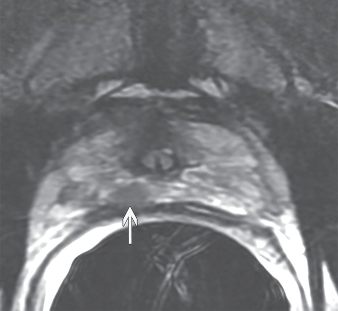
Figure 4a:

Prostate cancer in 59-year-old man. (a) Axial and (b) coronal T2-weighted MR images (6000/116 [effective]) demonstrate an area of low signal intensity in the right peripheral zone, apex, that was suspicious for tumor (arrows). (c) Representative image from whole-mount step-section pathologic specimen demonstrates a Gleason score of 7 (3 + 4) tumor in the right peripheral zone (outlined in green and black), with a volume of 0.6 cm3. (Hematoxylin-eosin stain; original magnification, ×1.05.)
