Abstract
Mutations in the human FOXP2 gene cause impaired speech development and linguistic deficits, which have been best characterised in a large pedigree called the KE family. The encoded protein is highly conserved in many vertebrates and is expressed in homologous brain regions required for sensorimotor integration and motor-skill learning, in particular corticostriatal circuits. Independent studies in multiple species suggest that the striatum is a key site of FOXP2 action. Here, we used in vivo recordings in awake-behaving mice to investigate the effects of the KE-family mutation on the function of striatal circuits during motor-skill learning. We uncovered abnormally high ongoing striatal activity in mice carrying an identical mutation to that of the KE family. Furthermore, there were dramatic alterations in striatal plasticity during the acquisition of a motor skill, with most neurons in mutants showing negative modulation of firing rate, starkly contrasting with the predominantly positive modulation seen in control animals. We also observed striking changes in the temporal coordination of striatal firing during motor-skill learning in mutants. Our results indicate that FOXP2 is critical for the function of striatal circuits in vivo, which are important not only for speech but also for other striatal-dependent skills.
Keywords: Foxp2, in vivo recording, KE family, motor-skill learning, speech and language, striatum
Introduction
Speech is one of the most complex and refined motor skills that we perform. Developmental disorders of speech and language affect up to 7% of 5- to 7-year-olds1 and are known to be highly heritable.2 However, the inheritance patterns are usually complex, indicating that multiple genes are likely involved, a difficulty that has hindered investigations into the underlying molecular aetiology.3 Identification of the genetic basis of a rare monogenic form of speech and language impairment has offered a unique opportunity to study these mechanisms.4 In the large multigenerational KE family, around half the members carry a point mutation in one copy of the FOXP2 gene, yielding an arginine-to-histidine substitution that disturbs the DNA-binding domain of the encoded transcription factor.4, 5 These people have difficulty mastering the sequences of orofacial motor movements necessary for fluent speech,6 a feature that is proposed to be a core deficit of the disorder.7 The speech motor deficits are accompanied by other expressive and receptive problems in both oral and written language.6 Additional families have been identified with speech and language problems caused by FOXP2 mutations,8 but the KE family remains the most well-studied example to date.
In the developing human brain, FOXP2 is expressed in neuronal subpopulations of the cortex, basal ganglia, thalamus and cerebellum; regions that are known to be important for motor-related functions.9 The striatum is the main recipient of cortical input to the basal ganglia, and neuroimaging studies of the KE family have implicated this structure in FOXP2-related speech and language deficits, uncovering changes in striatal grey-matter density,10 as well as altered striatal activation during language-based tasks.11, 12 Importantly, striatal lesions in humans, resulting from brain trauma or stroke, can result in verbal aphasia and amusica (a pitch-processing deficit).13, 14 The FoxP2 protein sequence and neural expression pattern are highly conserved in a number of other vertebrates, including songbirds and rodents,9, 15, 16, 17, 18 suggesting some common ancestral functions. (The standard nomenclature for FOX transcription factor proteins recommended by the winged helix/forkhead nomenclature committee19 is FOXP2 for humans, Foxp2 for mice and FoxP2 for all other species, with genes and RNA displayed in italics. To maintain brevity, we have used just FoxP2 when referring to several species.) For basic studies investigating the role of FoxP2 in skill learning and striatal function, these species are more amenable to experimentation.
We previously generated mutant mice (Foxp2-R552H line) that carry an identical mutation to that of the KE family.20 Homozygous mutants show severe developmental delay and motor impairment, dying 3–4 weeks after birth. Heterozygous mutants have motor-skill deficits, and slice recordings show impaired long-term depression at glutamatergic inputs into the striatum.20 Conversely, mice with a partially humanised version of Foxp2 have increased striatal long-term depression at corticostriatal synapses.21 Furthermore, in the zebra finch, FoxP2 knockdown in area X, a striatal nucleus essential for song development, impairs vocal learning.22
The evidence to date suggests that FoxP2 has a role in striatal function, with potential relevance for motor-skill learning. However, it is still unclear what impact the KE-family mutation has on striatal circuits during the learning of rapid motor sequences such as those required for speech. In this study, we show the consequences of the KE-family mutation on the ongoing activity of striatal circuits in vivo, and on the in vivo plasticity observed during skill learning, by performing multielectrode recordings in awake-behaving Foxp2-R552H/+ mice during training on the accelerating rotarod. This task engages the circuits required for the striatal-dependent learning of rapid motor sequences,23 which are likely to be functioning abnormally in this mouse line20 and which have been proposed to be dysfunctional in affected members of the KE family.7 We found that Foxp2-R552H/+ mice displayed abnormally high ongoing striatal activity. Moreover, during acquisition of a motor skill, most striatal neurons in Foxp2-R552H/+ mice showed negative modulation of firing rate unlike the positive modulation observed in controls. Changes in the temporal coordination of striatal firing were also evident during motor-skill learning in Foxp2-R552H/+ mice. These results show that Foxp2 is critical for the normal functioning of striatal circuits during the acquisition of motor skills.
Materials and methods
Animals
The Foxp2-R552H and Foxp2-S321X lines were maintained on a C57BL/6J background, and heterozygotes and wild-type littermates, of between 2 and 6 months of age, were used for behavioural and recording experiments. The generation, marker-assisted backcrossing and genotyping of this strain are fully described in the references.20, 24, 25 All procedures were approved by the National Institute on Alcohol Abuse and Alcoholism Animal Care and Use Committee.
Accelerating rotarod
A computer-interfaced rotarod (ENV-575M, Med-Associates, St Albans, VT, USA) was set to accelerate from 6 to 60 r.p.m. over a 300-s time period. Mice were trained for five consecutive days, with one daily session consisting of 10 trials separated by 300 s resting periods. Mice were placed on the rotarod and trials were deemed to have started when the rod began to turn. Trials ended when mice fell from the rod or after 300 s elapsed. Learning rate was calculated as follows (mean latency to fall trials 9 and 10)−(mean latency to fall trials 1 and 2)/9 (number of intertrial intervals).
In vivo extracellular recording
Implantation of multielectrode arrays and in vivo recording of neural activity in mice training on the accelerating rotarod were performed essentially as described in the reference.23 Arrays had two rows of eight polyamide-coated tungsten wires, which were 35 μm in diameter with 90° blunt cut tips. Wires were separated by a 250-μm gap, both within a row and across rows. The centre of each array was surgically positioned 2.2–2.4 mm below the surface of the brain, 0.5 mm anterior to and 1.7 mm lateral to bregma. Final electrode positioning in the dorsomedial striatum was determined by stereotaxic coordinates and by neural activity, which was monitored as the electrodes were lowered into the brain. Mice were allowed to recover for at least 10 days post surgery after which single-unit and multi-unit activity was recorded using the Plexon data acquisition system (Plexon Inc., Dallas, TX, USA). The start and finish of each trial were signalled to the MAP recording system (Plexon) as events. The recording cables were held by a custom-made pulley system, allowing mice to run and fall relatively freely. Mice remained at the bottom of the apparatus during intertrial periods, usually in a fairly immobile state. Neural activity was initially sorted using an online sorting algorithm and then resorted, after recording, using an offline sorting algorithm based on waveform, amplitude and interspike interval histogram (Plexon). The total number of units recorded on days 1–5 was 56, 78, 80, 72 and 70, respectively, for wild-type mice and 55, 54, 64, 58 and 48, respectively, for Foxp2-R552H/+ mice.
Neural data analyses
Data analyses were carried out using Neuroexplorer (Nex Technologies, Littleton, MA, USA) and custom-written programs in Matlab (The Mathworks Inc., Natick, MA, USA).
LFP power spectrum
The local field potential (LFP) power spectrum was estimated for 2 s sliding windows with 1-s steps by Welch's method; the parameters chosen allowed for a frequency resolution of 0.5 Hz. The percentage power for the following frequency bands was calculated; delta 1.5–4 Hz, theta 4.5–9 Hz and gamma 30–55 Hz.
Spike-triggered average
Spike-triggered averages of the LFP were calculated for −3 s to 3 s intervals. A spike-triggered average was considered significant when 20 or more 1 ms bins within the interval −0.1 s to 0.1 s lay outside the maximum or minimum values of the intervals −3 to −1 s and 1 s to 3 s.
Statistics
Statistical analyses were carried out using SPSS (SPSS Inc., Armonk, NY, USA) and Neuroexplorer. Task-related neurons were identified as having significant positive or negative modulation of firing rate during running. This was determined using a 99% confidence interval, which was calculated from the firing rate during the intertrial intervals. All results were averaged per subject, and subsequent analyses were performed on each subject's mean value. Data were analysed using repeated measures analysis of variance, and post hoc tests (Fisher's least significant difference) were applied when appropriate. Planned comparisons were used to analyse the LFP data.
Histology
Mice were perfused with 4% paraformaldehyde, and brains were removed and postfixed overnight in 4% paraformaldehyde. Electrode placement was verified by Nissl staining of 50 μm vibratome sections. For Foxp2 and interneuron immunohistochemistry, postfixed brains were transferred to 30% sucrose, and 40 μm coronal cryosections were cut on a sliding microtome. Immunohistochemistry was carried out using the Vectastain ABC or Vector MOM kit (both from Vector Laboratories, Peterborough, UK). Free-floating sections were incubated with Foxp2 (1:2000, goat N16, Santa Cruz Biotechnology Inc., Santa Cruz, CA, USA), ChAT (1:100, goat, Chemicon, Temecula, CA, USA) or PV (1:5000, mouse, Swant, Marly, Switzerland) antibodies. Proteins were visualised using the Vector VIP substrate purple kit (Vector Laboratories) or the DAB substrate kit (Fisher Scientific, Loughborough, UK). When looking at Foxp2 colocalisation with parvalbumin (PV) or cholineacetyltransferase (ChAT), nuclei were marked using methyl green counterstain (Vector Laboratories). To ascertain interneuron numbers, serial sections were taken throughout the striatum and stained alternatively with PV and ChAT antibodies. The top half of the striatum was considered to be dorsal, and this was subdivided into DM, DI and DL. The area of each region of interest and the number of positive cells within it were determined using ImageJ (http://rsbweb.nih.gov/ij/). Cell density (cells mm−2) was calculated by dividing the cell count by the area.
Results
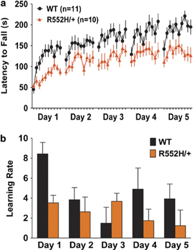
We first confirmed that mice carrying an aetiological Foxp2 mutation display impaired motor-skill learning.20 Mice were trained for five consecutive days on an accelerating rotarod, with one daily session consisting of 10 trials (running periods), separated by 300 s intervals. This intensive schedule uses a higher acceleration rate (6–60 r.p.m.) than previous experiments,20, 23, 26 and robustly shows the motor-skill learning deficits of Foxp2-R552H/+ animals. In line with previous findings,20 latency to fall was reduced in Foxp2-R552H/+ mice (F1,19=9.87, P<0.05; Figure 1a). A significant difference in performance was evident early in training (post hoc, P<0.05, comparison of third and fourth trials of session 1), and although Foxp2-R552H/+ mice improved, they never reached the fall times of wild-type animals (post hoc, P<0.05, comparison of final two trials of session 5). The early deficits observed in Foxp2-R552H/+ mice do not seem to stem from alterations in basal motor coordination, as performance during the first two trials of session 1 was not impaired (post hoc, P>0.05). Latency to fall from a fixed speed rat rotarod, a task that addresses motor coordination, was also not significantly different between Foxp2-R552H/+ and wild-type animals (Supplementary Figure 1a). Rather, the deficits appear to be due to a slower rate of learning during the first session (post hoc, P<0.05; Figure 1b). Notably, Foxp2-R552H/+ mice do not have generalised striatal-dependent deficits in learning or motivation, because they learn to press a lever to earn a food reward in the same way as wild-type animals in an operant task, which involves the striatum27 (Supplementary Figure 2). Furthermore, the severity of the deficits in rotarod skill learning appear to be related to the KE-family mutation, as the deficits observed in Foxp2-S321X/+ mice, which carry a premature stop codon and have a half dosage of Foxp2 protein, were less severe than those observed in Foxp2-R552H/+ mice (Supplementary Figure 3). Foxp2-S321X/+ animals also have no impairment in basal motor coordination (Supplementary Figure 1b).
Figure 1.
Foxp2-R552H/+ mice show impaired motor-skill learning. (a) Latency to fall of wild-type and Foxp2-R552H/+ mice during training on the accelerating rotarod. Mice received 10 trials a day for five consecutive days. (b) Rate of learning in wild-type and Foxp2-R552H/+ mice during training on the accelerating rotarod. Error bars represent s.e.m.
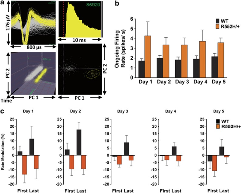
We therefore examined the impact of the KE-family mutation on striatal activity in vivo by performing multielectrode recordings in awake-behaving Foxp2-R552H/+ mice (n=5) and control wild-type littermates (n=6). As skill learning deficits in Foxp2-R552H/+ mice are apparent early in training, we targeted electrode arrays bilaterally to the dorsomedial striatum (Supplementary Figure 4), which is preferentially engaged during this initial phase of learning.26 After recovery from electrode implantation surgery, neuronal activity (Figure 2a) and LFP oscillations were measured during training on the accelerating rotarod.
Figure 2.
Foxp2-R552H/+ mice have abnormally high ongoing striatal activity and show negative modulation of firing rate during skill learning. (a) Example of a single-unit waveform (top left). Units were distinguished from each other and from noise using interspike interval histograms (top right) and two-dimensional cluster separation based on principal component analysis, where the circled yellow cluster represents the unit and the grey cluster noise (bottom right). The stability of a unit recording over the course of a session was assessed using three-dimensional cluster displays with a time axis (bottom left). (b) Average ongoing firing rate in wild-type and Foxp2-R552H/+ mice. (c) Average firing rate modulation during running compared with intertrial intervals, during the first and last trials of a session, in wild-type and Foxp2-R552H/+ mice. Error bars represent s.e.m.
We first investigated changes in ongoing striatal activity in Foxp2-R552H/+ mice by measuring the firing rate of striatal neurons during a 5-min period before the initial trial of each session, while mice were positioned at the bottom of the apparatus in a quiet but awake state (periods of no locomotion but occasional head movements). Intriguingly, we found that striatal neurons in Foxp2-R552H/+ mice showed higher ongoing firing rates than those in wild-type controls across the 5 days of recording (F1,9=5.54, P<0.05; Figure 2b). This is interesting because striatal medium spiny neurons (MSNs) usually display low in vivo firing rates,28 a condition that is thought to be critical for normal action selection and movement.29
We next investigated the effects of the Foxp2 mutation on the dynamic changes in striatal plasticity that are observed during motor-skill learning.26 We determined whether the firing rate of striatal neurons increased or decreased during running compared with the intertrial intervals by calculating the modulation in firing rate ((run−interval)/(run+interval) × 100), at the beginning and end of each session (first two trials of session—‘first' last two trials of session—‘last').23 Consistent with previous experiments,26 the average firing rate in wild-type dorsomedial striatum neurons increased during running compared with intertrial intervals in early training sessions, as revealed by positive modulation of firing rate (Figure 2c). However, striatal neurons in Foxp2-R552H/+ mice were substantially different (F1,9=7.68, P<0.05), showing predominantly negative modulation during running. This negative modulation was most evident at early stages and progressively diminished, with the last trials of day 5 being significantly different from the first trials of day 1 (post hoc, P<0.05), which is consistent with the dorsomedial striatum becoming less engaged with training.26 Furthermore, in wild-type mice the firing rate was significantly more modulated during the last trials of a session than the first (F1,5=27.88, P<0.05), revealing within-session plasticity. This plasticity was not observed in Foxp2-R552H/+ animals, where no significant differences between the first and last trials of a session were found (F1,4=3.96, P>0.05).
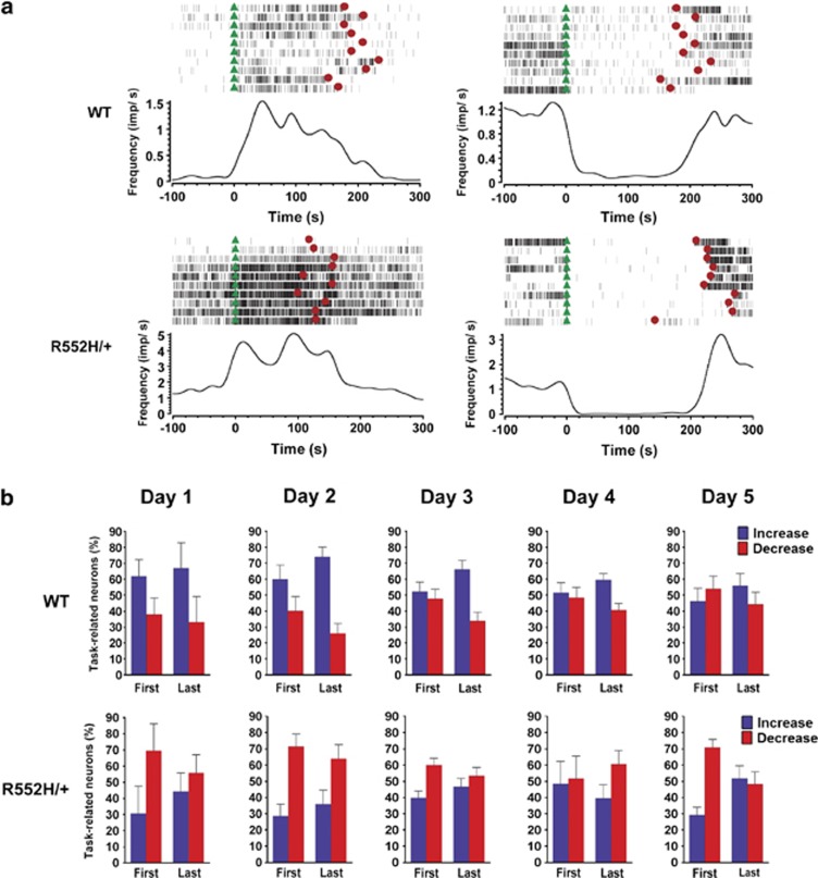
To examine how these differences in firing rate modulation emerge during skill learning, we determined the proportion of neurons whose firing rate changed significantly during running compared with the intertrial intervals (classified as task related, see Materials and methods and Figure 3a). The percentage of task-related neurons recorded was similar in wild-type and Foxp2-R552H/+ animals and remained relatively constant throughout the training (Supplementary Figure 5). However, when task-related neurons were divided into those that increased or decreased firing rate during running, more neurons were found to increase firing rate in wild-type mice (F1,10=22.25, P<0.05) and to decrease firing rate in Foxp2-R552H/+ mice (F1,8=7.12, P<0.05; Figure 3b). These results are consistent with the differences in firing rate modulation observed between Foxp2-R552H/+ mutants and controls (Figure 2c).
Figure 3.
A higher proportion of task-related neurons decrease firing rate during skill learning in Foxp2-R552H/+ mice. (a) Examples of neurons from wild-type (top) and Foxp2-R552H/+ (bottom) mice that increased (left) or decreased (right) firing rate (impulses per second) during running compared with intertrial intervals. Green triangles represent the start of a trial and red circles the end. (b) Percentage of task-related neurons that increased or decreased firing rate during running compared with intertrial intervals, during the first and last trials of a session, in wild-type (top panel) and Foxp2-R552H/+ (bottom panel) mice. Error bars represent s.e.m.
We next investigated whether the negative modulation of firing rate observed in Foxp2-R552H/+ during running on the rotarod was related to, or independent from, the higher ongoing activity of striatal neurons in these animals. It is plausible that neurons with high ongoing firing rates could be unable to increase firing further during running, and could even compensate by decreasing firing, hence resulting in the observed negative modulation of firing rate. However, we found that Foxp2-R552H/+ neurons with high ongoing firing rates were not modulated differently during running compared with those with lower firing rates (Supplementary Figure 6). Furthermore, one would expect that if a significant proportion of Foxp2-R552H/+ neurons were unable to modulate firing rate during running, then there would be a corresponding reduction in the number of task-related neurons, which is not observed (Supplementary Figure 5). Finally, if Foxp2-R552H/+ neurons with high baseline firing rates were not able to positively modulate firing during running, then the most prominent differences in rate modulation between genotypes would occur when the greatest levels of positive modulation are observed in wild-type mice and vice versa. However, as previously discussed, firing rate is significantly more modulated in the last trials of a session compared with the first in wild-type mice, but this within-session plasticity is clearly not evident in Foxp2-R552H/+ mice (Figure 2c). Taken together, these data indicate that the changes in striatal activity observed during skill learning in Foxp2-R552H/+ mice are not simply a direct consequence of the increased ongoing firing.
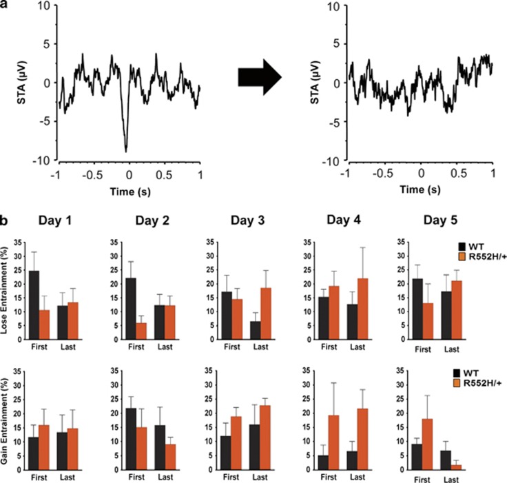
In addition to changes in firing frequency, temporal coordination of striatal activity has also been shown to be important for striatal functions, such as action selection and movement.29, 30 We therefore measured LFP oscillations, which mostly reflect the coordinated input into an area, in Foxp2-R552H/+ and control mice. We observed no significant differences in power at any of the frequencies examined, despite a tendency for higher power gamma oscillations in Foxp2-R552H/+ mice on day 1 (Supplementary Figure 7). We then investigated the temporal relationship between the oscillatory phase of the LFP (reflecting mostly input) and the firing of individual striatal neurons (reflecting output). We determined whether a neuron fired preferentially during a particular phase of the LFP (was ‘entrained' to the LFP) during running and intertrial intervals (Supplementary Figure 8), using spike-triggered averages of the LFP oscillations (see Materials and methods and refs 29,30; Figure 4a). We calculated how many neurons became entrained or lost entrainment to the LFP oscillations during running compared with the intertrial intervals. In wild-type mice, more neurons lost entrainment to the LFP oscillations during running in the first trials of a session compared with the last (F1,5=9.00, P<0.05), but in Foxp2-R552H/+ animals this within-session plasticity was not evident (F1,4=0.80, P>0.05) (Figure 4b). In addition, more neurons became entrained to the LFP during running in Foxp2-R552H/+ than in wild-type mice (F1,8=6.21, P<0.05), an effect that was particularly apparent during the late phase of training. These results show that the temporal coordination of striatal input and output differs between Foxp2-R552H/+ and wild-type mice.
Figure 4.
The temporal coordination of striatal activity is altered during skill learning in Foxp2-R552H/+ mice. (a) Spike-triggered average histograms showing a neuron that is entrained to the local field potential (LFP) during intertrial intervals (left) but loses entrainment during running periods (right). (b) Percentage of neurons that lost entrainment from (top panel) or gained entrained to (bottom panel) the LFP between intertrial interval and running during the first and last trials of a session. Error bars represent s.e.m.
Discussion
Previous neuroimaging studies of the KE family as well as experiments in mice and songbirds have highlighted possible roles for FoxP2 in the striatum, with potential relevance for motor-skill learning. However, the impact of the KE-family mutation on striatal function during the learning of rapid motor sequences was unclear. In this study, we demonstrate in vivo that this mutation dramatically alters the functioning of striatal circuits during motor-skill learning. We found that in contrast to control animals, which showed positive modulation of firing rate during skill acquisition, Foxp2-R552H/+ mice exhibited negative modulation of firing rate. We also found alterations in the temporal coordination of striatal activity in Foxp2-R552H/+ mice compared with controls.
We found Foxp2-R552H/+ mice to have abnormally high ongoing striatal activity. Striatal MSNs, which represent the vast majority of striatal neurons, usually display low in vivo firing rates.28 This property, together with appropriate levels of concurrent activity between MSNs, is thought to be critical for normal action selection.29 The high ongoing striatal firing rate in Foxp2-R552H/+ mice could therefore result in increased collateral inhibition, which may interfere with action selection and plasticity processes in this brain region. Our previous ex vivo data, which shows strong impairment of long-term depression at glutamatergic synapses onto MSNs in Foxp2-R552H/+ mice,20 is consistent with increased ongoing striatal activity. Nevertheless, it is important to note that besides glutamatergic input MSNs receive dopaminergic input from the mid-brain, and GABAergic input from interneurons and from other MSNs.28 Hence, elevated MSN activity could also result from changes in the strength of any of these other inputs (for example, increased dopaminergic input or decreased GABAergic input), as well as increased neuronal excitability. However, we found only modest colocalisation of Foxp2 with PV or ChAT, markers for the main subtypes of striatal interneurons, and there were equivalent numbers of PV- or ChAT-positive cells in the striata of wild-type and Foxp2-R552H/+ mice (Supplementary Figure 9), suggesting that striatal interneurons are unlikely to have a direct influence. We also showed that the increased ongoing striatal activity and the negative firing rate modulation during skill acquisition observed in Foxp2-R552H/+ mice are not correlated. This suggests that the underlying mechanisms of the two phenomena are dissociable and raises the possibility that they are mechanistically distinct. Although any of the mechanisms outlined above could contribute to the firing rate modulation differences that we observed, it seems unlikely that impaired long-term depression could directly give rise to an increase in negative modulation of firing rate. Selective deletion of Foxp2 will be a useful tool to investigate how specific cells types and brain regions contribute to changes in striatal activity as well as other phenotypes.31
Foxp2-S321X/+ and Foxp2-R552H/+ mice express a half dosage of normal wild-type Foxp2 protein, and thus both could potentially display haploinsufficiency. However, Foxp2-R552H/+ mice also simultaneously express a full-length mutant product with abnormal functional properties. In vitro studies indicate that the R552H mutation disrupts the DNA-binding and transactivation functions of Foxp2,5 suggesting that the mutant protein could act in a dominant-negative manner. Foxp2 has been shown to function as a homodimer, and also as a heterodimer with Foxp1 or Foxp4;32 hence dominant-negative effects could be exerted through protein–protein interactions or direct disturbance of transcription. In support of this proposal, we observed a less severe motor-skill learning phenotype in Foxp2-S321X/+ mice than in Foxp2-R552H/+ mice (Figure 1 and Supplementary Figure 3); further work will be necessary to determine whether this is indeed the case. Studies using chromatin immunoprecipitation, followed by microarray analysis, have been used to identify direct neural targets of FOXP2 and have indicated roles in neurodevelopment, neurite outgrowth, axon guidance, neurotransmission and synaptic plasticity.33, 34 The Foxp2-R552H mutation is likely to cause inappropriate or inefficient regulation of downstream targets mediating these processes,35 resulting in the dramatic changes in striatal neural activity that we observe.
In conclusion, the use of multielectrode recordings in awake-behaving mice with an aetiological Foxp2 mutation has allowed for a detailed assessment of its impact on the neuronal dynamics of striatal circuits during the learning of rapid motor sequences, beyond the resolution of human neuroimaging. The resulting data give novel insight into the mechanistic consequences of this mutation, which is important not only for speech but also for other striatal-dependent skills. It will be interesting to see whether the increased ongoing striatal activity and generalised motor-skill learning deficits observed in mice extend to people with disorders attributed to FOXP2 dysfunction. Although it is difficult to perform invasive electrophysiology studies on such people, our data suggest that resting state functional imaging and relatively demanding tests of motor-skill learning ability could be highly informative.
Acknowledgments
We thank Sonali Darbar, Rathi Puliyadi, Terrell D Holloway and Amber Luo for excellent technical assistance; John Burkhardt and Shih-Chieh Lin for help with Matlab; Emma Doran for running the operant learning task; and the staffs of the British Heart Foundation Functional Genetics Facility and the NIAAA vivarium for mouse husbandry. This work was supported by a Human Frontier Science Program short-term fellowship (to CAF); a Royal Society Research Fellowship (to SEF); the Wellcome Trust (075491/Z/04; 080971); the Division of Intramural Clinical and Basic Research of the National Institute on Alcohol Abuse and Alcoholism, US National Institutes of Health; the Champalimaud Neuroscience Programme; the European Research Council (STG 243393); and the Max Planck Society.
The authors declare no conflict of interest.
Footnotes
Supplementary Information accompanies the paper on the Molecular Psychiatry website (http://www.nature.com/mp)
Supplementary Material
References
- Tomblin JB, Records NL, Buckwalter P, Zhang X, Smith E, O'Brien M. Prevalence of specific language impairment in kindergarten children. J Speech Lang Hear Res. 1997;40:1245–1260. doi: 10.1044/jslhr.4006.1245. [DOI] [PMC free article] [PubMed] [Google Scholar]
- Bishop DV. Genetic and environmental risks for specific language impairment in children. Philos Trans R Soc Lond B Biol Sci. 2001;356:369–380. doi: 10.1098/rstb.2000.0770. [DOI] [PMC free article] [PubMed] [Google Scholar]
- Fisher SE, Lai CS, Monaco AP. Deciphering the genetic basis of speech and language disorders. Annu Rev Neurosci. 2003;26:57–80. doi: 10.1146/annurev.neuro.26.041002.131144. [DOI] [PubMed] [Google Scholar]
- Lai CS, Fisher SE, Hurst JA, Vargha-Khadem F, Monaco AP. A forkhead-domain gene is mutated in a severe speech and language disorder. Nature. 2001;413:519–523. doi: 10.1038/35097076. [DOI] [PubMed] [Google Scholar]
- Vernes SC, Nicod J, Elahi FM, Coventry JA, Kenny N, Coupe AM, et al. Functional genetic analysis of mutations implicated in a human speech and language disorder. Hum Mol Genet. 2006;15:3154–3167. doi: 10.1093/hmg/ddl392. [DOI] [PubMed] [Google Scholar]
- Watkins KE, Dronkers NF, Vargha-Khadem F. Behavioural analysis of an inherited speech and language disorder: comparison with acquired aphasia. Brain. 2002;125 (Pt 3:452–464. doi: 10.1093/brain/awf058. [DOI] [PubMed] [Google Scholar]
- Vargha-Khadem F, Gadian DG, Copp A, Mishkin M. FOXP2 and the neuroanatomy of speech and language. Nat Rev Neurosci. 2005;6:131–138. doi: 10.1038/nrn1605. [DOI] [PubMed] [Google Scholar]
- MacDermot KD, Bonora E, Sykes N, Coupe AM, Lai CS, Vernes SC, et al. Identification of FOXP2 truncation as a novel cause of developmental speech and language deficits. Am J Hum Genet. 2005;76:1074–1080. doi: 10.1086/430841. [DOI] [PMC free article] [PubMed] [Google Scholar]
- Lai CS, Gerrelli D, Monaco AP, Fisher SE, Copp AJ. FOXP2 expression during brain development coincides with adult sites of pathology in a severe speech and language disorder. Brain. 2003;126 (Pt 11:2455–2462. doi: 10.1093/brain/awg247. [DOI] [PubMed] [Google Scholar]
- Watkins KE, Vargha-Khadem F, Ashburner J, Passingham RE, Connelly A, Friston KJ, et al. MRI analysis of an inherited speech and language disorder: structural brain abnormalities. Brain. 2002;125 (Pt 3:465–478. doi: 10.1093/brain/awf057. [DOI] [PubMed] [Google Scholar]
- Vargha-Khadem F, Watkins KE, Price CJ, Ashburner J, Alcock KJ, Connelly A, et al. Neural basis of an inherited speech and language disorder. Proc Natl Acad Sci USA. 1998;95:12695–12700. doi: 10.1073/pnas.95.21.12695. [DOI] [PMC free article] [PubMed] [Google Scholar]
- Liegeois F, Baldeweg T, Connelly A, Gadian DG, Mishkin M, Vargha-Khadem F. Language fMRI abnormalities associated with FOXP2 gene mutation. Nat Neurosci. 2003;6:1230–1237. doi: 10.1038/nn1138. [DOI] [PubMed] [Google Scholar]
- Jarvis ED. Learned birdsong and the neurobiology of human language. Ann NY Acad Sci. 2004;1016:749–777. doi: 10.1196/annals.1298.038. [DOI] [PMC free article] [PubMed] [Google Scholar]
- Lieberman P. On the nature and evolution of the neural bases of human language. Am J Phys Anthropol. 2002;35 (Suppl:36–62. doi: 10.1002/ajpa.10171. [DOI] [PubMed] [Google Scholar]
- Ferland RJ, Cherry TJ, Preware PO, Morrisey EE, Walsh CA. Characterization of Foxp2 and Foxp1 mRNA and protein in the developing and mature brain. J Comp Neurol. 2003;460:266–279. doi: 10.1002/cne.10654. [DOI] [PubMed] [Google Scholar]
- Takahashi K, Liu FC, Hirokawa K, Takahashi H. Expression of Foxp2, a gene involved in speech and language, in the developing and adult striatum. J Neurosci Res. 2003;73:61–72. doi: 10.1002/jnr.10638. [DOI] [PubMed] [Google Scholar]
- Teramitsu I, Kudo LC, London SE, Geschwind DH, White SA. Parallel FoxP1 and FoxP2 expression in songbird and human brain predicts functional interaction. J Neurosci. 2004;24:3152–3163. doi: 10.1523/JNEUROSCI.5589-03.2004. [DOI] [PMC free article] [PubMed] [Google Scholar]
- Haesler S, Wada K, Nshdejan A, Morrisey EE, Lints T, Jarvis ED, et al. FoxP2 expression in avian vocal learners and non-learners. J Neurosci. 2004;24:3164–3175. doi: 10.1523/JNEUROSCI.4369-03.2004. [DOI] [PMC free article] [PubMed] [Google Scholar]
- Kaestner KH, Knochel W, Martinez DE. Unified nomenclature for the winged helix/forkhead transcription factors. Genes Dev. 2000;14:142–146. [PubMed] [Google Scholar]
- Groszer M, Keays DA, Deacon RM, de Bono JP, Prasad-Mulcare S, Gaub S, et al. Impaired synaptic plasticity and motor learning in mice with a point mutation implicated in human speech deficits. Curr Biol. 2008;18:354–362. doi: 10.1016/j.cub.2008.01.060. [DOI] [PMC free article] [PubMed] [Google Scholar]
- Enard W, Gehre S, Hammerschmidt K, Holter SM, Blass T, Somel M, et al. A humanized version of Foxp2 affects cortico-basal ganglia circuits in mice. Cell. 2009;137:961–971. doi: 10.1016/j.cell.2009.03.041. [DOI] [PubMed] [Google Scholar]
- Haesler S, Rochefort C, Georgi B, Licznerski P, Osten P, Scharff C. Incomplete and inaccurate vocal imitation after knockdown of FoxP2 in songbird basal ganglia nucleus Area X. PLoS Biol. 2007;5:e321. doi: 10.1371/journal.pbio.0050321. [DOI] [PMC free article] [PubMed] [Google Scholar]
- Costa RM, Cohen D, Nicolelis MA. Differential corticostriatal plasticity during fast and slow motor skill learning in mice. Curr Biol. 2004;14:1124–1134. doi: 10.1016/j.cub.2004.06.053. [DOI] [PubMed] [Google Scholar]
- Keays DA, Clark TG, Campbell TG, Broxholme J, Valdar W. Estimating the number of coding mutations in genotypic and phenotypic driven N-ethyl-N-nitrosourea (ENU) screens: revisited. Mamm Genome. 2007;18:123–124. doi: 10.1007/s00335-006-0065-z. [DOI] [PubMed] [Google Scholar]
- Coghill EL, Hugill A, Parkinson N, Davison C, Glenister P, Clements S, et al. A gene-driven approach to the identification of ENU mutants in the mouse. Nat Genet. 2002;30:255–256. doi: 10.1038/ng847. [DOI] [PubMed] [Google Scholar]
- Yin HH, Mulcare SP, Hilario MR, Clouse E, Holloway T, Davis MI, et al. Dynamic reorganization of striatal circuits during the acquisition and consolidation of a skill. Nat Neurosci. 2009;12:333–341. doi: 10.1038/nn.2261. [DOI] [PMC free article] [PubMed] [Google Scholar]
- Yin HH, Knowlton BJ. The role of the basal ganglia in habit formation. Nat Rev Neurosci. 2006;7:464–476. doi: 10.1038/nrn1919. [DOI] [PubMed] [Google Scholar]
- Kreitzer AC, Malenka RC. Striatal plasticity and basal ganglia circuit function. Neuron. 2008;60:543–554. doi: 10.1016/j.neuron.2008.11.005. [DOI] [PMC free article] [PubMed] [Google Scholar]
- Costa RM, Lin SC, Sotnikova TD, Cyr M, Gainetdinov RR, Caron MG, et al. Rapid alterations in corticostriatal ensemble coordination during acute dopamine-dependent motor dysfunction. Neuron. 2006;52:359–369. doi: 10.1016/j.neuron.2006.07.030. [DOI] [PubMed] [Google Scholar]
- Burkhardt JM, Jin X, Costa RM. Dissociable effects of dopamine on neuronal firing rate and synchrony in the dorsal striatum. Front Integr Neurosci. 2009;3:28. doi: 10.3389/neuro.07.028.2009. [DOI] [PMC free article] [PubMed] [Google Scholar]
- French CA, Groszer M, Preece C, Coupe AM, Rajewsky K, Fisher SE. Generation of mice with a conditional Foxp2 null allele. Genesis. 2007;45:440–446. doi: 10.1002/dvg.20305. [DOI] [PMC free article] [PubMed] [Google Scholar]
- Li S, Weidenfeld J, Morrisey EE. Transcriptional and DNA binding activity of the Foxp1/2/4 family is modulated by heterotypic and homotypic protein interactions. Mol Cell Biol. 2004;24:809–822. doi: 10.1128/MCB.24.2.809-822.2004. [DOI] [PMC free article] [PubMed] [Google Scholar]
- Spiteri E, Konopka G, Coppola G, Bomar J, Oldham M, Ou J, et al. Identification of the transcriptional targets of FOXP2, a gene linked to speech and language, in developing human brain. Am J Hum Genet. 2007;81:1144–1157. doi: 10.1086/522237. [DOI] [PMC free article] [PubMed] [Google Scholar]
- Vernes SC, Spiteri E, Nicod J, Groszer M, Taylor JM, Davies KE, et al. High-throughput analysis of promoter occupancy reveals direct neural targets of FOXP2, a gene mutated in speech and language disorders. Am J Hum Genet. 2007;81:1232–1250. doi: 10.1086/522238. [DOI] [PMC free article] [PubMed] [Google Scholar]
- Fisher SE, Scharff C. FOXP2 as a molecular window into speech and language. Trends Genet. 2009;25:166–177. doi: 10.1016/j.tig.2009.03.002. [DOI] [PubMed] [Google Scholar]
Associated Data
This section collects any data citations, data availability statements, or supplementary materials included in this article.


