Herein described is a simple, safe extracorporeal knotting technique.
Keywords: Laparoscopic surgery, Extracorporeal knotting technique
Abstract
Introduction:
Extracorporeal knotting in laparoscopic surgery can be used in certain situations or as a bridge to mastering more technically demanding intracorporeal suturing. We describe a simple, easy, and safe extracorporeal knotting technique.
Technique Description:
A simple knotting technique, borrowed from the art of tatting, is used.
Discussion:
This very simple and safe technique has been successfully followed in more than 50 cases for tying an extracorporeal knot. Its advantage is that any number of additional knots can be tied and easily slid down in a single maneuver.
INTRODUCTION
Minimal access surgery (MAS) has become the preferred technique for many surgeries, because it is less painful, permits earlier return to work, provides better cosmesis, and is more acceptable to the patient than traditional surgeries. Advanced MAS requires that the surgeon be adept at intracorporeal suturing and knotting. However, mastering this skill is a difficult process with a long and steep learning curve.1 Extracorporeal knots permit the knot to be tied outside and then, by using a knot pusher, applied snugly inside the body.
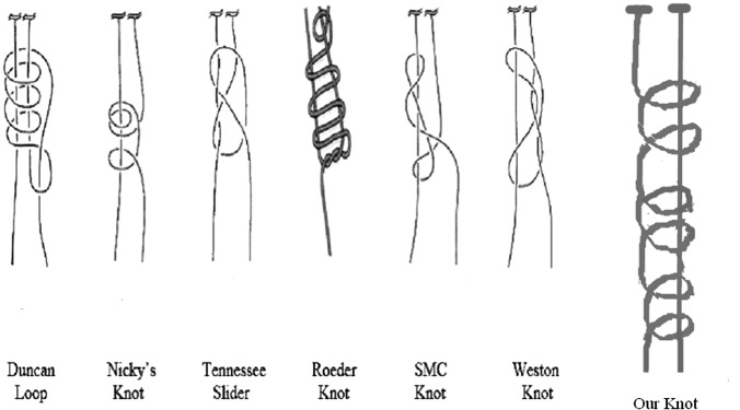
There are many variants of the extracorporeal slip knot: Roeder knot, Duncan loop, Nicky's knot, Tennessee slider, SMC knot, Weston knot, Meltzer, Tayside knot, and others (Figure 1). These knotting techniques are variations in turn around the axis or the number of reversed half hitches on alternating post. Each technique has its proponents and some have been modified for improvement, but there are constraints with these techniques in terms of size of suture material, the numbers of knots that can be applied at once, and the ease of sliding in the extracorporeal knot.2 We present a knot that is simple, easy, and fulfills all the qualities of an extracorporeal knot.
Figure 1.
Diagrammatic depiction of various extracorporeal knots used.
TECHNIQUE
Our technique of knot making is derived from traditional tatting work that has been used for ages for making lace and other handicraft items.3
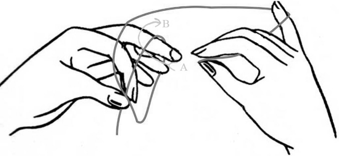
A long piece (at least 20cm) of suture material of choice (according to indication) is taken. It is divided into 2 equal limbs. Limb B is looped around the medial 3 fingers of the left hand, and the 2 limbs are crossed and held between the left thumb and index finger. The segment of suture running between the pinched thumb–index finger and the middle finger is considered the axis on which the knot is to be made. Limb B is then passed over the axis from left to right to create a loop, and its end is passed through this loop in a clockwise direction, from the front backwards, to create the first half hitch (Figure 2). The hitch is slid down along the axis to be pinched between the thumb and index finger.
Figure 2.
First step in tying the knot, A–B shows the direction of passing the thread through the loop.
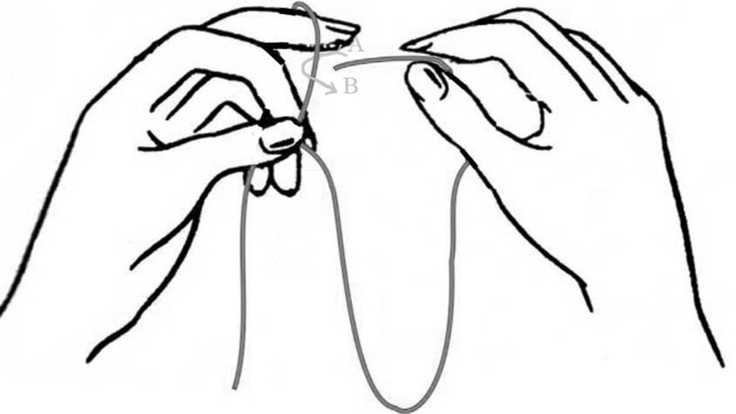
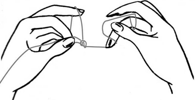
The same process is repeated by bringing limb B in front of the axis from right to left to create a loop, and its end is passed in a counterclockwise direction, through the loop from the back forward to create the second half hitch (Figure 3). This is also slid down along the axis to be pinched between the thumb and index finger. This constitutes the first complete knot (Figure 4).
Figure 3.
Second step in tying the knot, A–B shows the direction of passing the thread through the loop.
Figure 4.
First completed knot.
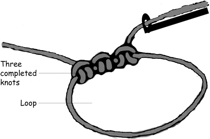
The above process is repeated twice more to fashion out 3 knots at the base of the loop. Care must be taken not to excessively pull limb B, because this would result in locking of the knot. The final configuration has 3 knots at the base of the loop, each with 2 half hitches thrown in opposite directions (Figure 5). Once the loop has been slid into the abdomen and placed at the desired site, traction is applied on both the limbs to tighten and lock the knot. This secures the knot and prevents it from slipping.
Figure 5.
Three knots at the base of the loop, each with 2 half hitches thrown in opposite directions and the knot pusher.
RESULTS
This easy and simple technique has been successfully followed in more than 50 cases.
DISCUSSION
Any surgeon's concern about the ease and safety of laparoscopic knotting is natural, and knots performed laparoscopically must be as safe as those traditionally performed. A knot should secure proper tissue approximation, and be simple, easy, quick, and reliable.
Most of the extracorporeal knots in use have 1 or 2 half hitches in the completed knot (Figure 5; modified from Khattab2). If required, in order to increase knot security,4 additional knots may be placed with these techniques; but they have to be individually placed extracorporeally and pushed into the abdomen, thereby increasing the mechanical stress on the suture and time spent. The major advantage in our technique is that any number of additional knots may be tied extracorporeally and pushed into the abdomen in a single maneuver. These knots slide down with equal ease, thus minimizing the risk of traction-induced trauma and of risk of stitches cutting through. Our technique can be used with sutures of different sizes and materials without affecting the knot security and loop security or the ability to slide the knot.
We tested the strength of the knot using 1-0 polyglactin braided, silk 1-0, and 1-0 polypropylene monofilament suture materials and compared them with that of the standard Roeder's knot using the same suture materials. Knot security was assessed by applying distracting force on the knot using a Universal Tensile Strength Testing Machine (Universal Textile Industries, Faridabad, India: Model no. CT 09-61-01-01). Using this technique with our knot, the suture broke on each occasion while the knot remained intact. For example, 1-0 polyglactin suture broke at a pressure of 80.25MPa or 818.1kg/cm2. On the contrary, Roeder's knot slipped and unraveled, while the suture material remained intact. Other studies have also shown, that under laboratory conditions, the ideal knot has 5 throws to maximize tensile strength and to reduce the risk of untying. This finding does not seem to vary with the type of suture material.4–6 Our knot has 6 half hitches that probably contribute to the security demonstrated with our knot.
The average time taken to create the final configuration of the knot was 20 seconds, while it was 30 seconds for the Roeder's knot. This knot is drawn from the art of tatting; which has been used throughout the ages. This shows that the technique is easy to learn, easy to teach, and does not have a long steep learning curve.
An additional advantage of our technique is, in addition to knot security, additional throws can be added to further increase the knot security in difficult locations without reducing the ease of sliding the knot or the tensile strength of the suture material. To our thinking, our technique has no specific limitations.
Laparoscopy has allowed surgeons to be innovative and many ideas have been incorporated from other fields. This idea originated from the art of tatting, which was learned by the first author (RK) as a child and has been used regularly in our unit in more than 50 cases.
CONCLUSION
This technique of the extracorporeal knotting is simple, easy, and reproducible with good knot and loop security and can be used with any suture material of any size.
Contributor Information
Reena Kothari, Department of Surgery, Government Netaji Subash Chandra Bose (NSCB) Medical College, Jabalpur (MP) India..
Uday Somashekar, Department of Surgery, Government Netaji Subash Chandra Bose (NSCB) Medical College, Jabalpur (MP) India..
Dhananjaya Sharma, Department of Surgery, Government Netaji Subash Chandra Bose (NSCB) Medical College, Jabalpur (MP) India..
Dileep Singh Thakur, Department of Surgery, Government Netaji Subash Chandra Bose (NSCB) Medical College, Jabalpur (MP) India..
Vinod Kumar, Department of Surgery, Government Netaji Subash Chandra Bose (NSCB) Medical College, Jabalpur (MP) India..
References:
- 1. Meilahn JE. The need for improving laparoscopic suturing and knot tying. J Laparoendosc Surg 1992;2(5):267. [DOI] [PubMed] [Google Scholar]
- 2. Khattab OS. Role of extracorporeal knots in laparoscopic surgery. http://www.laparoscopyhospital.com/extracorporael_knot.html Accessed May 19, 2011
- 3. http://www.allcrafts.net/tatting.htm; accessed May 19, 2011
- 4. Kadirkamanathan SS, Shelton JC, Hepworth CC, Laufer JG, Swain CP. A comparison of the strength of knots tied by hand and at laparoscopy. J Am Coll Surg. 1996;182(1):46–54 [PubMed] [Google Scholar]
- 5. Muffly TM, Kow N, Iqbal I, Barber MD. Minimum number of throws needed for knot security. J Surg Educ. 2011;68(2):130–133 [DOI] [PubMed] [Google Scholar]
- 6. Ivy JJ, Unger JB, Hurt J, Mukherjee D. The effect of number of throws on knot security with nonidentical sliding knots. Am J Obstet Gynecol. 2004;191(5):1618–1620 [DOI] [PubMed] [Google Scholar]


