Abstract
Darier-White disease (keratosis follicularis) is a rare disorder of keratinization involving the epidermis, mucous membranes, and nails. It is said to occur as a result of mutation in the ATP2A2 gene located on chromosome 12q23-24.1. In this article we present the case of two brothers with exacerbations of Darier-White disease who responded very well to systemic retinoids without any side effects within 2 weeks of commencing treatment.
Keywords: Darier-White disease, keratosis follicularis, isotretinoin
INTRODUCTION
Darier-White disease (keratosis follicularis) is a rare autosomal dominantly inherited disorder of keratinization involving the epidermis, mucous membranes, and nails.[1–3] Darier and White first described the condition independently in 1889.[4] It is said to occur as a result of mutation in the ATP2A2 gene located on chromosome 12q23-24.1, which encodes for the sarco/endoplasmic reticulum calcium ATPase isoform 2.[2] It has complete penetrance in adults, although with variable expression.[4] SERCA2 pump maintains a low cytosolic Ca2+ concentration required for desmosomal assembly[4] and thus it interferes with the adhesion and differentiation of keratinocytes, resulting in the disease.
Dariers disease usually begins during the first two decades of life[4] although can be overlooked until aggravated by sweating, summer, or heat.[2,5]
Clinically, it is characterized by the presence of symmetrically distributed hyperkeratotic papules with more inclination for seborrhoeic areas.[2] Palmar pitting, nail abnormalities like red and white longitudinal ridging and V-shaped notches of the free margin, and oral mucosal cobblestoning may be seen.[4]
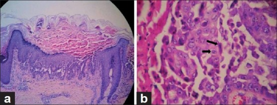
Histopathologically, Dariers disease is characterized by dyskeratosis in the form of premature keratinized large cells with basophilic nuclei (corps ronds) in the granular layer and grains more superficially.[4] Other features seen are suprabasal clefts with upward proliferation of papillae into clefts in the form of villi.
The basic principles in the control of Dariers disease is avoidance of ultraviolet (UV) light, sweating, and maintainence of personal hygiene.[4]
The beneficial effect of retinoids are well known for disorders of keratinization[1] and here we report two such cases where two brothers showed excellent response to oral isotretinoin.
CASE REPORT
A 30-year-old fisherman presented to our hospital with complaints of recurrent episodes of dark scaly raised lesions all over the body since the age of 10 years. At presentation, the lesions had increased over 2 weeks and during this period it had progressed to such an extent that it had incapacitated him due to generalized involvement. There was restriction in movements due to pain and difficulty in eating due to perioral lesions. The lesions were foul-smelling. There was characteristic photo-exacerbation of the lesions.
It was noteworthy that there had never been a complete clearance of the lesions throughout the years. His elder brother had similar complaints, which had commenced at the age of 9 years. Our patient had been receiving bland emollients, topical corticosteroids, and oral vitamin A during exacerbations but with no improvement.
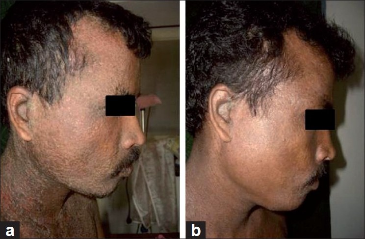
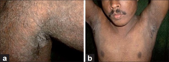
Examination revealed multiple dirty hyperpigmented papules that at places had coalesced to form plaques distributed symmetrically over the scalp, trunk, abdomen, back, groins, and scrotum. There were thick hyperpigmented greasy scales seen in both the external auditory canal [Figure 1a]. The lesions in the axillae had a verrucous appearance with a foul smell [Figure 2a]. Greasy scaling was seen over the involved sites and there was painful fissuring. A few vesicles with clear fluid were seen over the abdomen and the scapular area. The dorsal aspect of both hands had multiple tiny discrete skin-colored papules resembling acrokeratosis verruciformis of Hopf. Pits were seen over the palms and soles. The patient had even developed white nails with thinning of the nail plate and longitudinal striations. V-shaped scalloping was noted over the free edges of the finger nails. The oral mucosa had multiple white papules over the hard palate giving it a cobble-stone appearance.
Figure 1.

(a) Scaly papules on the face before treatment, (b) After treatment
Figure 2.

(a) Verrucous growth in the left axilla before treatment, (b) After treatment
With all the above findings, a clinical diagnosis of Darier-White disease was made in both the brothers. To confirm the diagnosis, a histopathological study of the involved skin was done, which showed profound acantholysis along with classical dyskeratosis and hyperkeratosis in the epidermis. Suprabasal acantholysis with formation of clefts was seen [Figure 3a] with mild lymphocytic infiltration in the dermis, thus confirming the diagnosis.
Figure 3.

(a) Hyperkeratosis, dyskeratosis with suprabasal acantholysis (b) corps ronds and grains
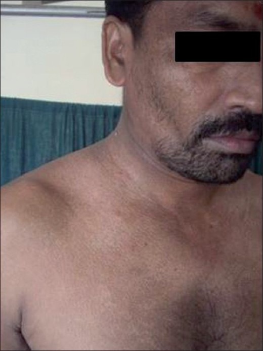
The patient was started on low-dose isotretinoin (20 mg/day) along with a tapering dose of oral corticosteroids and topical bland emollients. There was dramatic response within 2 weeks, with >50% improvement in the condition of our patient [Figure 1b,Figure 2b]. We had started the patient's elder brother [Figure 4] on high-dose isotretinoin (40 mg/day) at the time of presentation and once his lesions were under control this was reduced to low-dose isotretinoin.
Figure 4.

Brother after treatment
DISCUSSION
Many modalities of treatment have been tried and suggested for Darier-White disease, such as bland emollients, topical corticosteroids, course of oral antibiotics, topical retinoid, and microdermabrasion, but systemic retinoids have remained the mainstay of treatment.[2]
Retinoids have been found to have a beneficial role in Darier-White disease. The rationale for their use includes the promotion of exfoliation.[1] Topical retinoids may have synergistic effect with oral retinoids.[4] Conventional therapy for severe disease still relies in oral retinoids, with good response reported in 90% of patients. A study conducted by Kwok et al. at the University of California showed response in all five patients with Darier-White disease to oral isotretinoin given initially at a dose of 0.5 mg/kg/day, increasing to a maximum of 4 mg/kg/day for 16 weeks. There was more than 50% improvement in all the patients. However, discontinuation was marked by relapse. The use of retinoids in the treatment of Dariers disease maybe explained to some extent by the molecular basis of the disease.[5] The two patients we treated also showed >50% symptomatic improvement within 2 weeks of being started on oral isotretinoin.
Footnotes
Source of Support: Nil
Conflict of Interest: None declared
REFERENCES
- 1.Oostenbrink JH, Cohen EB, Steijlen PM, Van De Kerkhof PC. Oral Contraceptives in the treatment of Darier-White disease- A case report and review of literature. Clin Exp Dermatol. 1996;21:442–4. doi: 10.1111/j.1365-2230.1996.tb00152.x. [DOI] [PubMed] [Google Scholar]
- 2.Amerio P, Gobello T, Mazzanti C, Giaculli E, Ruggeri S, Sordi D, et al. Photodynamic therapy plus topical retinoids in Darier's disease. Photodiagnosis Photodyn Ther. 2007;4:36–8. doi: 10.1016/j.pdpdt.2006.09.002. [DOI] [PubMed] [Google Scholar]
- 3.Ramien ML, Prendiville JS, Brown KL, Cairns RA. Cystic bone lesions in a boy with Darier disease: A magnetic resonance imaging assessment. J Am Acad Dermatol. 2009;60:1062–6. doi: 10.1016/j.jaad.2008.10.049. [DOI] [PubMed] [Google Scholar]
- 4.Godic A. Darier disease.A review of pathophysiological mechanism. Acta Dermatoren. 2003;12:119–23. [Google Scholar]
- 5.Hulatt L, Burge S. Darier's disease: hopes and challenges. J R Soc Med. 2003;96:439–41. doi: 10.1258/jrsm.96.9.439. [DOI] [PMC free article] [PubMed] [Google Scholar]


