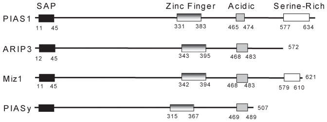
Fig. 8. Schematic representation of the primary structure of the PIAS family protein members used in this study.
Numbers indicate the amino acid residues limiting the different domains identified within the primary structure of the PIAS proteins. SAP: SAF-A/B, Acinus, PIAS domain. Notice that the differences in length observed among the different PIAS proteins are all due to variations in the C-terminal end of the proteins.

