Sir,
A 55-year-old male patient presented with numerous elevated facial swellings with ulceration of a single swelling over the right side of the scalp. According to the patient, he started developing small swellings on the face around the age of 15 years, which increased in number over the years. The patient started developing two new swellings over the forehead and the right side of the scalp around 7 years ago, which progressively became much larger. Around 2 months ago, the swelling on the right side of the scalp became painful and burst open to leave a wound that is increasing rapidly. He gave a history of similar facial swellings being present in his elder son who was 30 years old. On examination, a large fungating ulcer with raised edges was seen over the right parietal scalp [Figure 1]. The base was indurated and pale granulation tissue was observed in the floor of the ulcer. The ulcer bled on touch. A large skin-colored, firm swelling was observed on the left side of the forehead. In addition, numerous skin-colored to yellowish papules were found on the forehead, glabella, nose, naso-labial folds and upper lip [Figure 2]. Routine investigations were within normal limits. Two skin biopsies were performed, one from one of the smaller swellings in the upper lip and another from the margin of the ulcerated lesion on the scalp. The facial skin biopsy showed aggregations of basaloid cells. The tumor islands showed peripheral palisading of their cells [Figure 3]. Few horn cysts were also seen. [Figure 4] The skin biopsy from the scalp revealed irregular masses of epidermal cells proliferating downwards into the dermis. A large number of atypical squamous cells with variation in shape and size and hyperchromasia of the nuclei was seen [Figure 5]. Therefore, a diagnosis of multiple familial trichoepitheliomas in association with squamous cell carcinoma of scalp was made. The patient was referred to the surgical outpatient department for further management.
Figure 1.
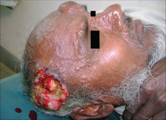
Fungating ulcer with raised edges is seen over the right parietal scalp along with skin-colored to yellowish papules on the forehead, nose and naso-labial folds
Figure 2.
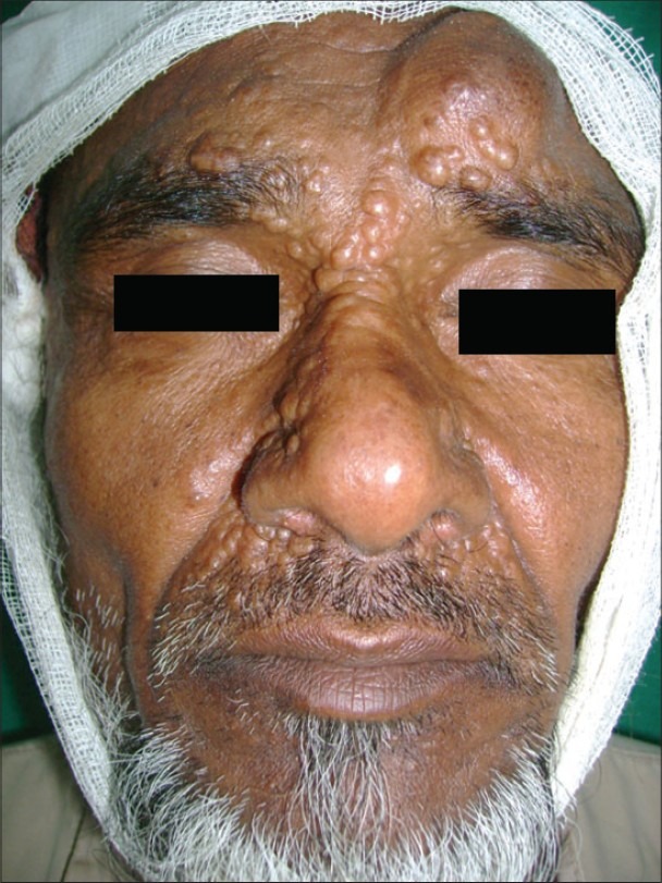
Skin-colored to yellowish papules on the forehead, glabella, nose, naso-labial folds and upper lip
Figure 3.
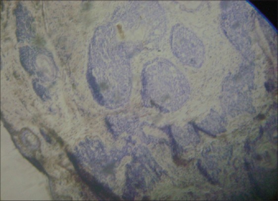
Basaloid epithelial formations (Haematoxylin and Eosin)
Figure 4.
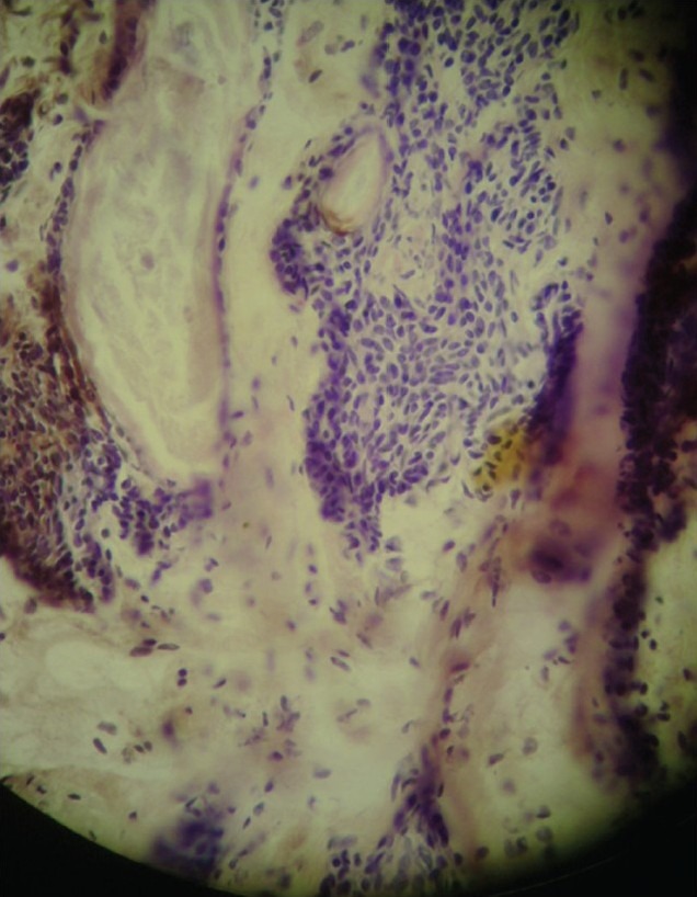
Horn cysts (Haematoxylin and Eosin)
Figure 5.
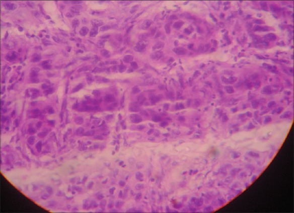
Atypical squamous cells with variation in shape, size and hyperchromasia of the nuclei (Haematoxylin and Eosin)
Trichoepitheliomas are hamartomas of hair germ. It can be solitary, multiple or desmoplastic type. Multiple trichoepitheliomas (Brooke-Fordyce disease) are mostly transmitted as an autosomal-dominant trait. Association of multiple trichoepitheliomas with malignancy is rare and most of the cases described are basal cell carcinomas.[1,2] Occasionally, trichoepitheliomas cannot be reliably differentiated from keratotic or morphea-like basal cell carcinomas. This difficulty could be the reason behind some of the apparent associations, whereas the present case describes an association between multiple trichoepitheliomas and squamous cell carcinoma, a rare occurrence.
Brooke-Spiegler syndrome consists of multiple trichoepitheliomas, cylindromas, spiradenomas and milia.[3] Brooke-Spiegler syndrome can also be rarely associated with cutaneous malignancies.[4] In the present case, the biopsy from the scalp lesion showed features of squamous cell carcinoma, but no features of cylindroma or spiradenoma. However, malignant transformation of a previous cylindroma or spiradenoma cannot be ruled out.
Familial trichoepithelioma is a rare condition. In this case, a very rare association of familial trichoepithelioma with squamous cell carcinoma was seen. This implies that such cases should be kept under long-term observation because of the possibility of malignant transformation.
REFERENCES
- 1.Carsuzaa F, Carloz E, Lebeuf M, Grobb JJ, Arnoux D. Multiple trichoepithelioma, cylindroma, miliaria and carcinomatous transformation. Ann Dermatol Venereol. 1992;119:746–8. [PubMed] [Google Scholar]
- 2.Johnson SC, Bennett RG. Occurrence of basal cell carcinoma among multiple trichoepitheliomas. J Am Acad Dermatol. 1993;28:322–6. doi: 10.1016/0190-9622(93)70046-v. [DOI] [PubMed] [Google Scholar]
- 3.Layegh P, Sharifi-Sistani N, Abadian M, Moghiman T. Brooke-Spiegler syndrome. Indian J Dermatol Venereol Leprol. 2008;74:632–4. doi: 10.4103/0378-6323.45109. [DOI] [PubMed] [Google Scholar]
- 4.Pizinger K, Michal M. Malignant cylindroma in Brooke-Spiegler syndrome. Dermatology. 2000;201:255–7. doi: 10.1159/000018499. [DOI] [PubMed] [Google Scholar]


