Abstract
Hypohidrotic ectodermal dysplasia (HED) is a rare genetic disorder characterized by the faulty development of the ectodermal structure, resulting in most notably anhydrosis/hypohydrosis, hypotrichosis and hypodontia. The condition is usually an X-linked recessive disorder affecting predominantly males. We are here reporting a classical case of hypohidrotic ectodermal dysplasia with a review of the literature.
Keywords: Ectodermal dysplasia, genetic disorder, hypohidrotic
INTRODUCTION
Hypohidrotic ectodermal dysplasia (HED) is a rare genetic disorder characterized by the faulty development of the ectodermal structure, resulting in most notably anhydrosis/hypohydrosis, hypotrichosis and hypodontia.[1] This condition is usually an X-linked recessive disorder affecting predominantly males.[2] Mutations in the gene encoding ligand ectodysplasin A (EDA) underlie classic, X-linked recessive HED, whereas mutations in the genes encoding the EDA receptor and (less frequently) the adaptor protein that associates with the EDA receptor's death domain result in autosomal dominant and autosomal recessive forms of HED. We are here reporting a classical case of HED with a review of the literature.
CASE REPORT
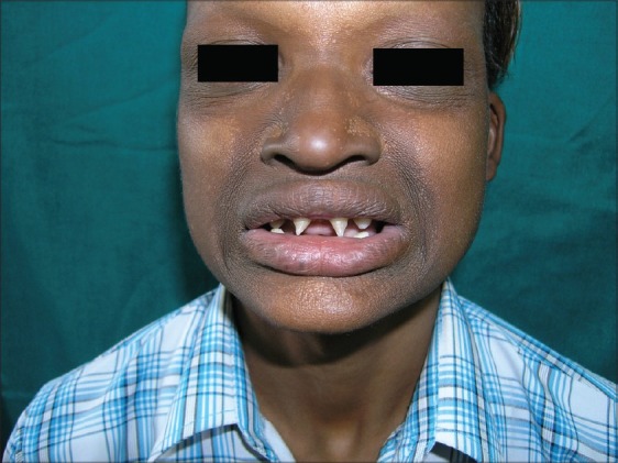
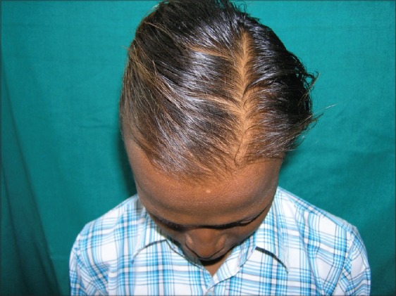
A 14-year-old male came to dermatology OPD for the consultation for recurrent itchy skin lesions over lower extremities for the past 4 years. On examination, he had discoid eczematous lesions suggestive of nummular eczema. Looking at his face, we noticed peculiar facies. There was loss of eyebrows and eyelashes. There was periorbital and perioral hyperpigmentation. The skin over these areas was dry and wrinkled. Lips were everted. There was hypodontia with peg-shaped incisors [Figure 1]. Sebaceous hyperplasia was also noted on the nose. The nasal bridge was depressed and frontal bossing was present. He had sparse, thin, lightly pigmented scalp hair [Figure 2]. There was mild palmoplantar hyperkeratosis. The oral mucosa, palate, nails were normal. There was a history of reduced sweating and heat intolerance. A clinical diagnosis of HED was made based on the history and cutaneous features. No other sibling had similar cutaneous features. None of the family members was involved in a similar manner in previous generations. There was no history of consanguinous marriage of parents. The systemic examination including otorhinolaryngological examination was normal. The physical development including external genitalia and mental development was normal. The routine biochemical tests were within normal limits.
Figure 1.

Characteristic facies of the patient with hypohidrotic ectodermal dysplasia with peg-shaped incisors
Figure 2.

The sparse, light-colored scalp hair
DISCUSSION
Ectodermal dysplasias are a group of inherited disorders that share common developmental defects involving at least two of the major structures classically hold to derive from the embryogenic ectoderms – hair, teeth, nails and sweat glands. HED is characterized by partial or complete absence of sweat glands, hypotrichosis, and hypodontia. The X-linked HED, otherwise called Christ–Siemens–Touraine Syndrome, was first described in 1848 by Thurnam. The incidence at birth is 1 in 100,000 males.[3] The complete syndrome does not occur in females but a female may show dental defects, sparse hair, reduced sweating, and dermatoglyphic abnormalities.
Clinically, HED is characterized by sparse or absent eccrine glands as well as by hypotrichosis and oligodontia with peg-shaped teeth as seen in the present case also.[4] The conical and pointed teeth are key features of the syndrome and may be the only obvious abnormality. Usually incisors and/or canines are characteristically affected. Because of their severely diminished ability to sweat, patients with HED have a propensity to develop hyperthermia with physical exertion or exposure to a warm environment, and affected infants often present with recurrent high fevers. The scalp hair, eyebrows, and eyelashes are sparse, fine, and oftentimes lightly pigmented. Our patient had loss of eyebrows and eyelashes, and sparse, thin, lightly pigmented scalp hair. In contrast to several other types of ectodermal dysplasia, nails were normal. Additional cutaneous features of HED include scaling or peeling of the skin during the neonatal period, periorbital hyperpigmentation and wrinkles, facial sebaceous hyperplasia, and eczematous dermatitis. Our present case also had nummular eczema for the past 4 years. HED patients have a characteristic facies with frontal bossing, a saddle nose, and full, everted lips as seen in this case also. Abnormal mucous glands result in extremely thick nasal secretions and a propensity to develop respiratory tract infections.
HED is inherited in an X-linked recessive, autosomal dominant, or autosomal recessive manner. Ninety-five percent of randomly selected individuals with HED have the X-linked recessive inheritance. The remainder (5%) have either the autosomal recessive or autosomal dominant inheritance. HED is caused by mutations in genes that encode several proteins with roles in the ectodysplasin signal transduction pathway. The activation of this cascade within epithelial cells at a critical time during embryogenesis results in the translocation of the transcription factor NF-κB into the nucleus and subsequent expression of target genes that are involved in the morphogenesis of eccrine glands, hair follicles, and teeth. Mutations in the EDA gene, which encodes the ectodysplasin ligand that initiates signaling through this pathway, cause the X-linked recessive form of HED that accounts for 75–95% of HED cases.[5] Mutations in the genes encoding the EDA receptor and (less frequently) the adaptor protein that associates with the EDA receptor's death domain result in autosomal dominant and autosomal recessive forms of HED, respectively.[6]
The management of children and adults with HED is a challenge because of their heat intolerance (especially during febrile illness or physical activities and in warm climate) and because of their susceptibility to pulmonary infections. During hot weather, affected individuals must have access to an adequate supply of water and a cool environment, which may mean “cooling vests,” air conditioning, a wet T-shirt, and/or a spray bottle of water. However, external cooling is less effective in these patients because their heat transfer from the core to the skin is also reduced, presumably due to poor capillary dilatation.[7] Affected individuals should learn to control their exposure to heat and to minimize its consequences. Early dental treatment that may range from simple restorations to dentures, in children over the age of 7 years, dental implants in the anterior portion of the mandibular arch, and replacement of dental prostheses as needed (often every 2.5 years) should be done to improve esthetics and chewing ability. Regular visits to an ENT physician may be necessary for the management of the nasal and aural concretions.
Footnotes
Source of Support: Nil
Conflict of Interest: None declared.
REFERENCES
- 1.Soloman LH, Kener EJ. The ectodermal dysplasia. Arch Dermatol. 1980;116:295–9. [Google Scholar]
- 2.Reed WB, Lopez DA, Lauding B. Clinical spectrum of anhydrotic ectodermal dysplasia. Arch Dermatol. 1970;102:134–43. [PubMed] [Google Scholar]
- 3.Stevenson AC, Kerr CB. On the distribution of the frequencies of mutation to genes determining harmful traits in man. Mutat Res. 1967;4:339–52. doi: 10.1016/0027-5107(67)90029-2. [DOI] [PubMed] [Google Scholar]
- 4.Cui CY, Schlessinger D. EDA signaling and skin appendage development. Cell Cycle. 2006;5:2477. doi: 10.4161/cc.5.21.3403. [DOI] [PMC free article] [PubMed] [Google Scholar]
- 5.Kere J, Srivastava AK, Montonen O, Zonana J, Thomas N, Ferguson B, et al. X-linked anhidrotic (hypohidrotic) ectodermal dysplasia is caused by mutation in a novel transmembrane protein. Nat Genet. 1996;13:409–16. doi: 10.1038/ng0895-409. [DOI] [PubMed] [Google Scholar]
- 6.Bal E, Baala L, Cluzeau C, El Kerch F, Ouldim K, Hadj-Rabia S, et al. Autosomal dominant anhidrotic ectodermal dysplasias at the EDARADD locus. Hum Mutat. 2007;28:703–9. doi: 10.1002/humu.20500. [DOI] [PubMed] [Google Scholar]
- 7.Brengelmann GL, Freund PR, Rowell LB, Olerud JE, Kraning KK. Absence of active cutaneous vasodilationassociated with congenital absence of sweat glands in humans. Am J Physiol. 1981;240:H571–5. doi: 10.1152/ajpheart.1981.240.4.H571. [DOI] [PubMed] [Google Scholar]


