Abstract
Varicella zoster virus causes both chicken pox and herpes zoster. The phenomenon of herpes zoster occurring concurrently in two non-contiguous dermatomes involving different halves of the body is termed herpes zoster duplex bilateralis (HZDB). Few cases, reported in the literature, were seen in either an immunosuppressed host or in the older age group. Here we present a case of HZDB in an immunocompetent host, probably the first in India.
Keywords: Bilateral herpes zoster, immunocompetent host, varicella zoster virus infection
INTRODUCTION
Herpes zoster duplex bilateralis (HZDB) is distinct from disseminated varicella zoster virus infections.[1] Its incidence is < 0.5%.[1,2] Few cases, reported in the literature, were seen in either an immunosuppressed host or in the older age group.[1–4] Noncontiguous multidermatomal herpes zoster occurring concurrently in remote dermatomes is very rare in an immunocompetent host.[1] Disseminated cutaneous zoster may be seen in 10-40% of immunosuppressed hosts and again is extremely rare in an immunocompetent host.[3]
After extensive research, to the best of our knowledge, till date no case of HZDB has been reported in an immunocompetent host of this or younger age group from India.
CASE REPORT
A 45-year-old male farmer presented with chief complaint of fever, malaise and vesico-bullous eruptions and crusted erosions over face and abdomen. Complaints started as painful erythematous fluid filled lesions over the left middle half of face about 10 days back and on the right side of abdomen 5 days back.
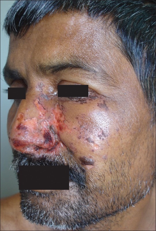
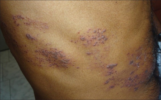
There was no past history of similar lesions, chicken pox in childhood, weight loss or any clinical features suggestive of diabetes mellitus/ asthma/ hypertension/ malignancy. He had not taken any medications for the last 6 months and never had an illness of any consequence. He had no risk factors for HIV, and denied any previous unusual infections or family history suggestive of an immune defect. The patient had no history of any mental, emotional, or financial stress. He was a non-alcoholic, non-smoker, and did not chew or snuff tobacco. His sleep was disturbed due to pain in the lesions but he denied insomna, prior to appearance of facial lesion. There was no history of trauma in immediate past. Systemic examination did not reveal any abnormalities. On cutaneous examination, multiple vesicobullous and crusted eruptions were seen in the maxillary division of trigeminal nerve (Figure 1 showing erosions and few healing bullae in the maxillary division of trigeminal nerve dermatome on the left side of the face) and T9 dermatomes (Figure 2 showing grouped vesicles in T9 dermatome on the right side of the abdomen). His complete blood picture, urine routine examination, blood sugar, chest radiograph, whole abdominal ultrasound, renal, and liver function tests were within normal limits. Tests for HIV/ Hepatitis A, B, and C were negative. The Mantoux test showed an induration of 4 mm and VDRL was nonreactive. Tzanck smear from abdominal eruptions showed multinucleated giant cells with intranuclear inclusion bodies. Tzanck smear from facial lesions was nonspecific.
Figure 1.

Erosions and few healing bullae in maxillary division of trigeminal nerve dermatome on the left side of face
Figure 2.

Grouped vesicles in T9 dermatome on the right side of the abdomen
The patient was treated with injection I/V acyclovir 10 mg/kg body weight 3 times/day along with other symptomatic management. The eruptions remained confined to the involved dermatome without any systemic involvement or spread. Cutaneous lesions healed gradually over a period of 3 weeks with scarring. A follow-up even after 1 year now has failed to reveal any recurrence of herpes zoster or any other symptom suggestive of an immunocompromised status.
DISCUSSION
Varicella zoster virus (VZV) is a neurotropic human herpes virus. It causes both, chicken pox and herpes zoster (shingles).[3] After causing chicken pox, it remains latent for decades in cranial nerves, dorsal root and autonomic nervous system ganglia. An effective immune system maintains the viral genome in latently infected cell and prevents viral replication and spread via retrograde axonal flow to the skin.
Reactivation of HZ is usually confined to a single episode and a single dermatome, though the latent viral genomes are present in many peripheral sensory ganglia.[4] This implies the confounding of some local factors, e.g., the number of viral copies present in cell or a local trauma to nerve root ganglion, along with the systemic immunocompromised conditions play a role.[4] Reactivation of the varicella zoster virus is thought to be due to the waning of VZV-specific T-cell immunity as one age.[3] The context that enables VZV reactivation bilaterally in the absence of systemic immunocompromised condition makes this extremely rare phenomenon. Beside second attack of HZ is rare in an immunocompetent host.[2]
In this case the diagnosis of herpes zoster is supported by clinical picture and cytology findings. Tzanck smear from facial lesions was inconclusive as the patient presented late and most of the lesions had stared crusted and healing. Though viral culture and antibody studies could not be done due to financial constraints, VZV IgM titers are not very high during reactivation and sensitivity of VZV culture is approximately 20%.[5] However in our patient it is unclear whether the reactivation occurred independently in two separate ganglia or if it was caused by viral spread from one ganglion to another. Incidentally the patient is healthy even after 1 year with no additional herpes zoster or any other significant infection or any symptoms indicative of immunocompromised state.
This case emphasizes the need for further studies and observations regarding the mechanism for VZV reactivation as it can occur in two remote dermatomes without any systemic immunosupression.
Footnotes
Source of Support: Nil
Conflict of Interest: None declared.
REFERENCES
- 1.Vu AQ, Radonich MA, Heald PW. Herpes Zoster in seven disparate dermatomes (Zoster Multiplex): Report of a case and review of the literature. J Am Acad Dermatol. 1999;40:868–9. doi: 10.1053/jd.1999.v40.a96944. [DOI] [PubMed] [Google Scholar]
- 2.Yoo KH, Park JH, Kim BJ, Kim MN, Song KY. Herpes Zoster Duplex Bilateralis in a patient with breast cancer. Cancer Res Treat. 2009;41:50–2. doi: 10.4143/crt.2009.41.1.50. [DOI] [PMC free article] [PubMed] [Google Scholar]
- 3.Burdett C, Mendoza N, Arora A, Bartlett B, Gewirtzman A, Tremaine AM, et al. A rare case of disseminated shingles in an immunocompetent patient following a 7-days treatment with oral valacyclovir. J Clin Virol. 2008;43:233–5. doi: 10.1016/j.jcv.2008.06.018. [DOI] [PubMed] [Google Scholar]
- 4.Peretz A, Nowatzky J, Steiner I. Herpes Zoster Duplex Bilateralis. J Neurol Neurosurg Psychiatry. 2007;78:818. doi: 10.1136/jnnp.2006.114116. [DOI] [PMC free article] [PubMed] [Google Scholar]
- 5.Sauerbrei A, Sommer M, Wutzler P. Virologic diagnosis of herpes zoster. Hautarzt. 1990;50:873–8. doi: 10.1007/s001050051002. [DOI] [PubMed] [Google Scholar]


