Abstract
Hemangiomas are neoplastic proliferations of endothelial cells, characterized by a period of growth after birth, and eventual spontaneous involution. The course can be uneventful with spontaneous resolution; or it may be marked by complications such as infection, bleeding, ulceration, visual defects and feeding difficulties. Apart from these, rare life-threatening complications such as congestive heart failure and consumption coagulopathy may also be seen. Although hemangiomas commonly occur in the head and neck region, intraoral occurrence is relatively rare. A port wine stain is defined as a macular telangiectatic patch which is present at birth and remains throughout life. They may be localized or extensive, affecting a whole limb. This article reports a rare case of co-occurrence of port wine stain with intraoral hemangioma.
Keywords: Hemangioma, intraoral, port wine stain, vascular malformations
INTRODUCTION
Hemangiomas are defined as benign neoplasms composed of proliferative and hyperplastic vascular endothelium. They exhibit rapid postnatal growth followed by slow involution, often leading to complete regression. Although most of these tumors are small and innocuous, associated structural congenital anomalies have been reported.[1]
A port wine stain is defined as a macular telangiectatic patch which is present at birth and remains throughout life. They may be localized or extensive, affecting a whole limb. They are often associated with an underlying disorder. These are best considered as low-flow vascular malformations which may occur on any part of the body but commonly affect the face in the distribution of the trigeminal nerve. Initially, the lesions are pale pink patches, eventually they evolve into a violaceous color, remain static or even lighten. Facial port wine stain typically evolve into thicker areas with vascular blebs, occasionally pyogenic granulomas and underlying tissue hypertrophy. This may include bony overgrowth and eruption of early dentition.[2]
CASE REPORT
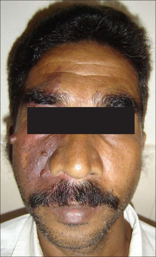
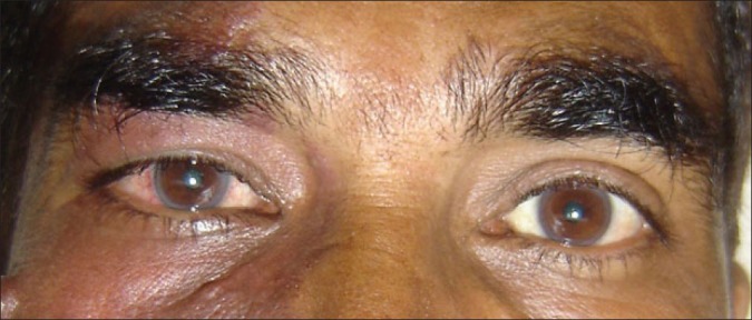
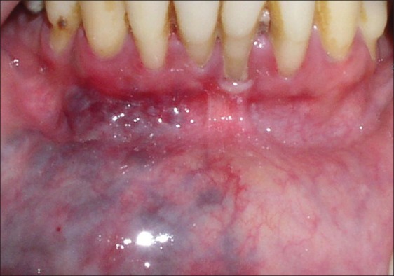
A 40- year-old male reported to the dental clinic with complaints of stains on the teeth and bleeding gums. On examination a violaceous patch on the right side of the face was seen, extending about 2 cm below the hairline superiorly to the angle of the mouth inferiorly [Figure 1] laterally from the right tragus, medially to the midline. Four papulo-nodular lesions were found overlying the lesion and distributed over the right malar area. Bulbar conjunctiva on the right side was also involved [Figure 2]. Intraorally bluish elevated lesions were observed on the mucosal aspect of lower lip [Figure 3] on the right side. Blanching was observed on diascopy [Figure 4]. The patient reported that the lesion was present since birth. No such lesions were seen else were in the body. No history of CNS disorders were reported by the patient. Based on the classical clinical features a provisional diagnosis of port wine stain with intraoral hemangioma was made. Nevus of ota was considered as differential diagnoses, which were ruled out due to their characteristic color difference. CT scan ruled out intracranial extension that eliminated possibilities of a syndromic association.
Figure 1.

Showing unilateral distribution of port wine stain on the right side of the face
Figure 2.

Reddish discoloration of right eye in comparison to the normal left eye
Figure 3.

Showing intraoral hemangioma on the mucosal aspect of lower lip
Figure 4.

Showing blanching of the intraoral hemangioma during diascopy test
DISCUSSION
Vascular anomalies are common birthmarks.[3] A classification system, first proposed by Mulliken and Glowacki was revised in 1996 by the International Society for the Study of Vascular Anomalies based on clinical, radiological and hemodynamic characteristics, into vascular malformations and vascular tumors.[4] Vascular malformations are errors of morphogenesis whereas hemangiomas and other vascular tumors grow by cellular proliferation.
Hemangiomas usually appear within the first month of life although they present as a precursor lesion in the immediate perinatal period. This can be very subtle, with a faint telangiectatic patch or even a patch of nevus anemicus-like pallor. They may occur anywhere on the skin or mucosal surface.[5] The overall clinical appearance is dependent on how deep into the skin they extend. They may be superficial as in majority of the cases appearing reddish in color. When they are located in deeper areas, they have more bluish color.[6] Hemangiomas usually occur in the head and neck region but are less common in the oral cavity. Majority of the hemangiomas involute with time but 10-20% of them fail to involute completely and may require post adolescent ablative treatment.[7]
Hemangiomas are the most common tumors of infancy, occurring in as many as 2.6% of neonates and 12% of children aged 1 year.[8,9] Up to 30% of preterm infants with low birth weight (1000 gm) may have hemangiomas.[10] In the oral cavity, the bones and the muscles are affected as well as the mucosa and the skin. Hemangiomas are approximately 3-5 times more common in females than in males.[11] Our patient had hemangioma on the lip.
The most commonly affected facial bones are the mandible, the maxilla and the nasal bones. Intraosseous lesions affect the mandible more often than the maxilla, with a ratio of 2:1 reported in one study.[12] Involvement of the zygoma is rare.[13] Intramuscular hemangiomas in the oral region are most commonly seen in the masseter, compromising 5% of all intramuscular hemangiomas.[14]
A port wine stain is defined as a macular telangiectatic patch which is present at birth and remains throughout life. They may be localized or extensive, affecting a whole limb. These are best considered as low-flow vascular malformations which may occur on any part of the body but commonly affect the face in the distribution of the trigeminal nerve. Initially, the lesions are pale pink patches, but with time they may mature into a violaceous color, remain static or even lighten and may become nodular[2] because of vascular ectasia.[15] Our patient presented with similar nodular swelling.
Port wine stains are associated with the following syndromes, Sturge-Weber-Dimitri syndrome characterized by noninherited and nonfamilial, port wine stain, leptomeningeal angiomas and Klippel-Trenaunay syndrome characterized by port wine stain, angiomatosis of the extremities. These syndromes were ruled out in our case.[16]
A wide range of treatment options have been proposed for port wine stain which includes pulsed tuneable dye laser (PDL) which has become the treatment of choice. Laser therapy has been the most successful at eliminating port wine stains. It is the only method that can destroy the tiny blood vessels in the skin without significantly damaging the skin.[17] Hemangiomas can be treated with corticosteroids, interferon-α, vincristine, PDL and surgery.[18] Cryosurgery may be used to correct lip and other soft tissue deformities.[19]
Our patient presented with hemangioma of the lower lip with ipsilateral port wine hemangioma. We ruled out possibility of any intracranial extension by imaging studies and referred for a laser ablasion.
CONCLUSIONS
Coexistence of port wine stain with intraoral cavernous hemangioma of the lip on the same side has been rarely reported in literature. The present case has highlighted the coexistence of both these vascular lesions.
Footnotes
Source of Support: Nil
Conflict of Interest: None declared.
REFERENCES
- 1.Mendiratta V, Jabeen M. Infantile hemangioma: An update. Indian J Dermatol Venereol Leprol. 2010;76:469–75. doi: 10.4103/0378-6323.69048. [DOI] [PubMed] [Google Scholar]
- 2.Klapman MH, Yao F. Thickening and nodules in port-wine stains. J Am Acad Dermatol. 2001;44:300–2. doi: 10.1067/mjd.2001.111353. [DOI] [PubMed] [Google Scholar]
- 3.Mulliken JB, Fishman SJ, Burrows PE. Vascular anomalies. Curr Probl Surg. 2000;37:517–84. doi: 10.1016/s0011-3840(00)80013-1. [DOI] [PubMed] [Google Scholar]
- 4.Enjolras O, Mulliken JB. Vascular tumours and vascular malformations (new issues) Adv Dermatol. 1997;13:375–423. [PubMed] [Google Scholar]
- 5.Margileth AM, Museles M. Cutaneous hemangiomas in children: Diagnosis and conservative management. JAMA. 1965;194:523–6. [PubMed] [Google Scholar]
- 6.Esterly NB. Cutaneous hemangiomas, vascular stains and malformations, and associated syndromes. Curr Probl Dermatol. 1995;7:69–107. doi: 10.1016/s0045-9380(96)80023-5. [DOI] [PubMed] [Google Scholar]
- 7.Waner M, Suen JY, Dinehart S. Treatment of hemangiomas of the head and neck. Laryngoscope. 1992;102:1123–32. doi: 10.1288/00005537-199210000-00007. [DOI] [PubMed] [Google Scholar]
- 8.Stal S, Hamilton S, Spira M. Hemangiomas, lymphangiomas, and vascular malformations of the head and neck. Otolaryngol Clin North Am. 1986;19:769–96. [PubMed] [Google Scholar]
- 9.Jacobs AH, Walton RG. The incidence of birthmarks in the neonate. Pediatrics. 1976;58:218–22. [PubMed] [Google Scholar]
- 10.Amir J, Metzker A, Krikler R, Reisner SH. Strawberry hemangioma in preterm infants. Pediatr Dermatol. 1986;3:331–2. doi: 10.1111/j.1525-1470.1986.tb00535.x. [DOI] [PubMed] [Google Scholar]
- 11.Marchuk DA. Pathogenesis of hemangioma. J Clin Invest. 2001;107:665–6. doi: 10.1172/JCI12470. [DOI] [PMC free article] [PubMed] [Google Scholar]
- 12.Hayward JR. Central cavernous hemangioma of the mandible: Report of four cases. J Oral Surg. 1981;39:526–32. [PubMed] [Google Scholar]
- 13.Cuesta Gil M, Navarro-Vila C. Intraosseous hemangioma of the zygomatic bone.A case report. Int J Oral Maxillofac Surg. 1992;21:287–91. doi: 10.1016/s0901-5027(05)80739-8. [DOI] [PubMed] [Google Scholar]
- 14.Wolf GT, Daniel F, Krause CJ, Kaufman RS. Intramuscular hemangioma of the head and neck. Laryngoscope. 1985;95:210–3. doi: 10.1288/00005537-198502000-00018. [DOI] [PubMed] [Google Scholar]
- 15.Neville, Damm, Allen, Bouquot . Chap 12. 2nd ed. 2004. Oral and Maxillofacial Pathology; p. 168. [Google Scholar]
- 16.Enjolras O, Chapot R, Merland JJ. Vascular anomalies and the growth of limbs: A review. J Pediatr Orthop B. 2004;13:349–57. doi: 10.1097/01202412-200411000-00001. [DOI] [PubMed] [Google Scholar]
- 17.Lanigan SW, Taibjee SM. Recent advances in laser treatment of port-wine stains. Br J Dermatol. 2004;151:527–33. doi: 10.1111/j.1365-2133.2004.06163.x. [DOI] [PubMed] [Google Scholar]
- 18.Batta K, Goodyear HM, Moss C, Williams HC, Hiller L, Waters R. Randomized controlled study of early pulsed dye laser treatment of uncomplicated childhood haemangiomas: Results of a 1-year analysis. Lancet. 2002;360:521–7. doi: 10.1016/S0140-6736(02)09741-6. [DOI] [PubMed] [Google Scholar]
- 19.Stewart RE, Barber TK, Trontman KC. Scientific foundations and clinical practice. St. Louis: CV Mosby; 1982. Pediatric Dentistry; pp. 502–3. [Google Scholar]


