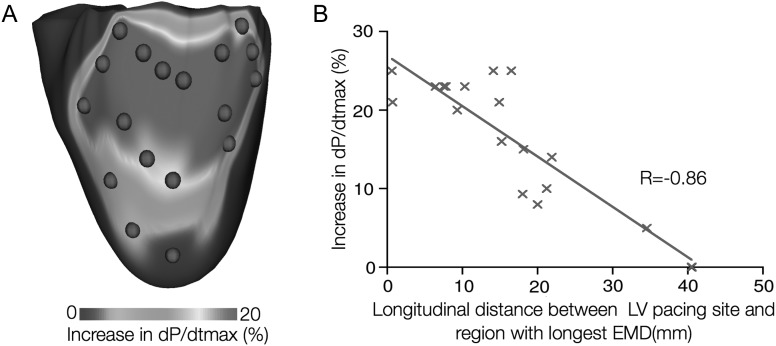
Figure 3.
Optimizing the placement of the Left Ventricle (LV) pacing electrode for cardiac resynchronization therapy in the failing canine heart. (A) Map of the percentage increase in dP/dtmax as a function of the LV pacing site (P is pressure). Red dots denote LV pacing sites. (B) Correlation of longitudinal distance between LV pacing site and region with longest EMD, and percentage increase in dP/dtmax.

