Abstract
The use of microarrays as a multiple analytic system has generated increased interest and provided a powerful analytical tool for the simultaneous detection of pathogens in a single experiment. A wide array of applications for this technology has been reported. A low density oligonucleotide microarray was generated from the genetic sequences of Y. pestis and B. anthracis and used to fabricate a microarray chip. The new generation chip, consisting of 2,240 spots in 4 quadrants with the capability of stripping/rehybridization, was designated as “Y-PESTIS/B-ANTHRACIS 4x2K Array.” The chip was tested for specificity using DNA from a panel of bacteria that may be potentially present in food. In all, 37 unique Y. pestis-specific and 83 B. anthracis-specific probes were identified. The microarray assay distinguished Y. pestis and B. anthracis from the other bacterial species tested and correctly identified the Y. pestis-specific oligonucleotide probes using DNA extracted from experimentally inoculated milk samples. Using a whole genome amplification method, the assay was able to detect as low as 1 ng genomic DNA as the start sample. The results suggest that oligonucleotide microarray can specifically detect and identify Y. pestis and B. anthracis and may be a potentially useful diagnostic tool for detecting and confirming the organisms in food during a bioterrorism event.
1. Introduction
Microarray technology has great potential for use in diagnostics, and DNA microarrays have received considerable attention due to the ability to simultaneously analyse a very large number of nucleic acid sequence targets and detect multiple genetic targets or genomes from multiple pathogens on a single slide [1]. The technology has played an increasingly important role in genomics and has generated increased interest in the last decade.
DNA microarrays consist of several oligonucleotide probes that have been immobilized on a solid glass support, and the technique has great potential to be used for the discrimination of closely related strains by employing oligonucleotides specific for each target organism. Hence, the design of a suitable probe set is the key in the development of microarrays as all probes on a microarray should be highly specific for their target genes. The probes should be able to bind efficiently to target sequences to allow the detection of very low abundance targets in complex mixtures with high sensitivity [2]. The use of DNA microarrays has been shown to be effective for the high-throughput detection of pathogenic microorganisms in clinical, environmental, food, and water samples [3–8]. However, the application for the detection of biothreat agents in food has not been documented. Food is considered a vulnerable target for bioterrorist attack, and events related to the deliberate contamination of food using conventional foodborne pathogens such as Salmonella [9] make foodborne bioterrorism involving Y. pestis and B. anthracis a possibility. Foodborne bioterrorism response preparedness involving Y. pestis and B. anthracis is required to deal with any potential threat involving the food supply. The development of a species-specific method for simultaneous detection of biothreat agents from food is essential.
In this study, we describe an approach that involves the use of a new generation microarray to allow the simultaneous detection and identification of Y. pestis and B. anthracis from food. We designed and tested probes based on the virulence genes from the two biothreat agents and demonstrate that this microarray approach has the potential to be used for the specific detection of B. anthracis and Y. pestis in a foodborne application.
2. Materials and Methods
2.1. Microarray Probe Design and Chip Fabrication
Whole genome sequences of B. anthracis Ames [10] and Y. pestis CO92 [11] from GenBank were selected for the custom design of around 35-mer probes and used to fabricate a low density custom array. The oligonucleotide probes were generated from the virulence plasmids, and a 4x2K chip array (four identical arrays of 2,000+ spots), designated as “Y-PESTIS/B-ANTHRACIS 4x2K Array,” was custom-designed (CustomArray Inc. USA, formerly CombiMatrix). In all, 533 and 1,707 probes were generated for Y. pestis and B. anthracis, respectively. With this 4x2K chip design, eight identical experiments can be run on a single chip. The oligonucleotide probes are electrochemically synthesised directly onto the chip and does not require the need to order them separately for resuspension and spotting using a robot. The above described features are lacking in the design of the old microarray chips and make this a new generation microarray. Another unique feature of this new generation chip is that it can be stripped and reused for up to three times. Each CustomArray 4x2K microarrays can be stored in a cool dry place for up to 4 months.
2.2. Bacterial Strains and DNA Extraction
The B. anthracis strains used in this study were kindly provided by Dr. Elizabeth Golsteyn Thomas (Canadian Food Inspection Agency) and Mr. Doug Bader (Defence Research Development Canada). The sources of all the other bacterial strains used are provided in a previous paper [12] and listed in Table 1. Bacteria were grown on Tryptic Soy Agar (Becton, Dickinson and Company, USA) supplemented with 5% sheep blood, and single colonies were transferred subsequently into Tryptic Soy Broth at 37°C overnight. Genomic DNA was extracted as previously described [12].
Table 1.
Strains used in this study.
| Species | Strain | Remarks |
|---|---|---|
| Foodborne pathogen mix | ||
|
| ||
| Proteus vulgaris | ATCC 8427 | |
| Klebsiella pneumoniae | ATCC 13883 | |
| Shigella dysenteriae | ATCC 11835 | |
| Escherichia coli O157H7 | EDL933 | |
| Mannheimia haemolytica | Z13 | |
| Vibrio vulnificus | Z86 | |
| Citrobacter braakii | ATCC 12012 | |
| Salmonella typhimurium | 71-471 | |
| Aeromonas hydrophila | Z22 | |
| Pseudomonas aeruginosa | ATCC 27853 | |
| Listeria monocytogenes | ATCC 15313 | |
| Streptococcus pyogenes | ATCC 19615 | |
| Enterococcus faecalis | ATCC 29212 | |
| Micrococcus lysodeikticus | Z9 | |
| Staphylococcus aureus | Z13 | |
| Campylobacter jejuni | ATCC 11168 | |
|
| ||
| Yersinia spp. mix | ||
|
| ||
| Yersinia pseudotuberculosis | ATCC 29833 | |
| Yersinia pseudotuberculosis | Turku | |
| Yersinia enterocolitica | ATCC 23715 | |
| Yersinia kristensenii | ATCC 33638 | |
| Yersinia frederiksenii | ATCC 33641 | |
| Yersinia intermedia | ATCC 29909 | |
|
| ||
| Bacillus spp. mix | ||
|
| ||
| Bacillus cereus | ATCC 14579 | |
| Bacillus subtilis | NWBL 0060 | |
| Bacillus thuringiensis | ATCC 10792 | |
| Bacillus coagulans | ATCC 7050 | |
|
| ||
| Yersinia pestis strains | ||
|
| ||
| Yersinia pestis | Pp 1967 | Wild-type isolate |
| Yersinia pestis | 195/P | Wild-type isolate |
| Yersinia pestis | 6/69H+ | Wild-type isolate |
| Yersinia pestis | M23 | Wild-type isolate |
| Yersinia pestis | Yokohama | Wild-type isolate |
| Yersinia pestis | CO92 | NCBI Genome Ref Seq: NC_003143.1 [11] |
|
| ||
| Bacillus anthracis strains | ||
|
| ||
| Bacillus anthracis | Ames | NCBI Genome Ref Seq: NC_003997.3 [10] |
| Bacillus anthracis | Ba 44 | isolate from moose, NWT Canada in Aug 1993 |
| Bacillus anthracis | Ba 56 | isolate from cattle, ON Canada in Aug 1996 |
| Bacillus anthracis | Ba 79 | isolate from bear, NWT Canada in Jul 2000 |
| Bacillus anthracis | Ba 127 | isolate from caprine, BC Canada in Dec 2001 |
| Bacillus anthracis | Ba 131 | isolate from soil, Canada, date N/A |
| Bacillus anthracis | Ba 252 | isolate from cattle, SK Canada in Jul 2006 |
| Bacillus anthracis | Ba 59 | isolate from bison, MB Canada in Jul 1998 Cap- |
| Bacillus anthracis | Ba 158 | ATCC 4229 Tox–(pXO1-) |
| Bacillus anthracis | Sterne | NCBI Genome Ref Seq: NC_005945.1(pXO2-) |
2.3. DNA Amplification and Labeling
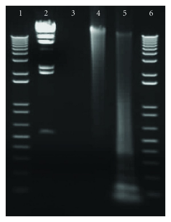
Genomic DNA was amplified using REPLI-g Mini Kit (Qiagen GmBH, Hilden, Germany) (Figure 1), and amplified DNA provided consistent hybridization results (data not shown). For labeling, 2 μg of amplified DNA was digested with RsaI (Life Technologies, Carlsbad, CA, USA) at 37°C for 4 hours and subsequently labeled with Alexa Fluor 555 and/or 647 florescent dyes using BioPrime Plus Array CGH Genomic Labeling Kit (Life Technologies, Carlsbad, CA, USA) following the manufacturer's instructions. Concentration and labeling efficiency of DNA was determined using the NanoDrop ND-1000 fluorometer (Thermo Fisher Scientific, Waltham, MA, USA). Labeled DNA was stored at −20°C in amber microcentrifuge tubes unless used for hybridization immediately. For specificity studies, genomic DNA of Y. pestis CO92 or a wild-type B. anthracis isolate number 179 was mixed with the genomic DNAs extracted from a panel of bacteria listed in Table 1 at different ratios (1 : 1, 1 : 3, 1 : 9, and 1 : 19 in weight). DNA amplification and labeling were done as described above.
Figure 1.

1% agarose gel loaded with B. anthracis DNA before and after amplification using REPLI-g genomic DNA amplification kit and RsaI digestion. Lanes 1 and 6, : 1 kb molecular weight markers, lane 2: Hind III molecular weight marker (23130, 9416, 6557, 4361, 2322, 2027, and 546 bp), lane 3, : 5 μL of 20 ng/μL B. anthracis Ba 179 genomic DNA, lane 4, : 5 μL of REPLI-g amplified DNA (355 ng/μL), and lane 5: RsaI digested DNA.
2.4. DNA Hybridization to Microarray and Chip Stripping
Hybridization of labeled genomic DNA to the CustomArray 4x2K format slide was performed according to the manufacturer's protocol (CustomArray Inc. Bothell, WA, USA). Each microarray consisting of 4 identical array sectors was individually loaded with different DNA samples. By using two florescent dyes, eight samples were tested simultaneously on one microarray slide. Briefly, 30 μL of hybridization solution containing 100 ng of two fluorochrom-labeled (Alexa Fluor 555 or 647) DNA was pipetted into each of the four chambers and covered with foil adhesive tape to avoid light. The covered slide was incubated in a humid hybridization rotisserie oven (UVP, LLC, Upland, CA, USA) for 16 hours at 50°C with gentle rotation. All labeled DNA samples were hybridized in duplicate with either Alexa Fluor 555 or 647 to ensure consistency of the results.
Following hybridization and imaging, microarrays were submerged in 0.5 M sodium hydroxide at room temperature for 15 minutes and stripped using the CombiMatrix CustomArray Stripping Kit (CustomArray Inc.). Stripped slides were scanned before rehybridization to make sure there were no more signals and stored in a slide holder containing PBS at 4°C. Rehybridization for a single slide was repeated a maximum of two times.
2.5. Microarray Data Analysis
Hybridized microarrays were imaged using the Axon 4000B Microarray Scanner (Axon Instruments, Molecular Devices, LLC, Sunnyvale, CA, USA). Hybridization of each sample was done in duplicate, and scanning was done in triplicate. The TIFF images were analyzed using the GenePix Pro software Version 5.0 (Axon Instruments), and the data was extracted for further analysis of the total intensity of each spot. All data were transferred to Microsoft Excel for Cluster and TreeView analysis (Stanford University, CA, USA), and then heat maps were generated following the instructions of the software [13].
2.6. Application of Microarray for the Analysis of Spiked Food Samples
To investigate the use of the microarray for a foodborne application, Yersinia pestis strain Pp1967 was cultured overnight in TSB at 28°C, and about 106 CFU/mL was inoculated into 25 mL of 1% skimmed milk purchased from a local grocery store. A 225 mL volume of buffered peptone water was added, and the spiked milk sample was stomached for 2 minutes using a stomacher (Seward Ltd., West Sussex, UK). The samples were aliquoted into 10 mL volumes in 15 mL of Falcon tubes, boiled for 10 minutes to kill cells, and centrifuged. Pellets were collected, and DNA was extracted using the DNeasy Blood and Tissue Kit (Qiagen GmBH, Germany). The extracted DNA was labeled, amplified, digested and hybridized to the chip as described above.
3. Results
3.1. Hybridization of Genomic DNA from Yersinia pestis and Bacillus anthracis Strains
Six Y. pestis strains and two Y. pseudotuberculosis strains were examined to evaluate the positive spots. All DNA samples were tested in duplicate, and the total fluorescent intensity (TFI) data for all 2,240 probes were assembled after subtracting the average intensity of ten negative spots. Spots showing abnormality on the microarray slides due to hybridization failures were filtered out. The TFI values higher than 20,000 were selected as positive signals. This number was determined based on the results from normalized data from Y. pestis CO92 or B. anthracis Ames, showing 0 (the median) of log-transformed (log2) values. For B. anthracis, the Ames strain and 7 wild-type isolates from animals in Canada from 1996 to 2006 and 4 Bacillus species (B. cereus, B. thuringiensis, B. coagulans, and B. subtilis) were also examined (Table 1). The data obtained from genomic DNA cocktails of either Yersinia spp. or Bacillus spp. and foodborne pathogens were analyzed against positive probes of the two bacterial agents. About 300 probes were found to be positive for each Y. pestis strain and out of this, 72 of them gave positive values in three or more strains. Subsequently, positive probes from Y. pseudotuberculosis ATCC 29833 and Turku strains, and genomic DNA cocktails of Yersinia spp. or foodborne pathogens were compared to the 72 probes, and finally 37 were selected as Y. pestis-specific probes. For B. anthracis, about 800 targets were analyzed and after analyzing the positive probes from the DNA cocktails of 4 Bacillus species and foodborne pathogens, 83 were selected as B. anthracis-specific.
The sequences of the 37 Y. pestis-specific probes are shown in Table 2a and the 83 probe sequences for B. anthracis in Table 2b (see Supplementary Material available online doi:10.1155/2012/627036).
3.2. Specificity and Sensitivity of the Specific Probes for Y. pestis and B. anthracis
To examine the specificity of the 37 specific probes for Y. pestis and 83 for B. anthracis, genomic DNA from the two organisms was mixed with those from a panel of foodborne bacterial pathogens and subsequently amplified with REPLI-g before loading onto the microarray slides. This was to mimic the detection of Y. pestis and B. anthracis from food samples if contaminated with other pathogens. The results showed that all the probes had strong positive signals (data not shown).
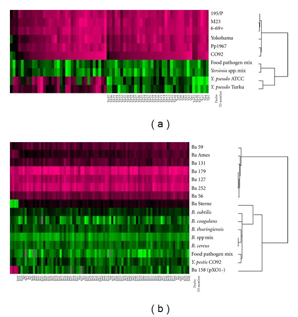
Figures 2(a) and 2(b) show the heat map and clustering results for Y. pestis and B. anthracis. The heat maps show unique patterns to each bacterial species and are easily distinguished from closely related organisms. Additionally, for Y. pestis, another 35 probes shown on the heat map with positive intensity levels in Y. pseudotuberculosis as well could be potentially considered as Y. pestis-specific probes since the signals were significantly higher than the ones from Y. pseudotuberculosis.
Figure 2.

Unique patterns of the heat map and clustering images generated from the data of Y. pestis (a) and B. anthracis (b) specific probes. The total intensity of the positive spots for either Y. pestis or B. anthracis was normalized using microarray data analysis software (Cluster and Treeview [5]) after converting to log2 scale. Probes with significant intensity are shown in pink. The 37 probes for Y. pestis and 83 for B. anthracis have probe ID numbers underneath the heat maps.
3.3. Milk Spiked with Y. pestis
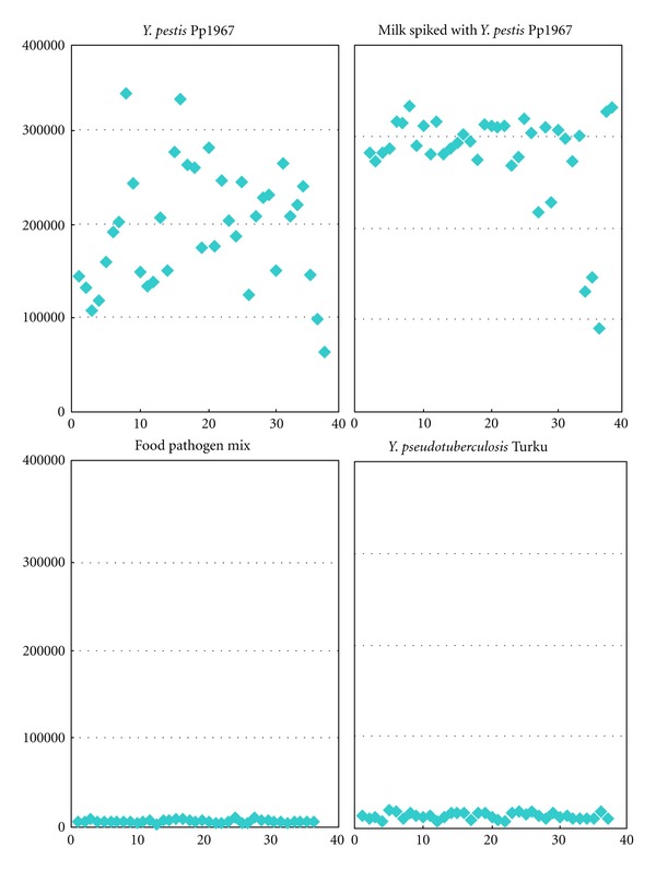
To demonstrate the direct application of this CustomArray DNA hybridization technique for detection in food, we extracted DNA from milk samples spiked with 106 CFU/mL Y. pestis Pp1967. Strong signal intensities were shown in all 37 probe spots (Figure 3). These spots did not show any signal intensity in negative control milk samples indicating that the probes were specific for Y. pestis.
Figure 3.

Total fluorescent intensity (TFI) of 37 Y. pestis-specific probes for amplified genomic DNA extracted from spiked milk samples. The X-axis numbers represent Y. pestis probe ID number. The Y-axis shows averaged TFI for each probe.
4. Discussion
Detection and confirmation of biothreat agents such as Yersinia pestis and Bacillus anthracis in food are very important for the effective protection of the public from any potential foodborne bioterrorism threat. To date, the simultaneous detection of these two biothreat agents in food using microarray such as described in this study has not been reported. Traditional methods used for the confirmation of pathogens such as enrichment culture, microscopy, serology, and biochemical assays have several limitations, in which they are laborious and time consuming and hence inefficient in addressing potential foodborne bioterrorism concerns.
Rapid and specific identification of foodborne pathogens and biothreat agents is the key for early detection and quick response in the event of foodborne disease outbreak. Real-time PCR has recently been developed as a rapid and specific method for detecting biothreat agents in food [12, 14, 15]. Even though multiplex real-time PCR assay can amplify several different target regions of genomic DNA in a single reaction, there is limited information available from this technique; thus, a combination of different methods should be considered as an alternative.
DNA microarray technology has been applied to foodborne pathogen detection [16, 17]. This approach has a strong potential not only to identify multiple pathogens with very high specificity in a single experiment, but also to be able to demonstrate genetic differences and similarities among bacterial strains [18, 19] and provide further characterization of bacterial isolates. To obviate the use of PCR which requires the design of specific primers and optimization of the reaction conditions, we amplified genomic DNA using QIAGEN REPLI-g whole genome amplification system. REPLI-g technology has been successfully used to amplify genomic DNA [20] and according to the report, the intensity values of REPLI-g amplified DNA can provide relevant information about copy numbers of the gene targets in the genome as amplification is uniform. The report showed that the higher the intensity values, the higher the copy number of the gene target. The high intensities observed with targets on plasmid pPCP1 in the present work confirm that this plasmid has a high copy number. The sensitivity of DNA microarrays is usually poor when total genomic DNA is used for hybridization [21]; however, this can be enhanced to allow detection of low level concentration of pathogens using labeling and hybridization of amplified products [2, 5, 22, 23]. The application of a random whole genome amplification step prior to labeling in the current study enabled the use of as low as 1 ng of DNA as the start material for the microarray analysis. This led to the successful simultaneous detection and identification of Y. pestis and B. anthracis in a single assay. This did not require the design of PCR primers to generate materials for labeling and hybridization. Furthermore, the stripping and reuse of the microarray chip including the high-throughput ability to run 4 separate experiments simultaneously on a single chip offers a unique new generation microarray and allows experiments to be replicated under identical conditions, thus ensuring high reproducibility of results. This is not the case with the use of the conventional microarrays where a chip is used once, and replication of experiments is done using different chips.
Several reports have demonstrated the use of DNA microarrays for the detection of pathogens in food [24–27]. The technology has also been developed for biodefence applications involving B. anthracis and Y. pestis [28–30]. However, to our knowledge, the use of the technology for the simultaneous detection and identification of biothreat agents in food has not been documented. Here we demonstrate the first successful application in food biodefence involving the detection of Y. pestis in spiked milk samples.
The high specificity of the species-specific probes is demonstrated by the lack of cross-reactivity with a panel of closely related and distantly related strains that may be potentially present in food. The 37 and 83 species-specific probes generated for Y. pestis and B. anthracis, respectively, should enable the use of microarray for the direct detection and identification from contaminated food without the need for isolating the biothreat agent. However, this number can be expanded to about 300 and 800 probes for Y. pestis and B. anthracis, respectively, and used for the confirmation of the bacteria if isolated in pure culture from contaminated food. The careful analysis of genetic sequence information and selection of probes contributed to this. In particular, the targeting of virulence genes from the virulence plasmids that are unique to the two organisms contributed to the high specificity observed. Previous studies involving the use of virulence genes in the design of microarrays for the detection of pathogenic bacteria have been reported [31–33]. The use of several virulence genes as detection markers offers more information on the virulence potential of strains implicated and adds more value to the use of microarray as a platform for the detection and confirmation of biothreat agents.
In conclusion, the new generation microarray developed in this study is novel for the simultaneous detection and identification of Y. pestis and B. anthracis in food, and demonstrates the usefulness of microarray as a potential diagnostic tool for biodefence applications.
Supplementary Material
Table 2a. Yersinia pestis specific probes.
Table 2b. B. anthracis specific probes.
Acknowledgments
The authors thank Dr. Elizabeth Golsteyn Thomas, OIE Reference Laboratory for Anthrax, Canadian Food Inspection Agency, and Mr. Doug Bader, Defence Research Development Canada, for providing the Bacillus anthracis strains used in this study. The authors thank Drs. Soren Alexandersen and Aruna Ambagala and Mr. Matthew Thomas for reviewing the paper. This work was funded by the Public Security and Anti-Terrorism (PSAT) Program of the Ministry of Defence, Government of Canada.
References
- 1.Seidel M, Niessner R. Automated analytical microarrays: a critical review. Analytical and Bioanalytical Chemistry. 2008;391(5):1521–1544. doi: 10.1007/s00216-008-2039-3. [DOI] [PMC free article] [PubMed] [Google Scholar]
- 2.Call DR. Challenges and opportunities for pathogen detection using DNA microarrays. Critical Reviews in Microbiology. 2005;31(2):91–99. doi: 10.1080/10408410590921736. [DOI] [PubMed] [Google Scholar]
- 3.Baxi MK, Baxi S, Clavijo A, Burton KM, Deregt D. Microarray-based detection and typing of foot-and-mouth disease virus. Veterinary Journal. 2006;172(3):473–481. doi: 10.1016/j.tvjl.2005.07.007. [DOI] [PubMed] [Google Scholar]
- 4.Bekal S, Brousseau R, Masson L, Prefontaine G, Fairbrother J, Harel J. Rapid identification of Escherichia coli pathotypes by virulence gene detection with DNA microarrays. Journal of Clinical Microbiology. 2003;41(5):2113–2125. doi: 10.1128/JCM.41.5.2113-2125.2003. [DOI] [PMC free article] [PubMed] [Google Scholar]
- 5.González SF, Krug MJ, Nielsen ME, Santos Y, Call DR. Simultaneous detection of marine fish pathogens by using multiplex PCR and a DNA microarray. Journal of Clinical Microbiology. 2004;42(4):1414–1419. doi: 10.1128/JCM.42.4.1414-1419.2004. [DOI] [PMC free article] [PubMed] [Google Scholar]
- 6.Jin DZ, Wen SY, Chen SH, Lin F, Wang SQ. Detection and identification of intestinal pathogens in clinical specimens using DNA microarrays. Molecular and Cellular Probes. 2006;20(6):337–347. doi: 10.1016/j.mcp.2006.03.005. [DOI] [PubMed] [Google Scholar]
- 7.Maynard C, Berthiaume F, Lemarchand K, et al. Waterborne pathogen detection by use of oligonucleotide-based microarrays. Applied and Environmental Microbiology. 2005;71(12):8548–8557. doi: 10.1128/AEM.71.12.8548-8557.2005. [DOI] [PMC free article] [PubMed] [Google Scholar]
- 8.Wilson WJ, Strout CL, DeSantis TZ, Stilwell JL, Carrano AV, Andersen GL. Sequence-specific identification of 18 pathogenic microorganisms using microarray technology. Molecular and Cellular Probes. 2002;16(2):119–127. doi: 10.1006/mcpr.2001.0397. [DOI] [PubMed] [Google Scholar]
- 9.Török TJ, Tauxe RV, Wise RP, et al. A large community outbreak of salmonellosis caused by intentional contamination of restaurant salad bars. Journal of the American Medical Association. 1997;278(5):389–395. doi: 10.1001/jama.1997.03550050051033. [DOI] [PubMed] [Google Scholar]
- 10.Read TD, Peterson SN, Tourasse N, et al. The genome sequence of Bacillus anthracis Ames and comparison to closely related bacteria. Nature. 2003;423(6935):81–86. doi: 10.1038/nature01586. [DOI] [PubMed] [Google Scholar]
- 11.Parkhill J, Wren BW, Thomson NR, et al. Genome sequence of Yersinia pestis, the causative agent of plague. Nature. 2001;413(6855):523–527. doi: 10.1038/35097083. [DOI] [PubMed] [Google Scholar]
- 12.Amoako KK, Goji N, Macmillan T, et al. Development of multitarget real-time PCR for the rapid, specific, and sensitive detection of Yersinia pestis in milk and ground beef. Journal of Food Protection. 2010;73(1):18–25. doi: 10.4315/0362-028x-73.1.18. [DOI] [PubMed] [Google Scholar]
- 13.Eisen MB, Spellman PT, Brown PO, Botstein D. Cluster analysis and display of genome-wide expression patterns. Proceedings of the National Academy of Sciences of the United States of America. 1998;95(25):14863–14868. doi: 10.1073/pnas.95.25.14863. [DOI] [PMC free article] [PubMed] [Google Scholar]
- 14.Kawasaki S, Fratamico PM, Horikoshi N, et al. Multiplex real-time polymerase chain reaction assay for simultaneous detection and quantification of Salmonella species, Listeria monocytogenes, and Escherichia coli O157:H7 in ground pork samples. Foodborne Pathogens and Disease. 2010;7(5):549–554. doi: 10.1089/fpd.2009.0465. [DOI] [PubMed] [Google Scholar]
- 15.Woubit A, Yehualaeshet T, Habtemariam T, Samuel T. Novel genomic tools for specific and real-time detection of biothreat and frequently encountered foodborne pathogens. Journal of Food Protection. 2012;75:660–670. doi: 10.4315/0362-028X.JFP-11-480. [DOI] [PMC free article] [PubMed] [Google Scholar]
- 16.Kostić T, Stessl B, Wagner M, Sessitsch A, Bodrossy L. Microbial diagnostic microarray for food- and water-borne pathogens. Microbial Biotechnology. 2010;3(4):444–454. doi: 10.1111/j.1751-7915.2010.00176.x. [DOI] [PMC free article] [PubMed] [Google Scholar]
- 17.Suo B, He Y, Paoli G, Gehring A, Tu SI, Shi X. Development of an oligonucleotide-based microarray to detect multiple foodborne pathogens. Molecular and Cellular Probes. 2010;24(2):77–86. doi: 10.1016/j.mcp.2009.10.005. [DOI] [PubMed] [Google Scholar]
- 18.Wang X, Han Y, Li Y, et al. Yersinia genome diversity disclosed by Yersinia pestis genome-wide DNA microarray. Canadian Journal of Microbiology. 2007;53(11):1211–1221. doi: 10.1139/W07-087. [DOI] [PubMed] [Google Scholar]
- 19.Zhou D, Han Y, Dai E, et al. Identification of signature genes for rapid and specific characterization of Yersinia pestis . Microbiology and Immunology. 2004;48(4):263–269. doi: 10.1111/j.1348-0421.2004.tb03522.x. [DOI] [PubMed] [Google Scholar]
- 20.Treff NR, Su J, Tao X, Northrop LE, Scott RT. Single-cell whole-genome amplification technique impacts the accuracy of SNP microarray-based genotyping and copy number analyses. Molecular Human Reproduction. 2011;17(6):335–343. doi: 10.1093/molehr/gaq103. [DOI] [PMC free article] [PubMed] [Google Scholar]
- 21.Rhee SK, Liu X, Wu L, Chong SC, Wan X, Zhou J. Detection of genes involved in biodegradation and biotransformation in microbial communities by using 50-mer oligonucleotide microarrays. Applied and Environmental Microbiology. 2004;70(7):4303–4317. doi: 10.1128/AEM.70.7.4303-4317.2004. [DOI] [PMC free article] [PubMed] [Google Scholar]
- 22.Kim DH, Lee BK, Kim YD, Rhee SK, Kim YC. Detection of representative enteropathogenic bacteria, Vibrio spp., pathogenic Escherichia coli, Salmonella spp., Shigella spp., and Yersinia enterocolitica, using a virulence factor gene-based oligonucleotide microarray. Journal of Microbiology. 2010;48(5):682–688. doi: 10.1007/s12275-010-0119-5. [DOI] [PubMed] [Google Scholar]
- 23.Loy A, Bodrossy L. Highly parallel microbial diagnostics using oligonucleotide microarrays. Clinica Chimica Acta. 2006;363(1-2):106–119. doi: 10.1016/j.cccn.2005.05.041. [DOI] [PubMed] [Google Scholar]
- 24.Kim HJ, Park SH, Lee TH, Nahm BH, Kim YR, Kim HY. Microarray detection of food-borne pathogens using specific probes prepared by comparative genomics. Biosensors and Bioelectronics. 2008;24(2):238–246. doi: 10.1016/j.bios.2008.03.019. [DOI] [PubMed] [Google Scholar]
- 25.Myers KM, Gaba J, Al-Khaldi SF. Molecular identification of Yersinia enterocolitica isolated from pasteurized whole milk using DNA microarray chip hybridization. Molecular and Cellular Probes. 2006;20(2):71–80. doi: 10.1016/j.mcp.2005.09.006. [DOI] [PubMed] [Google Scholar]
- 26.Panicker G, Call DR, Krug MJ, Bej AK. Detection of pathogenic Vibrio spp. in shellfish by using multiplex PCR and DNA microarrays. Applied and Environmental Microbiology. 2004;70(12):7436–7444. doi: 10.1128/AEM.70.12.7436-7444.2004. [DOI] [PMC free article] [PubMed] [Google Scholar]
- 27.Wang XW, Zhang L, Jin LQ, et al. Development and application of an oligonucleotide microarray for the detection of food-borne bacterial pathogens. Applied Microbiology and Biotechnology. 2007;76(1):225–233. doi: 10.1007/s00253-007-0993-x. [DOI] [PubMed] [Google Scholar]
- 28.Burton JE, Oshota OJ, North E, et al. Development of a multipathogen oligonucleotide microarray for detection of Bacillus anthracis . Molecular and Cellular Probes. 2005;19(5):349–357. doi: 10.1016/j.mcp.2005.06.004. [DOI] [PubMed] [Google Scholar]
- 29.Sergeev N, Distler M, Courtney S, et al. Multipathogen oligonucleotide microarray for environmental and biodefense applications. Biosensors and Bioelectronics. 2004;20(4):684–698. doi: 10.1016/j.bios.2004.04.030. [DOI] [PubMed] [Google Scholar]
- 30.Tomioka K, Peredelchuk M, Zhu X, et al. A multiplex polymerase chain reaction microarray assay to detect bioterror pathogens in blood. Journal of Molecular Diagnostics. 2005;7(4):486–494. doi: 10.1016/S1525-1578(10)60579-X. [DOI] [PMC free article] [PubMed] [Google Scholar]
- 31.Chizhikov V, Rasooly A, Chumakov K, Levy DD. Microarray analysis of microbial virulence factors. Applied and Environmental Microbiology. 2001;67(7):3258–3263. doi: 10.1128/AEM.67.7.3258-3263.2001. [DOI] [PMC free article] [PubMed] [Google Scholar]
- 32.Hong BX, Jiang LF, Hu YS, Fang DY, Guo HY. Application of oligonucleotide array technology for the rapid detection of pathogenic bacteria of foodborne infections. Journal of Microbiological Methods. 2004;58(3):403–411. doi: 10.1016/j.mimet.2004.05.005. [DOI] [PubMed] [Google Scholar]
- 33.Sergeev N, Distler M, Vargas M, Chizhikov V, Herold KE, Rasooly A. Microarray analysis of Bacillus cereus group virulence factors. Journal of Microbiological Methods. 2006;65(3):488–502. doi: 10.1016/j.mimet.2005.09.013. [DOI] [PubMed] [Google Scholar]
Associated Data
This section collects any data citations, data availability statements, or supplementary materials included in this article.
Supplementary Materials
Table 2a. Yersinia pestis specific probes.
Table 2b. B. anthracis specific probes.


