Abstract
Atrial myxoma is the most common primary cardiac tumor. Its clinical presentation varies greatly from asymptomatic incidental mass to serious life-threatening cardiovascular complications. We herein describe the clinicopathological and imaging features of a huge left atrial myxoma protruding into the left ventricle during diastole and obstructing diastolic filling of the left ventricle thus causing drop attacks by prolapsing into the mitral valve. The patient (a 56-year-old female) underwent emergency surgery with complete removal of a 74 g weighing myxoma from the left atrium. She recovered without any complications. Awareness of this uncommon acute presentation of atrial myxoma is necessary for timely diagnosis and prompt surgical intervention to avoid irreversible cardiovascular complications.
Keywords: Cardiac tumors, myxoma, echocardiography, computed tomography, surgery, pathology
Introduction
Compared to other cardiac diseases, primary tumors of the heart are very rare. Their frequency in old autopsy series ranged from 0.001 to 0.03% [1]. About 75% of cardiac tumors are benign, atrial myxomas being the most common [1]. Atrial myxoma may present with a wide variety of non-specific clinical symptoms and some may remain asymptomatic [2]. However, myxomas may cause life-threatening cardiac symptoms thus necessitating emergency surgery [3]. Here we present the diagnostic evaluation and successful surgical resection of a huge atrial myxoma in a 56-year-old woman that caused drop attacks as a consequence of prolapsing into the mitral valve.
Case description
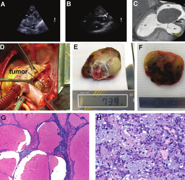
A 56-year-old Caucasian female was referred to the emergency department of our hospital soon after she collapsed at home. She noted progressive chest pain associated with dyspnea, fatigue and edema of the lungs. The patient’s clinical history included hypothyroidism and hypercholesterolemia. At admission to hospital the patient presented a blood pressure of 100/55 mmHg and a heart rate of 60 beats per minute (bpm). Trans-thoracic and transesophageal echocardiography revealed a huge mobile left atrial mass protruding into the left ventricle during diastole and obstructing diastolic filling of the left ventricle (Figure 1A and 1B). Subsequently, a contrast-enhanced ECG-gated 64-slice spiral computed tomography (CT) confirmed the presence of a very large globular mass filling the left atrium (Figure 1C). The patient was taken to the operating theatre, where a median sternotomy was performed and cardiopulmonary bypass was installed through aorto-right atrium cannulation (Figure 1D). The huge gelatinous tumor mass was successfully excised (Figure 1E). Before leaving the hospital on day 9 after surgery, echocardiography showed a normal left and right ventricular function with an ejection fraction of 60%, well-functioning heart valves, and no relevant pericardial effusion.
Figure 1.
A: Transthoracic echocardiography (modified 4-chamber view) showed a large mobile left atrial mass (68 × 52 mm) attached to the atrial septum protruding into the left ventricle during diastole (LV, left ventricle; LA, left atrium; RV, right ventricle; RA, right atrium); B: Transesophageal echocardiography (long-axis view) revealed large heterogeneous, lobulated echogenic mass with a short stalk (arrow) attached to the interatrial septum, obstructing diastolic filling of the left ventricle (Ao, Aorta); C: Computed tomographic scan showing large globular mass filling left atrium with narrow base of attachment to the interatrial septum; D: Intraoperative photograph showing the giant left atrial tumor mass; E: The gelatinous tumor formation weight 74 mg; F: The cut section of the resection specimen showed extensive peliosis-like cystic hemorrhagic spaces within otherwise tan-yellow to obviously myxoid tissue; G: Histological examination of Hematoxylin and Eosin (H&E)-stained sections confirmed the presence of large cystic spaces lined by compressed tumorous tissue, but lacking true endothelial lining; H: The tumor showed otherwise typical features of atrial myxoma with ovoid to spindled or rounded bland-looking tumor cells arranged in cords, microtrabeculae, nests and perivascular ribbons within a strikingly myxoid edematous background rich in small-sized capillaries with occasional hemosiderin pigments indicating recurrent stromal bleeding.
Pathological findings
The resection specimen was composed of a large soft polypoid mass measuring 6.5 cm in maximum diameter and weighing 74 g, attached to endomyocardial tissue at the resection base. The resection margins were unremarkable. The cut section showed extensive peliosis-like cystic hemorrhagic spaces within otherwise tan-yellow to gelatinous tissue (Figure 1F). Histological examination of Hematoxylin and Eosin (H&E)-stained sections confirmed the presence of large cystic spaces lined by compressed tumorous tissue, but lacking true endothelial lining (Figure 1G). The tumor showed otherwise typical features of atrial myxoma with ovoid to spindled or rounded bland-looking tumor cells arranged in cords, microtrabeculae, nests and perivascular ribbons within a strikingly myxoid edematous background rich in small-sized capillaries with occasional hemosiderin pigments indicating recurrent stromal bleeding (Figure 1H). The surrounding endomyocardial tissue at the tumor base showed evidence of reactive chronic inflammation with prominent lymphoid aggregates.
Discussion
The clinical presentation and the histological appearance of primary cardiac tumors varies greatly. While a majority of them presents with non-specific or insidious symptoms or as incidental findings, still others may present with acute symptoms necessitating emergency surgery. For an appropriate surgical planning, a precise preoperative diagnosis of cardiac tumors and their distinction from thrombi and other tumor-like lesions is mandatory. Echocardiography, CT and MRT are the most useful diagnostic tools in the assessment of cardiac tumors, which in almost all cases precisely locates the tumor and defines its extent and thus resectability [4]. Atrial myxoma usually measures a few centimeters when diagnosed. However, several cases of huge atrial myxomas have been reported [5-9]. Most of these unusually giant myxomas have been associated with mechanical complications that resulted in acute and life-threatening cardiac symptoms. On the other hand, huge but asymptomatic atrial myxoma is uncommon [5]. Huge myxomas commonly undergo variable degrees of regressive changes with evidence of old and recent haemorrhage. Occasional cases my display unusual regressive changes including osseous metaplasia. These changes might be so extensive that the lesion closely mimics a large organizing thrombus and the neoplastic nature of the lesion might be recognized only after thorough sampling of the specimen. On the contrary, large mural atrial thrombi my occasionally closely mimic myxomas [10].
In summary, we described the clinical features, imaging characteristics and histopathological findings in an unusual case of huge atrial myxoma that caused acute cardiac symptoms as a result of prolapsing into the mitral valve. This unusual presentation of atrial myxoma needs be included in the differential diagnosis of atrial thrombi and other causes of acute-onset cardiac symptoms associated with mass-occupying lesions in the heart.
References
- 1.Burke AP, Virmani R. Tumors of the heart and great vessels. In: Rosai J, Sobin LH, editors. Atlas of Tumor Pathology. Third series, fascicle 16. Washington, DC: Armed Forces Institute of Pathology; 1996. [Google Scholar]
- 2.Selkane C, Amahzoune B, Chavanis N, Raisky O, Robin J, Ninet J, Obadia JF. Changing management of cardiac myxoma based on a series of 40 cases with long-term follow-up. Ann Thorac Surg. 2003;76:1935–8. doi: 10.1016/s0003-4975(03)01245-1. [DOI] [PubMed] [Google Scholar]
- 3.Van der Mieren G, Duchateau J, Herijgers R. Left atrial myxoma: presentation with acute aortic occlusion and ‘resolution’ of the primary tumor. Acta Chir Belg. 2007;107:687–9. doi: 10.1080/00015458.2007.11680147. [DOI] [PubMed] [Google Scholar]
- 4.Strecker T, Rösch J, Weyand M, Agaimy A. Primary and metastatic cardiac tumors: imaging characteristics, surgical treatment, and histopathological spectrum: a 10-year-experience at a German heart center. Cardiovasc Pathol. 2012 Jan 31;21:436–43. doi: 10.1016/j.carpath.2011.12.004. [DOI] [PubMed] [Google Scholar]
- 5.Yuce M, Dagdelen S, Ergelen M, Eren N, Caglar N. A huge obstructive myxoma located in the right heart without causing any symptom. Int J Cardiol. 2007;114:405–6. doi: 10.1016/j.ijcard.2005.11.086. [DOI] [PubMed] [Google Scholar]
- 6.Yang TY, Tsai JP, Chang CH, Kuo JY, Hung CL. Giant right atrial myxoma with pulmonary trunk dislodgement causing intermittent tricuspid obliteration and clinical manifestations of right heart failure. Echocardiography. 2011;28:E183–6. doi: 10.1111/j.1540-8175.2011.01508.x. [DOI] [PubMed] [Google Scholar]
- 7.Affronti A, Di Bella I, Prontera P, Da Col U, Ramoni E, Donti E, Paris M, Ragni T. Obstruction of the tricuspid valve orifice by a huge right atrial myxoma associated with the Carney complex: a case report. J Card Surg. 2010;25:674–6. doi: 10.1111/j.1540-8191.2010.01114.x. [DOI] [PubMed] [Google Scholar]
- 8.Dobarro D, Gómez-Rubín Mdel C, Sánchez-Recalde A, López-Fernández T, García-Fernández E, Viana A, López-Sendón JL. A huge atrial myxoma causing severe double mitral lesions. Heart Lung Circ. 2009;18:131–2. doi: 10.1016/j.hlc.2007.11.143. [DOI] [PubMed] [Google Scholar]
- 9.Leonard S, Ryan J. A heavy heart; A massive right atrial myxoma causing fatigue and shortness of breath. Ir Med J. 2010;103:83–4. [PubMed] [Google Scholar]
- 10.Kim MS, Park JH, Kang SK, Na MH, Lee JH, Choi SW, Jeong JO, Seong IW. Acute ST-segment elevation myocardial infarction due to a huge floating thrombus mimicking a myxoma in the left atrium. J Am Soc Echocardiogr. 2009;22:1085.e1–3. doi: 10.1016/j.echo.2009.04.003. [DOI] [PubMed] [Google Scholar]


