

Abstract
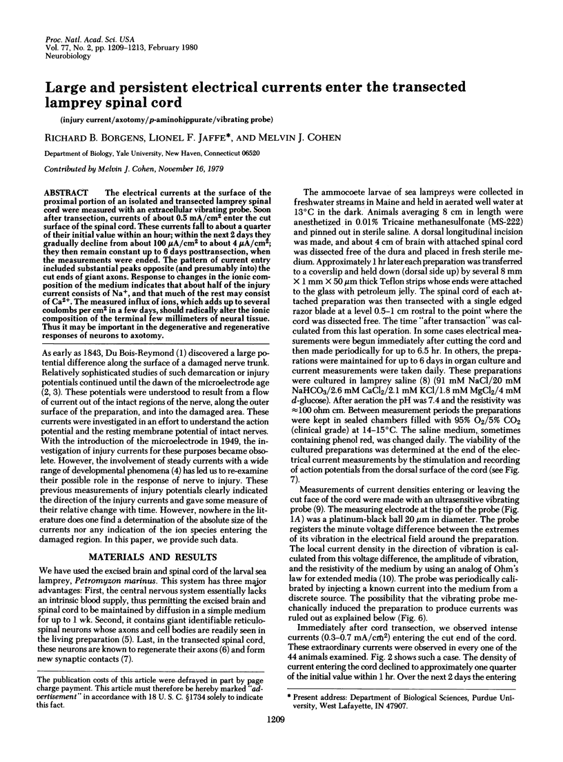
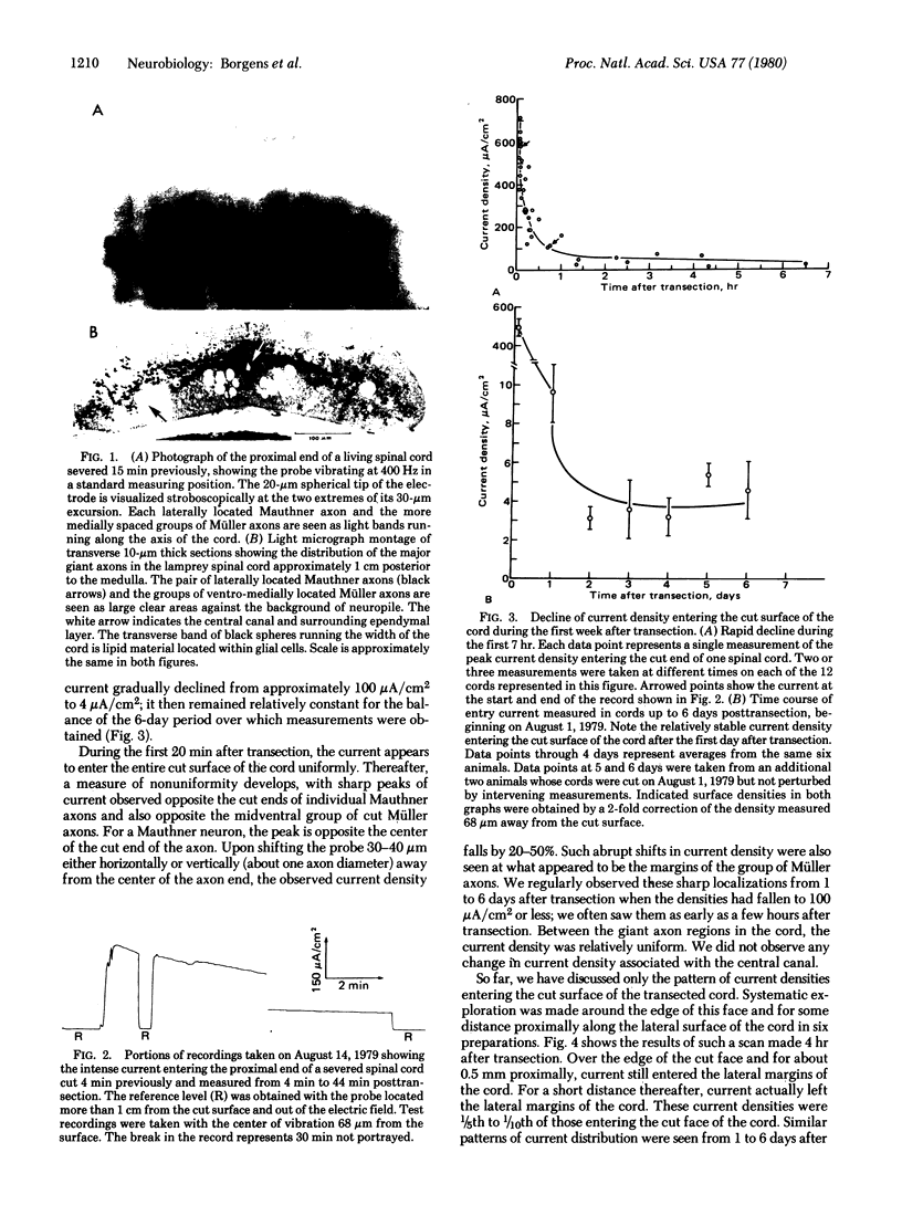
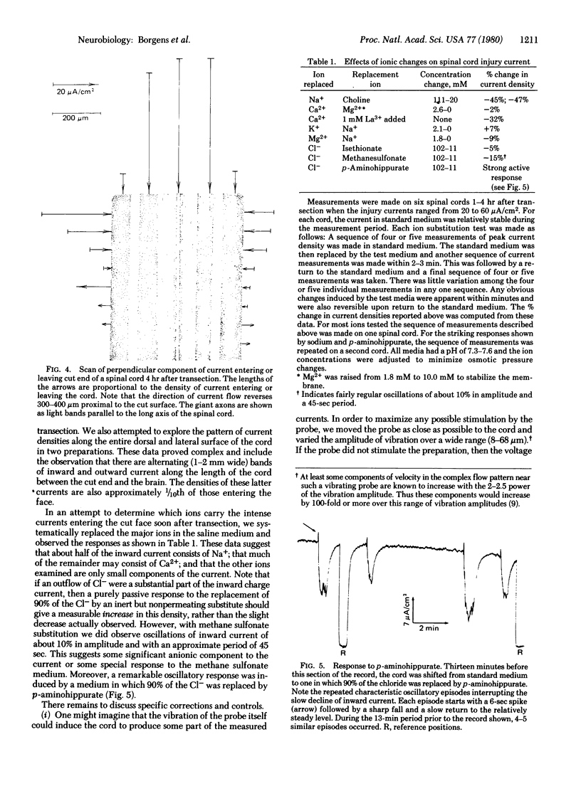
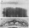
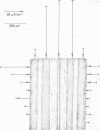
The electrical currents at the surface of the proximal portion of an isolated and transected lamprey spinal cord were measured with an extracellular vibrating probe. Soon after transection, currents of about 0.5 mA/cm2 enter the cut surface of the spinal cord. These currents fall to about a quarter of their initial value within an hour; within the next 2 days they gradually decline from about 100 microA/cm2 to about 4 microA/cm2; they then remain constant up to 6 days posttransection, when the measurements were ended. The pattern of current entry included substantial peaks opposite (and presumably into) the cut ends of giant axons. Response to changes in the ionic composition of the medium indicates that about half of the injury current consists of Na+, and that much of the rest may consist of Ca2+. The measured influx of ions, which adds up to several coulombs per cm2 in a few days, should radically alter the ionic composition of the terminal few millimeters of neural tissue. Thus it may be important in the degenerative and regenerative responses of neurons to axotomy.
Full text
PDF
Images in this article
Selected References
These references are in PubMed. This may not be the complete list of references from this article.
- Jaffe L. F., Nuccitelli R. An ultrasensitive vibrating probe for measuring steady extracellular currents. J Cell Biol. 1974 Nov;63(2 Pt 1):614–628. doi: 10.1083/jcb.63.2.614. [DOI] [PMC free article] [PubMed] [Google Scholar]
- Jaffe L. F., Nuccitelli R. Electrical controls of development. Annu Rev Biophys Bioeng. 1977;6:445–476. doi: 10.1146/annurev.bb.06.060177.002305. [DOI] [PubMed] [Google Scholar]
- Rovainen C. M. Physiological and anatomical studies on large neurons of central nervous system of the sea lamprey (Petromyzon marinus). I. Müller and Mauthner cells. J Neurophysiol. 1967 Sep;30(5):1000–1023. doi: 10.1152/jn.1967.30.5.1000. [DOI] [PubMed] [Google Scholar]
- Rovainen C. M. Regeneration of Müller and Mauthner axons after spinal transection in larval lampreys. J Comp Neurol. 1976 Aug 15;168(4):545–554. doi: 10.1002/cne.901680407. [DOI] [PubMed] [Google Scholar]
- Schlaepfer W. W., Bunge R. P. Effects of calcium ion concentration on the degeneration of amputated axons in tissue culture. J Cell Biol. 1973 Nov;59(2 Pt 1):456–470. doi: 10.1083/jcb.59.2.456. [DOI] [PMC free article] [PubMed] [Google Scholar]
- Schlaepfer W. W., Hasler M. B. Characterization of the calcium-induced disruption of neurofilaments in rat peripheral nerve. Brain Res. 1979 May 25;168(2):299–309. doi: 10.1016/0006-8993(79)90171-9. [DOI] [PubMed] [Google Scholar]
- Wickelgren W. O. Physiological and anatomical characteristics of reticulospinalneurones in lamprey. J Physiol. 1977 Aug;270(1):89–114. doi: 10.1113/jphysiol.1977.sp011940. [DOI] [PMC free article] [PubMed] [Google Scholar]
- Wood M. R., Cohen M. J. Synaptic regeneration in identified neurons of the lamprey spinal cords. Science. 1979 Oct 19;206(4416):344–347. doi: 10.1126/science.482943. [DOI] [PubMed] [Google Scholar]
- Zelená J., Lubińska L., Gutmann E. Accumulation of organelles at the ends of interrupted axons. Z Zellforsch Mikrosk Anat. 1968;91(2):200–219. doi: 10.1007/BF00364311. [DOI] [PubMed] [Google Scholar]