

Abstract
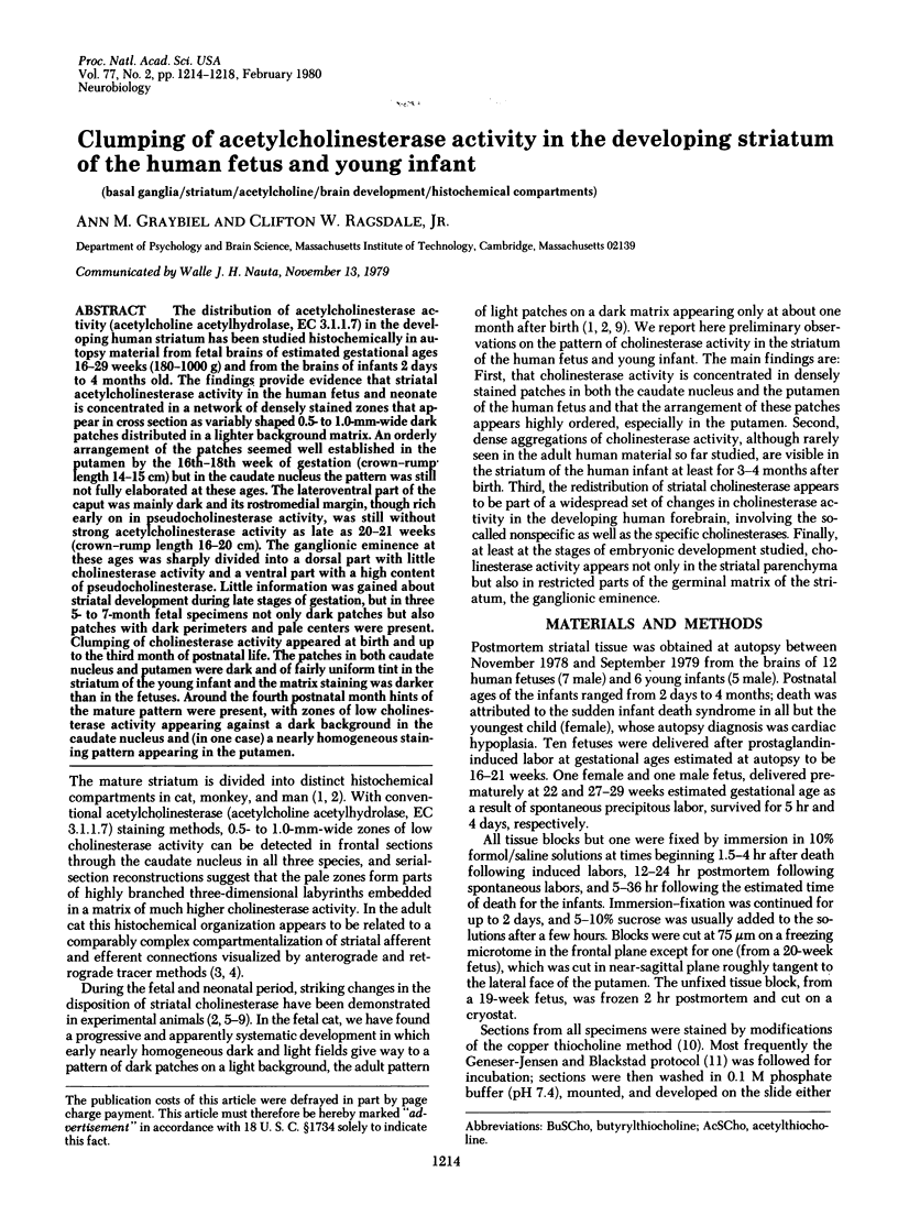
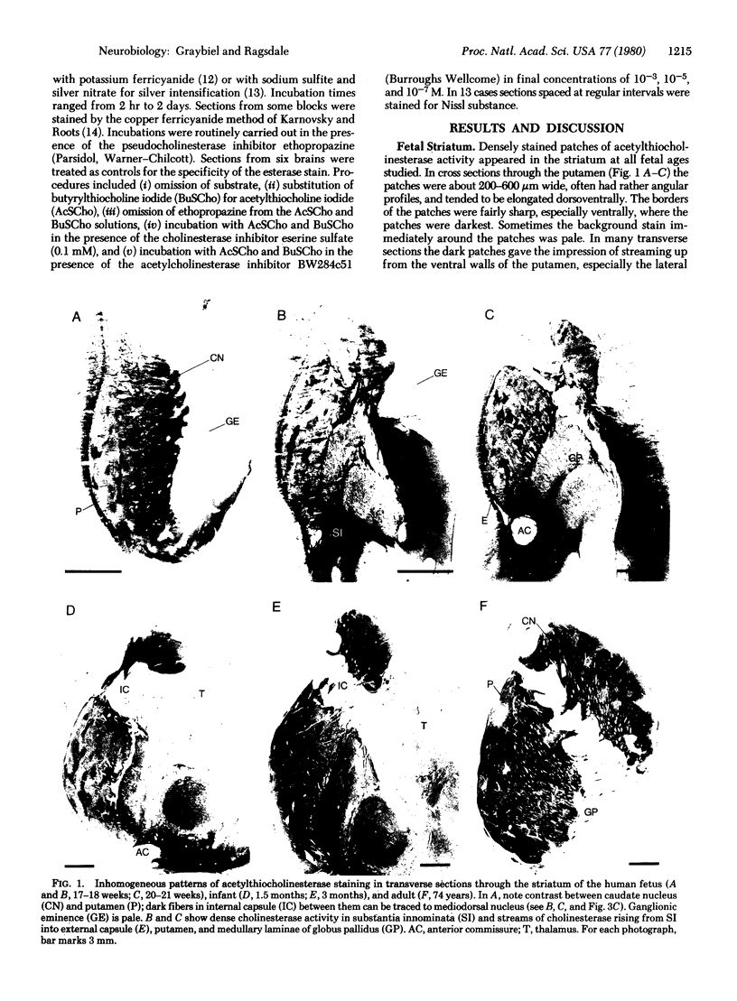
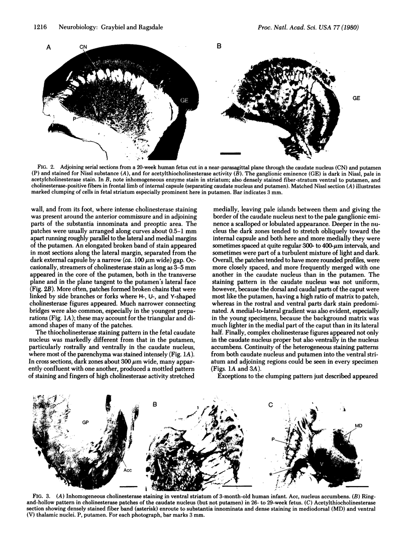
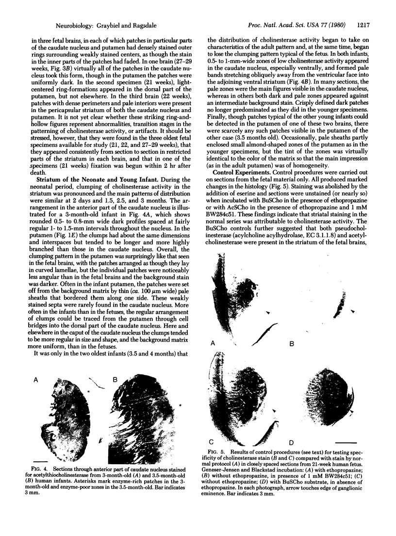
The distribution of acetylcholinesterase activity (acetylcholine acetylhydrolase, EC 3.1.1.7) in the developing human striatum has been studied histochemically in autopsy material from fetal brains of estimated gestational ages 16-29 weeks (180-1000 g) and from the brains of infants 2 days to 4 months old. The findings provide evidence that striatal acetylcholinesterase activity in the human fetus and neonate is concentrated in a network of densely stained zones that appear in cross section as variably shaped 0.5- to 1.0-mm-wide dark patches distributed in a lighter background matrix. An orderly arrangement of the patches seemed well established in the putamen by the 16th-18th week of gestation (crown-rump length 14-15 cm) but in the caudate nucleus the pattern was still not fully elaborated at these ages. The lateroventral part of the caput was mainly dark and its rostromedial margin, though rich early on in pseudocholinesterase activity, was still without strong acetylcholinesterase activity as late as 20-21 weeks (crown-rump length 16-20 cm). The ganglionic eminence at these ages was sharply divided into a dorsal part with little cholinesterase activity and a ventral part with a high content of pseudocholinesterase. Little information was gained about striatal development during late stages of gestation, but in three 5- to 7-month fetal specimens not only dark patches but also patches with dark perimeters and pale centers were present. Clumping of cholinesterase activity appeared at birth and up to the third month of postnatal life. The patches in both caudate nucleus and putamen were dark and of fairly uniform tint in the striatum of the young infant and the matrix staining was darker than in the fetuses. Around the fourth postnatal month hints of the mature pattern were present, with zones of low cholinesterase activity appearing against a dark background in the caudate nucleus and (in one case) a nearly homogeneous staining pattern appearing in the putamen.
Keywords: basal ganglia, striatum, acetylcholine, brain development, histochemical compartments
Full text
PDF
Images in this article
Selected References
These references are in PubMed. This may not be the complete list of references from this article.
- Butcher L. L., Hodge G. K. Postnatal development of acetylcholinesterase in the caudate-putamen nucleus and substantia nigra of rats. Brain Res. 1976 Apr 23;106(2):223–240. doi: 10.1016/0006-8993(76)91022-2. [DOI] [PubMed] [Google Scholar]
- Geneser-Jensen F. A., Blackstad T. W. Distribution of acetyl cholinesterase in the hippocampal region of the guinea pig. I. Entorhinal area, parasubiculum, and presubiculum. Z Zellforsch Mikrosk Anat. 1971;114(4):460–481. doi: 10.1007/BF00325634. [DOI] [PubMed] [Google Scholar]
- Graybiel A. M., Ragsdale C. W., Jr Fiber connections of the basal ganglia. Prog Brain Res. 1979;51:237–283. [PubMed] [Google Scholar]
- Graybiel A. M., Ragsdale C. W., Jr Histochemically distinct compartments in the striatum of human, monkeys, and cat demonstrated by acetylthiocholinesterase staining. Proc Natl Acad Sci U S A. 1978 Nov;75(11):5723–5726. doi: 10.1073/pnas.75.11.5723. [DOI] [PMC free article] [PubMed] [Google Scholar]
- Graybiel A. M., Ragsdale C. W., Jr, Moon Edley S. Compartments in the striatum of the cat observed by retrograde cell labeling. Exp Brain Res. 1979 Jan 2;34(1):189–195. doi: 10.1007/BF00238352. [DOI] [PubMed] [Google Scholar]
- KARNOVSKY M. J., ROOTS L. A "DIRECT-COLORING" THIOCHOLINE METHOD FOR CHOLINESTERASES. J Histochem Cytochem. 1964 Mar;12:219–221. doi: 10.1177/12.3.219. [DOI] [PubMed] [Google Scholar]
- Krnjević K., Silver A. A histochemical study of cholinergic fibres in the cerebral cortex. J Anat. 1965 Oct;99(Pt 4):711–759. [PMC free article] [PubMed] [Google Scholar]
- Krnjević K., Silver A. Acetylcholinesterase in the developing forebrain. J Anat. 1966 Jan;100(Pt 1):63–89. [PMC free article] [PubMed] [Google Scholar]
- Mesulam M. M., Van Hoesen G. W. Acetylcholinesterase-rich projections from the basal forebrain of the rhesus monkey to neocortex. Brain Res. 1976 Jun 4;109(1):152–157. doi: 10.1016/0006-8993(76)90385-1. [DOI] [PubMed] [Google Scholar]
- Nobin A., Björklund A. Topography of the monoamine neuron systems in the human brain as revealed in fetuses. Acta Physiol Scand Suppl. 1973;388:1–40. [PubMed] [Google Scholar]
- Olson L., Boréus L. O., Seiger A. Histochemical demonstration and mapping of 5-hydroxytryptamine- and catecholamine-containing neuron systems in the human fetal brain. Z Anat Entwicklungsgesch. 1973 Apr 16;139(3):259–282. doi: 10.1007/BF00519968. [DOI] [PubMed] [Google Scholar]