

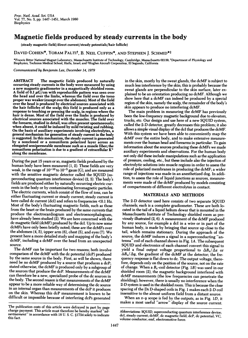
Abstract
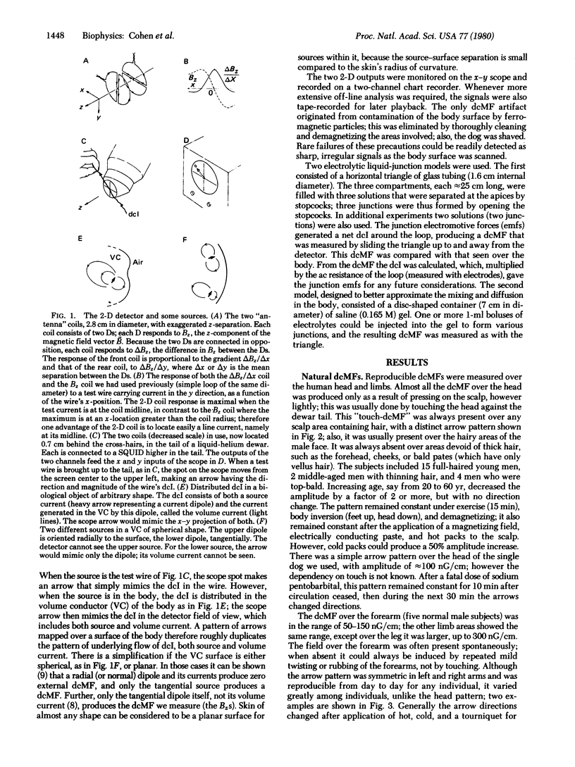
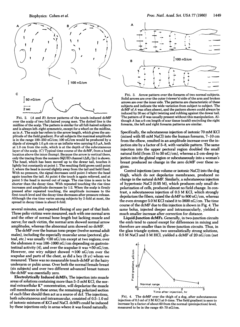
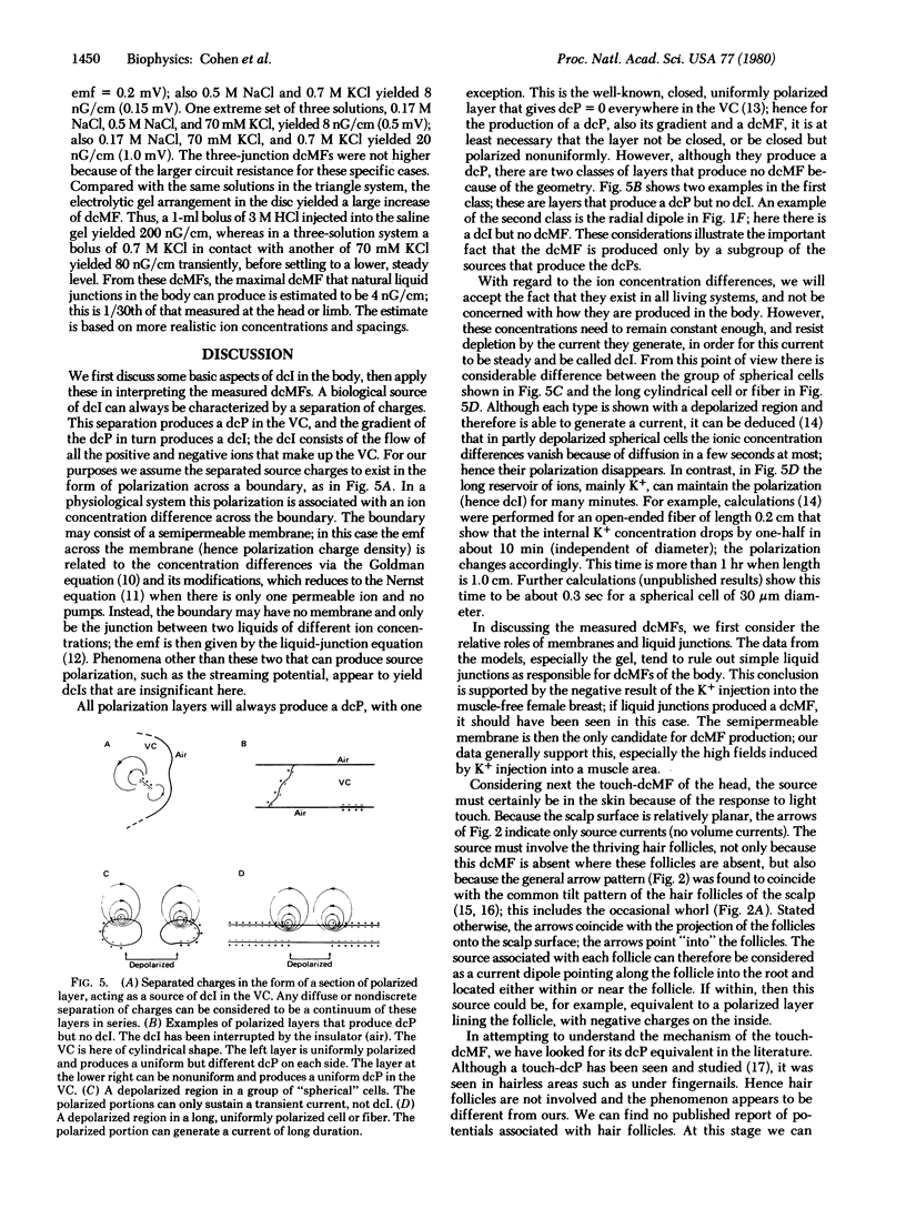
The magnetic fields produced by naturally occurring steady currents in the body were measured by using a new magnetic gradiometer in a magnetically shielded room. A field of 0.1 micro G/cm with reproducible pattern was seen over the head and over the limbs, whereas the field over the torso proper was weaker (except over the abdomen). Most of the field over the head is produced by electrical sources associated with the hair follicles of the scalp; this field is produced only as a response to touching or pressing the scalp, in regions where the hair is dense. Most of the field over the limbs is produced by electrical sources associated with the muscles. The field over the forearm, studied in detail, was often present spontaneously; when absent, it could be induced by mild twisting and rubbing. On the basis of auxiliary experiments involving electrolytes, a general mechanism for generation of steady current in the body is suggested. In this mechanism, the steady current is generated by a nonclosed or a nonuniform polarized layer across an elongated semipermeable membrane such as a muscle fiber; the nonuniform polarization is due to a gradient of extracellular K+ along the membrane.
Full text
PDF

Selected References
These references are in PubMed. This may not be the complete list of references from this article.
- Cohen D., Hosaka H. Part II: magnetic field produced by a current dipole. J Electrocardiol. 1976;9(4):409–417. doi: 10.1016/s0022-0736(76)80041-6. [DOI] [PubMed] [Google Scholar]
- Edelberg R. Local electrical response of the skin to deformation. J Appl Physiol. 1973 Mar;34(3):334–340. doi: 10.1152/jappl.1973.34.3.334. [DOI] [PubMed] [Google Scholar]
- Grynszpan F., Geselowitz D. B. Model studies of the magnetocardiogram. Biophys J. 1973 Sep;13(9):911–925. doi: 10.1016/S0006-3495(73)86034-5. [DOI] [PMC free article] [PubMed] [Google Scholar]
- LASSEN N. A. MUSCLE BLOOD FLOW IN NORMAL MAN AND IN PATIENTS WITH INTERMITTENT CLAUDICATION EVALUATED BY SIMULTANEOUS XE-133 AND NA24 CLEARANCES. J Clin Invest. 1964 Sep;43:1805–1812. doi: 10.1172/JCI105054. [DOI] [PMC free article] [PubMed] [Google Scholar]
- PARNELL J. P. Hair pattern and distribution in mammals. Ann N Y Acad Sci. 1951 Mar;53(3):493–497. doi: 10.1111/j.1749-6632.1951.tb31951.x. [DOI] [PubMed] [Google Scholar]
- Palti Y., Gold R., Stämpfli R. Diffusion of ions in myelinated nerve fibers. Biophys J. 1979 Jan;25(1):17–31. doi: 10.1016/S0006-3495(79)85275-3. [DOI] [PMC free article] [PubMed] [Google Scholar]
- Picton T. W., Hillyard S. A. Cephalic skin potentials in electroencephalography. Electroencephalogr Clin Neurophysiol. 1972 Oct;33(4):419–424. doi: 10.1016/0013-4694(72)90122-8. [DOI] [PubMed] [Google Scholar]
- Sano K., Miyake H., Mayanagi Y. Steady potentials in various stress conditions in man. Electroencephalogr Clin Neurophysiol. 1967;(Suppl):264+–264+. [PubMed] [Google Scholar]