

Abstract
Injuries due to bear mauling are rarely reported in the literature. Bears are strong and agile wild animals, potentially dangerous, unpredictable and can inflict serious injuries. Mammalian attack injuries have a special place in traumatology because of their high complication rate when compared with similar soft tissue wounds otherwise caused. Bites from attacking animals may lead to local infection, and wounds that are potentially contaminated with a variety of pathogens. The excellent blood supply of the face makes infection a rare occurrence, however, the injury may cause sufficient disfigurement to require extensive reconstruction. Treatment of mammalian bite wounds must address both the management of soft tissue deformities and then prevention of post treatment infection. Although generally considered to be dirty or contaminated they could be successfully treated by surgical cleansing and primary suture with a favourable outcome. The bony injuries also have to be addressed. Management of such injuries often need multidisciplinary approach and multiple secondary surgeries to treat the secondary defects.
Introduction
Injuries due to bear mauling are rarely reported in the literature. Bears are strong and agile wild animals, potentially dangerous, unpredictable and can inflict serious injuries. Mammalian attack injuries have a special place in traumatology because of their high complication rate when compared with similar soft tissue wounds otherwise caused. Animal bite wounds are more common on upper extremities but a large percentage are also located on the face and head [1]. Bear maul cases are rarely reported in the literature. The commonly involved injury sites being the face (80.57%) and head (54.67%). Bite wounds by large animals can present in a more serious fashion with bone injuries. All the bony injuries will be associated with soft-tissue injuries namely punctures, lacerations and avulsions with or without an actual tissue loss [2]. The facial bones (27.09%) were commonly involved, followed by the skull (5.75%). Predominance of zygoma in the face and frontal bone in the skull explains the fact that the most prominent parts are easily targeted [3]. The management of facial bite wounds often is a challenge because of unforeseeable infections that can occur because of primary bacterial invasion. The reported incidence of wound infection varies from 13 to 30% and their optimal management is controversial [1, 4]. Extensive soft tissue defects should be treated according to the criteria of an esthetic reconstructive surgery. The purpose of this article is to report two cases of bear maul injury and their subsequent management.
Case Reports
Case 1
A 50 year old male patient reported to our department with a history of bear maul injury. There was no history of vomiting, loss of consciousness. Patient gives history of bleeding from mouth and nose for about 10 min. On examination there was a deep laceration on the face extending from left side of the forehead starting at about a centimeter below the hairline running obliquely towards the right side lateral to eyebrow till the zygomatic arch. One more laceration was evident below the right eye extending mediolaterally from the bridge of nose laterally involving the lower eyelid and running over the zygomatic arch and connected to the previous laceration. The skin covering this defect was hanging lose laterally. Ocular dystopia and enophthalmos were evident on right eye (Figs. 1, 2). Fractured segments were evident over right zygomatic bone as well as in the arch. Occlusion was satisfactory. Patient sustained minor lacerations over his right and left upper limbs which were sutured in primary health centres in their place.
Fig. 1.
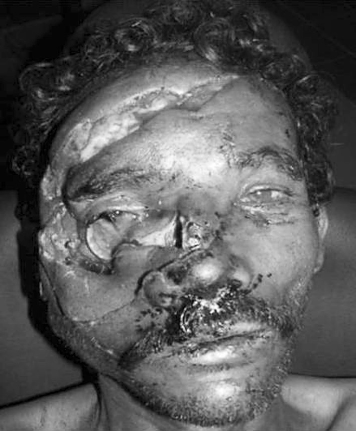
Pre-operative photograph
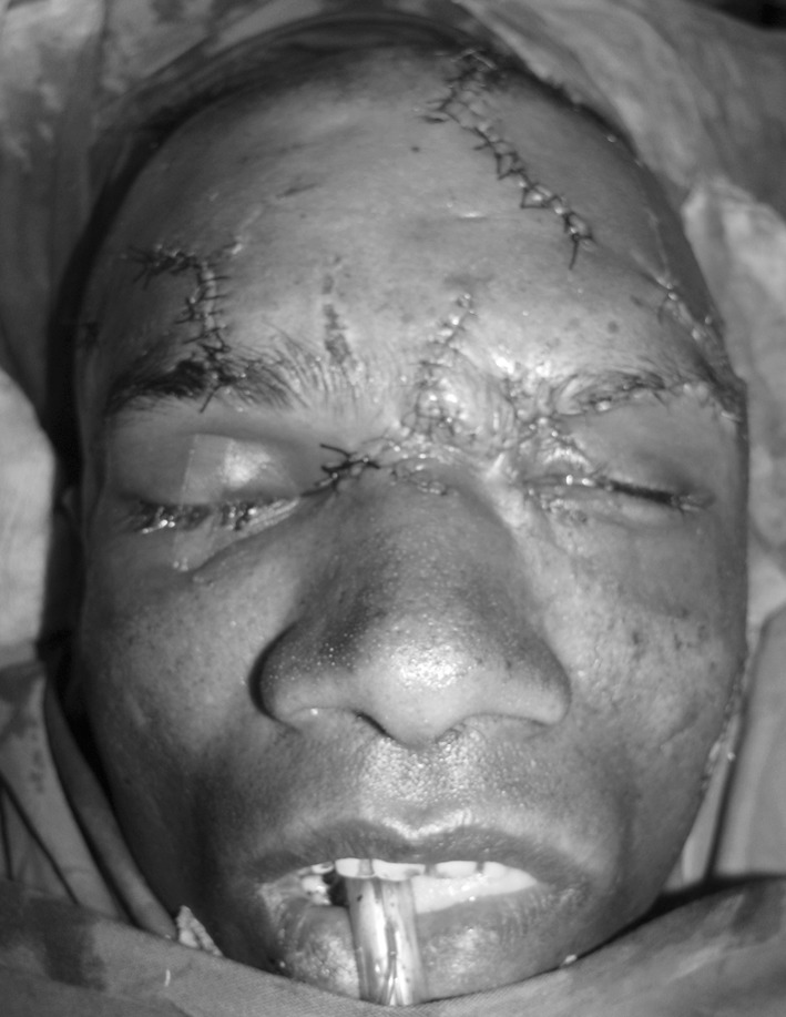
Fig. 2.
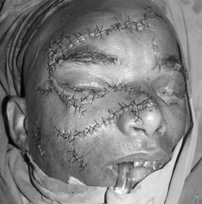
Immediate post operative photograph of case 1
Water’s view revealed the fracture of infraorbital rim, zygomatic bone, zygomatic arch and the nasal bone on the right side.
Under general anesthesia after thorough irrigation with normal saline and debridement of the wound, fractures were reduced. Arch was elevated under direct vision, nasal fractures reduced by closed method, miniplate fixation at F-Z suture and infraorbital rim was carried out. As there was comminuted fracture over anterior wall of the sinus the two major segments were fixed with 26 G stainless steel wire. Wound was closed in layers. Post operative recovery of the patient was uneventful. 1 month post operatively he noticed blurring of vision in right eye and ophthalmology opinion was obtained, diagnosed as cicatricial medial lower lid ectropion with canalicular fibrosis, (Fig. 3) advised ectropion correction with conjunctival DCR with jones tube. Patient was not willing for further treatment and was lost to follow up.
Fig. 3.
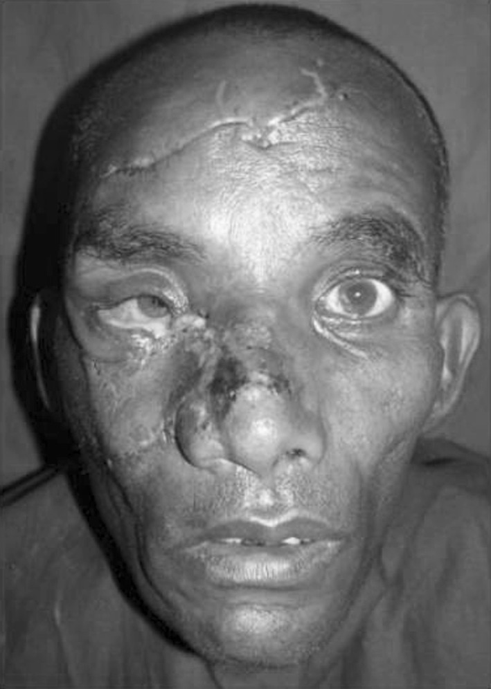
One month post operative photograph showing rightlower lid ectropion
Case 2
A 36 year old male patient reported with the history of bear maul injury. There was no history of vomiting, loss of consciousness. Bleed from the nose and left ear for about 15 min was observed by the patient. On examination there was evidence of subconjunctival haemorrhage and chemosis on left eye. Deep laceration of about 11 cm was evident on left side of face extending from nasal bridge involving the left upper eyelid, supraorbital region and forehead. One more vertical laceration of about 6 cm was extending from the right forehead region at about 2 cm below the hairline running downward to involve the right eyebrow. One more laceration was seen on left side of the face extending from the zygomatic arch, about a cm anterior to tragus running obliquely over the cheek and terminating at the level of commissure (Figs. 4, 5, 6). On palpation tenderness elicited over nasal dorsum and left preauricular region, step deformity was evident on left infraorbital margin. There was blurring of vision in the left eye. Occlusion was satisfactory. There were other associated lacerated wounds over the scalp, left ear and abdomen which were treated at a local primary health centre by sutures.
Fig. 4.
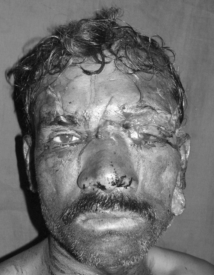
Pre-operative photograph
Fig. 5.
Immediate post operative photograph of case 2
Fig. 6.
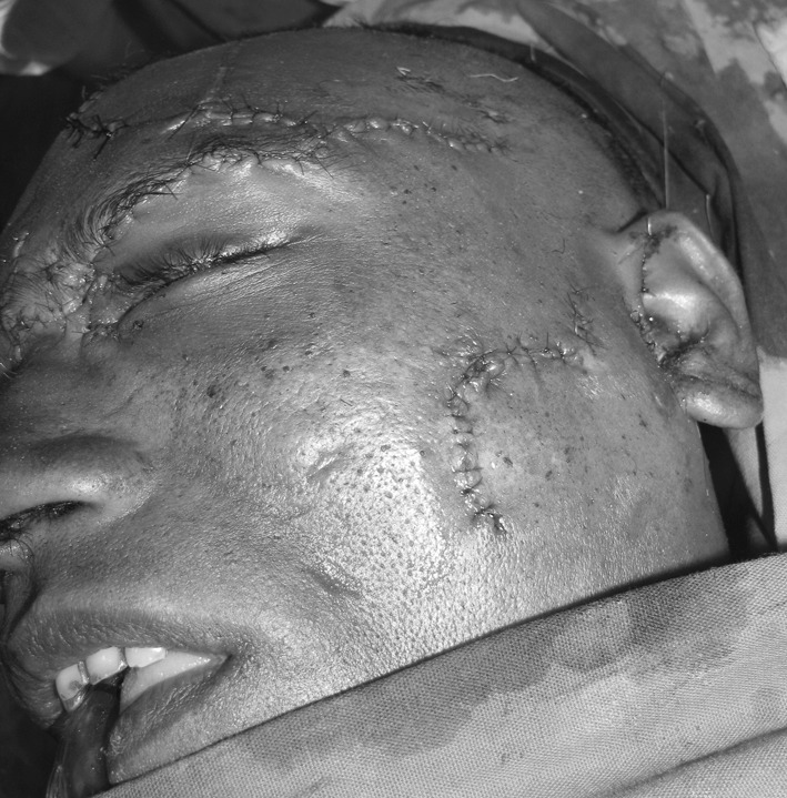
Immediate post operative photograph of case 2
Water’s view revealed fracture of infraorbital rim of left orbit and lateral wall of the nose on left side. Ophthalmological clearance was obtained. Under general anesthesia after thorough irrigation of the wounds with normal saline and debridement, reduction of fracture fragments done and fixation of nasal bone and fractured segment, carrying medial canthal ligament to the frontal process of maxilla was done using miniplate. Layered closure of the wound was carried out. Post recovery of the patient was uneventful. He reported after 5 months with a complaint of watery eye on left side, ophthalmologist’s opinion was obtained and it was diagnosed as dacrocystitis and was put on ofloxacin eye drops. Patient reported only after one and half year with a complaint of pus discharge and swelling in the left medial canthal region (Fig. 7). Plate removal in that region was done and was operated for dacrocystorhinostomy.
Fig. 7.
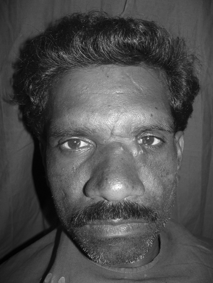
One and half year post operative photograph showing dacrocystitis on left eye
Discussion
The actual incidence of injuries sustained by humans in animal attacks is unknown, as most of the cases are fatal or approach after a certain duration. Animal injuries inflicted on to humans are distinct in nature causing tearing, cutting, penetrating and crush injuries of which having a combination of sharp and blunt trauma. To complicate matters, many animal attacks occur in remote and wilderness areas and there are substantial delays before notification, rescue and presentation to definitive care Mammalian bite injuries can have distressing physical and psychological consequences. Wild animal injuries are contaminated from claws and teeth and provide two distinct significant sources of contamination. Although death from animal bites is uncommon, injuries inflicted by black bears should be treated as a major trauma [5].
Bite wounds have always been considered complex injuries contaminated with a unique polymicrobial inoculum. Facial bite wounds generally display low infection rates, commonly attributed to the rich blood supply of the area. Significant delay beyond 6–12 h—in seeking medical attention increases the likelihood of infection [1, 2, 6].
The microbiology of bite wounds will consist of oral microflora of the offending animal. The bacteriology of animal wounds is varied and it is multibacterial with pasteurella being the most common species. Pasteurellamultocida organism are associated with a rapid onset of infection, whereas the latency period is more than 24 h, staphylococci, streptococci or anaerobes are more likely etiologic agents. Cultures are most useful in case initial antibiotic therapy fails [2, 7, 8]. The bacteriology of bear bite wounds is limited by the absence of systematic study and the paucity of data obtained from cultures prior to, or even after the initiation of, antimicrobial therapy. In a report by Kunimoto et al. [9] they isolated mainly the aerobic gram negative bacilli and enterococcus from their cases but they do not deny the presence of anaerobes in bear bite wounds. So it seems prudent to administer broad-spectrum antimicrobial agents.
The gold standard antibiotic is amoxicillin with clavulanic acid, or be a combination of penicillin and cephalosporin, in penicillin allergic patients moxifloxacin or clindamycin with ciprofloxacin covers most bite wound isolates. Azithromycin can be used in penicillin allergic pregnant women and in children. Duration of antibiotics can range from 3 to 5 days in cases of prophylaxis, 7–14 days in soft tissue infections and minimum of 3 weeks in infections involving the joints and bones [7, 8]. Bite wounds are considered tetanus prone, so appropriate immunization should be administered if the patient has had fewer than three doses of tetanus toxoid in their lifetime or more than 5 years have passed since the last dose [2, 4]. In our cases we went for amoxicillin in one case and cephalosporin in other case along with metronidazole. Antibiotics were administered for a period of 7–10 days.
Mammalian bite wounds are associated with an infection rate between 10 and 20%. Prophylactic antibiotics are recommended for high risk wounds namely those more than 8 h old, deep punctures, wounds that require surgical repair, hand and lower extremity wounds and wounds in medically compromised patients [4, 8].
The treatment of these lacerations should include thorough physical examination along with radiographic examination must warranted. These wounds should be irrigated with hydrogen peroxide and saline thoroughly, meticulously debrided and closed. For bite wounds presenting beyond the golden 24 h period primary closure is controversial. Facial bite wounds presenting as late as 4 days post injury can be attempted with an acceptable risk of infection. In these cases delayed closure is a time honoured practice. This implies a waiting period of 4–5 days during which time the wound is kept open, usually with moist gauze dressings providing drainage, while edema is allowed to subside. In severely crushed or mangled wounds, besides being at increased risk for infection, tend to become very edematous within hours. Delayed primary closure is indicated in these cases to avoid dehiscence [2, 8]. We have achieved acceptable results by closing the lacerations primarily. In our cases it was not necessary to go for v–y or z platy. In cases of unacceptable scars z platy or v–y plasties can be considered.
In most of the bear attack cases soft tissue injuries will be associated with underlying bony injuries. So radiographic examination should be carried out to rule out the fractures before attempting to close the wounds. The bony injuries should be addressed accordingly. Close follow up is necessary in these cases to avoid any complications and to treat any such complications as early as possible. In our cases the management of residual deformities such as lower lid ectropion with canalicular fibrosis seen after 5 months in the first case and dacrocystitis in the second case who reported after one and half year needed treatment.
Conclusion
Bear attack injuries require a team approach, as the injuries are not restricted to one area. Careful evaluation of bite wounds with physical as well as radiological examination should be carried out prior to definitive management. Proper surgical toilet with wound irrigation followed by careful debridement along with addressing the bony injuries remains the mainstay of treatment of all bite wounds. Most of these cases though treated with primary closure at the earliest, come with residual deformities that require multiple surgeries in a staged manner. In all these cases clinical judgement should be used and close follow up is recommended for the early management of residual deformities.
References
- 1.Wolff K-D. Management of animal bite injuries of the face: experience with 94 patients. J Oral Maxillofac Surg. 1998;56:838–843. doi: 10.1016/S0278-2391(98)90009-X. [DOI] [PubMed] [Google Scholar]
- 2.Stefanopoulos PK. Management of facial bite wounds. Oral Maxillofac Surg Clin N Am. 2009;21:247–257. doi: 10.1016/j.coms.2008.12.009. [DOI] [PubMed] [Google Scholar]
- 3.Rasool A, Wani AH, Darzi MA, Inam Zaroo M, Iqbal S, Bashir SA, et al. Incidence and pattern of bear maul injuries in Kashmir. Injury. 2010;41:116–119. doi: 10.1016/j.injury.2009.07.077. [DOI] [PubMed] [Google Scholar]
- 4.Kesting MR, Holzle F, Pox C, Thurmuller P, Wolff K-D. Animal bite injuries to the head: 132 cases. Br J Oral Maxillofac Surg. 2006;44:235–239. doi: 10.1016/j.bjoms.2005.06.015. [DOI] [PubMed] [Google Scholar]
- 5.Dhar SA, Butt MF, Farooq M, Mir MR, Wani ZA, Afzal Suhail, et al. Pattern of orthopaedic injuries in bear attacks: report from a tertiary care centre in Kashmir. Injury. 2008;39:249–255. doi: 10.1016/j.injury.2007.07.028. [DOI] [PubMed] [Google Scholar]
- 6.Ruskin JD, Laney TJ, Wendt SV, Markin RS. Treatment of mammalian bite wounds of the maxillofacial region. J Oral Maxillofac Surg. 1993;51:174–176. doi: 10.1016/S0278-2391(10)80017-5. [DOI] [PubMed] [Google Scholar]
- 7.Bahram R, Burke JE, Lanzi GL. Head and neck injury from a leopard attack: case report and review of the literature. J Oral Maxillofac Surg. 2004;62:247–249. doi: 10.1016/j.joms.2003.04.015. [DOI] [PubMed] [Google Scholar]
- 8.Stefanopoulos PK, Tarantzopoulou AD. Facial bite wounds: management update. Int J Oral Maxillofac Surg. 2005;34:464–472. doi: 10.1016/j.ijom.2005.04.001. [DOI] [PubMed] [Google Scholar]
- 9.Kunimoto D, Rennie R, Citron DM, Goldstein EJC. Bacteriology of a bear bite wound to a human: case report. J Clin Microbiol. 2004;42:3374–3376. doi: 10.1128/JCM.42.7.3374-3376.2004. [DOI] [PMC free article] [PubMed] [Google Scholar]