Abstract
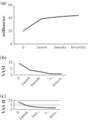
The following study was conducted in Meenakshi Ammal Dental College on 32 patients. The objective of our study was to assess the efficacy of arthrocentesis for the management of acute closed lock of TMJ. All patients presented with a history of sudden and persistent difficulty in mouth opening and associated TMJ pain. A total of 27 female and 5 male patients were treated using arthrocentesis and lavage under local anesthesia. Patients were assessed for pain and jaw function using visual analogue scales, VAS I (Fig. 4b), VAS II (Fig. 4c), respectively and mouth opening MMO (Fig. 4a) for a period of 6 months. Our results showed satisfactory relief in pain and improved mouth opening in 30 patients. The results proved that arthrocentesis is a very useful technique for treatment of acute closed lock of TMJ. However to arrive at a definitive conclusion a long term evaluation is required.
Keywords: Arthrocentesis, TMJ disorders, Lavage, Acute closed lock group
Introduction
The temporomandibular joint (TMJ) is a unique diarthroidal synovial joint with highly complex functions. Success of management of temporomandibular joint disorders depends on accurate diagnosis and appropriate treatment planning.
Arthrocentesis is a simple and minimally invasive technique for the management of temporomandibular disorders. Murakami et al. [2–4] were the first to offer a systematic description of temporomandibular joint arthrocentesis, which they called “Manipulation Technique”, followed by pumping and hydraulic pressure. Sanders motivated this procedure, with excellent results in releasing closed lock by arthroscopic lavage and lysis, a procedure, which was initially, used as diagnostic maneuver. However, it was first developed in Florida, USA by Nitzan et al. [5, 7]. The concept was based on the observation that simple lavage and lysis of the upper joint space using arthroscopy was highly successful in patients with closed lock and re-established normal range of mouth opening. In this text we share with you our experience of 32 cases of internal derangement (acute closed lock) treated by this simple technique at the Department of Oral and Maxillofacial Surgery, Meenakshi Ammal Dental College and Hospitals, Chennai.
Materials and Methods
This study was conducted in the division of Oral and Maxillofacial Surgery—Meenakshi Ammal Dental College and Hospital, Chennai. The study was undertaken on 32 patients who underwent arthrocentesis and lavage for the treatment of acute closed lock of the TMJ.
Twenty seven females and five males with TMJ disorders were treated with arthrocentesis and lavage with manipulation. The age range of this group of patients was 18 to 27 years (average 23 years) and the duration of the presenting symptoms ranged from 1 to 3 months. Patients selected for this study were those who presented with the chief complaint of sudden and persistent limited mouth opening often associated with pain in the affected TMJ.
Prior to each procedure, baseline data in the form of Maximum Mouth Opening (MMO) and 2 Visual Analogue Scales (VAS) relating to the degree of pain and mandibular dysfunction (chewing) were collected. The data was again collected after one week and on subsequent follow-up in order to gauge the effectiveness.
MMO was measured as the distance in mm between the incisal edges of the central incisors at maximum pain free mouth opening. One VAS (VAS-I) ranging from 0 to 10, was used to assess the level of pain experienced by the patient and the second VAS (VAS-II) ranging from 0 to 10, was used to document the level of disturbed jaw function (i.e., chewing ability). Zero (0) on each VAS was taken to mean no pain (VAS-I) or no impairment in chewing ability (VAS-II) and Ten (10) was most intense pain imaginable (VAS-I) or total inability to chew, (VAS-II).
Evaluation of the patients involved a history and clinical examination as well as a radiographic examination. The radiographic examination included panoramic view for all patients to rule out any bony changes or pathology.
Diagnosis of acute closed lock was made through detailed case history and clinical examination. All the patients were then followed up at one week, three weeks, three months, and six months after the procedure.
Technique for TMJ Arthrocentesis and Lavage
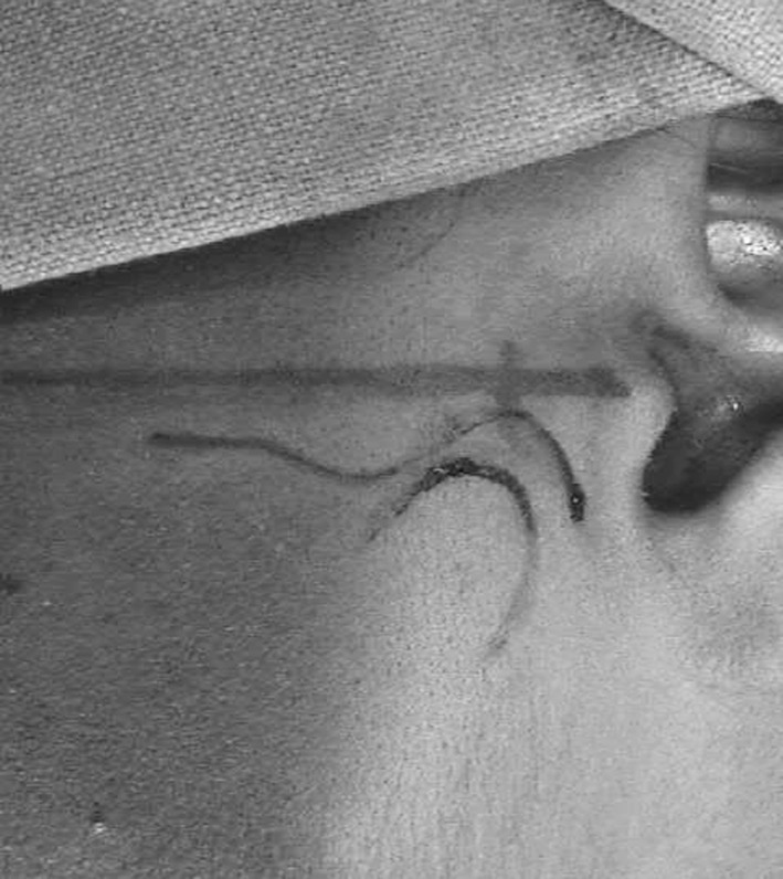
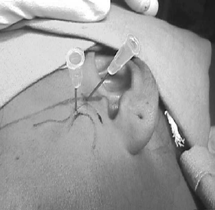
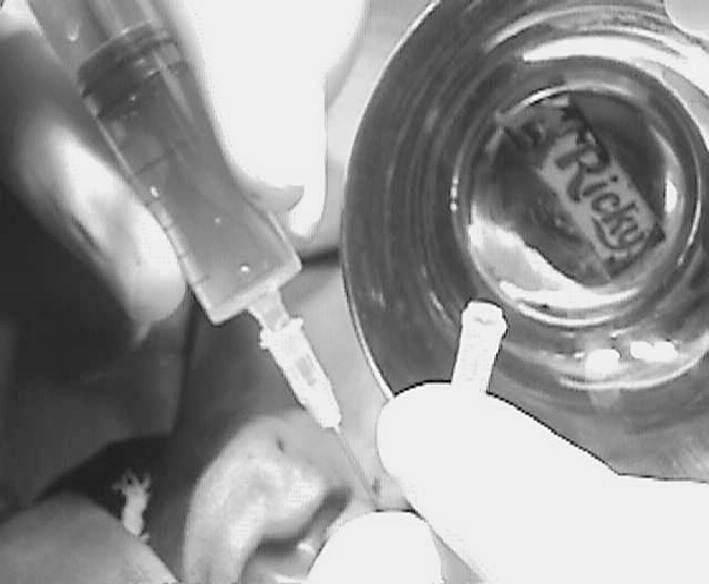
After the patients were positioned, the ear and pre auricular skin over the TMJ were prepared. The condylar and zygomatic processes and the articular eminence were palpated and outlined. This is to provide approximate anatomic landmarks for the placement of the needles. Two points were then marked over the articular fossa and eminence, 1 and 2 cm in front of the tragus along the trago-canthal line (Fig. 1). This is similar to the entry points used for arthroscopic procedures. Next, the auriculo temporal nerve was blocked with about 2 ml of local anesthetic. Once the anesthesia had been secured, a 19-gauge needle was introduced into the superior joint space at the glenoid fossa (posterior mark). This was followed by the injection of 2 to 3 ml of normal saline solution, to distend the joint space. Another needle was inserted into the distended compartment in the area of the articular eminence to establish a free flow of the solution through the superior joint space (Fig. 2). A syringe filled with normal saline solution was then connected to one of the needles and the solution was injected into the superior joint space. The second needle provides on outflow for the solution, which was drained in a kidney dish. A total of 50 to 100 ml was used to lavage (Fig. 3) the superior joint space. During the procedure, the outlet needle was momentarily blocked with finger pressure 2 or 3 times to help distend and lyse the joint adhesions. Once the needles were removed the patients jaw was gently manipulated in the vertical, protrusive and lateral excursions to help further free up of the disc.
Fig. 1.

Points marked on the trago canthal line
Fig. 2.

Insertion of needles
Fig. 3.

Onset of lavage
Post-Operative Phase
All the patients were administered anti-inflammatory/muscle relaxants. Post operatively Ibuprofen 400 mg + Methocarbamol 750 mg (Ibugesic M) was prescribed 3 times daily for 3 days.
Course of physiotherapy was commenced immediately following the procedure to promote and maintain an improved range of mandibular movement.
All patients were followed up at 1, 3 weeks, 3 and 6 months postoperatively for analyzing maximum mouth opening and pain (VAS-I) and masticatory efficiency (VAS-II).
Results
A total of 32 patients underwent arthrocentesis under LA at our centre. All 32 patients achieved considerable improvement for a period of 3 months at least. However 3 patients reported back in the 3 to 6 months period with recurrence of pain and pre procedural symptoms, following which they underwent a second sitting of arthrocentesis, out of which, 2 patients only showed marginal improvement and were taken up for surgical intervention after 6 months.
Discussion
Frost et al. [1] reported that arthrocentesis is the first line procedure for the treatment of acute and chronic closed lock of the TMJ in internal derangement. It is a simple minimally invasive and effective procedure with proven long-term results and minimal potential complications.
In our study on 32 patients with TMJ internal derangement, clinical diagnosis of acute closed lock was made. The diagnosis was made based on a detailed case history, clinical examination and use of radiograph (OPG) to rule out any bony pathology. Almost all the patients reported with sudden pain and difficulty in mouth opening over a period of 1 to 2 months.
We achieved very good results in all 32 patients with immediate improvement in mouth opening (Fig. 4a) and pain relief (<2 weeks post procedure) (Fig. 4b) which corroborates with the findings of Nitzan et al. [6]. All patients were counseled and advised isometric jaw exercises after the procedure. In a period of 1 week to 1 month jaw function also improved considerably (Fig. 4c). All patients were followed up at intervals of 1, 3 weeks, 3 and 6 months after the procedure for evaluation. Except for the 2 patients mentioned earlier all the 30 patients were quite happy with the improvement with emphasis on pain (Fig. 4b), mouth opening (Fig. 4a) and jaw function (Fig. 4c). Therefore at the end of the 6 month follow up period we had successfully treated 30 patients reported with acute closed lock of the TMJ, with a success rate of 93.75%.
Fig. 4.
a Maximum mandibular opening. b Pain. c Jaw function
Arthrocentesis has become a viable alternative to arthrotomy and other arthroscopic invasive procedures under general anaesthesia for certain conditions involving the TMJ particularly for the treatment of acute closed lock. It seems to be very effective in eliminating symptoms of TMJ pain and restoring mandibular function. However, further long-term evaluation is required to arrive at a definitive conclusion. In our experience, we found arthrocentesis to be a very useful procedure for the treatment of acute closed lock of the TMJ, as it is a very minimally invasive, cost effective technique with very low morbidity. However for the success of the treatment patient cooperation is of paramount importance.
Summary and Conclusion
This study was conducted in the division of Oral and Maxillofacial Surgery, Meenakshi Ammal Dental College and Hospital. 32 cases of acute closed lock of the TMJ were chosen and symptomatically treated by arthrocentesis and lavage. The results obtained have been fairly gratifying.
The merits and demerits of this procedure have been discussed.
Based on the study the following conclusion was made.
This is a simple technique, which can be carried out on an outpatient basis.
Less invasive, hence easily acceptable to the patients.
Cost effective.
Low morbidity.
Highly effective in treatment of acute closed lock patients.
Procedure may be repeated if necessary.
It may be tried before arthrotomy in cases of internal derangement.
However, a long-term follow up and evaluation is required to arrive at a statistically definitive conclusion.
References
- 1.Frost DE, Kendell BD. The use of arthrocentesis for treatment of temporomandibular joint disorders. J Oral Maxillofac Surg. 1999;57:583–587. doi: 10.1016/S0278-2391(99)90080-0. [DOI] [PubMed] [Google Scholar]
- 2.Murakami K, Hosaka H, Moriya Y, Segami N. Short-term treatment outcome study for the management of TMJ closed lock. A comparison of arthrocentesis to non-surgical therapy and arthroscopic lysis and lavage. Oral Med Oral Pathol Oral Surg. 1995;80:253–257. doi: 10.1016/S1079-2104(05)80379-8. [DOI] [PubMed] [Google Scholar]
- 3.Murakami K, Moriya Y, Goto K, Segami N. Four-year follow-up study of TMJ arthroscopic surgery for advanced stage internal derangements. J Oral Maxillofac Surg. 1996;54:285–290. doi: 10.1016/S0278-2391(96)90742-9. [DOI] [PubMed] [Google Scholar]
- 4.Nitzan DW, Dolwick MF, Martinez A. TMJ arthrocentesis—a simplified treatment for severe limited mouth opening. J Oral Maxillofac Surg. 1991;49:1163–1167. doi: 10.1016/0278-2391(91)90409-F. [DOI] [PubMed] [Google Scholar]
- 5.Nitzan DW, Mahier Y, Simkin A. Intra articulator pressure measurements in patients suddenly developing, severely limited mouth opening. J Oral Maxillofac Surg. 1992;50:1038–1042. doi: 10.1016/0278-2391(92)90485-I. [DOI] [PubMed] [Google Scholar]
- 6.Nitzan DW. Arthrocentesis for management of severe closed lock of the TMJ. J Oral Maxillofac Surg Clin N Am. 1994;6(2):245. [Google Scholar]
- 7.Nitzan DW, et al. Reported that the etiology for acute closed lock was an anterior displacement of the articular disc and the “anchored disc phenomenon”. J Oral Maxillofac Surg. 1999;58:342. [Google Scholar]


