



Abstract
A facultative iron-reducing [Fe(III)-reducing] Paenibacillus sp. strain was isolated from Hanford 300A subsurface sediment biofilms that was capable of reducing soluble Fe(III) complexes [Fe(III)-nitrilotriacetic acid and Fe(III)-citrate] but unable to reduce poorly crystalline ferrihydrite (Fh). However, Paenibacillus sp. 300A was capable of reducing Fh in the presence of low concentrations (2 μM) of either of the electron transfer mediators (ETMs) flavin mononucleotide (FMN) or anthraquinone-2,6-disulfonate (AQDS). Maximum initial Fh reduction rates were observed at catalytic concentrations (<10 μM) of either FMN or AQDS. Higher FMN concentrations inhibited Fh reduction, while increased AQDS concentrations did not. We also found that Paenibacillus sp. 300A could reduce Fh in the presence of natural ETMs from Hanford 300A subsurface sediments. In the absence of ETMs, Paenibacillus sp. 300A was capable of immobilizing U(VI) through both reduction and adsorption. The relative contributions of adsorption and microbial reduction to U(VI) removal from the aqueous phase were ∼7:3 in PIPES [piperazine-N,N′-bis(2-ethanesulfonic acid)] and ∼1:4 in bicarbonate buffer. Our study demonstrated that Paenibacillus sp. 300A catalyzes Fe(III) reduction and U(VI) immobilization and that these reactions benefit from externally added or naturally existing ETMs in 300A subsurface sediments.
INTRODUCTION
The uranium (U) contamination in 11 of the 18 U.S. Department of Energy (DOE) sites causes significant remediation costs due to its presence in soils and groundwater (57, 62). Hanford 300A is one of the U.S. DOE sites in which soils, sediments, and groundwater are heavily contaminated with U (50, 57). Physical and chemical methods have been developed to remediate U in contaminated sites; unfortunately, to date, these methods have not proven cost-effective or sustainable (48, 62). Biological processes harnessing indigenous microbial activities in the field, such as the in situ microbial reduction of soluble, mobile U(VI) to insoluble, immobile U(IV), provide an alternative approach that has been considered to be one of the prominent remediation strategies for U-contaminated sites (2, 29, 48). Dissimilatory metal-reducing bacteria (DMRB) can directly reduce soluble U(VI) to insoluble U(IV) (27, 41, 44, 65). Shewanella and Geobacter have been extensively studied as model DMRB organisms to understand their roles in influencing the fate of U at contaminated sites (25, 27, 37, 38, 44). Reductive products from DMRB respiring on minerals can further interact with U(VI), influencing U bioremediation processes (5, 36, 70). Fe(III) oxides and Fe(III)-bearing clay minerals are significant components in soils and sediments (33, 76). In situ biostimulation of DMRB favors microbial Fe(III) over U(VI) reduction for anaerobic respiration, since the potential energy gain is higher for Fe(III) respiration than for U(VI) respiration (24). Microbial Fe(III) respiration results in Fe(II) or Fe(II)-bearing minerals which can enhance abiotic U(VI) reduction and can even outcompete enzymatic U(VI) reduction (5, 70). Therefore, it is critical to correlate U reduction with solid-phase iron reduction.
Recent studies have shown that microorganisms isolated from U-contaminated sites play significant roles in both Fe(III) reduction and U immobilization (22, 26, 60, 62, 63, 74). Fredrickson et al. (22) examined the Fe(III) and U(VI) reduction capability of Deinococcus radiodurans R1. Fe(III) reduction by D. radiodurans R1 was limited to Fe(III) nitrilotriacetic acid [Fe(III)-NTA] and solid-phase hydrous ferric oxide (HFO), and goethite was reduced only in the presence of a high concentration (100 μM) of anthraquinone-2,6-disulfonate (AQDS) as an electron transfer mediator (ETM). However, U(VI) reduction was slow [∼95 to 100% of initial U(VI) in 21 days with 5 × 10⁷ CFU/ml] even in the presence of 100 μM AQDS. Sani et al. (60) studied Fe(III), Cr(VI), and U(VI) reduction by Cellulomonas species isolated from an enrichment culture from Hanford 300A sediments. A Cellulomonas species was capable of reducing Fe(III) and U(VI) but required a longer time [∼56% of initial Fe(III)-NTA and ∼40% of initial U(VI) in 100 h with 500 mg/liter total cell proteins] in bicarbonate-buffered non-growth medium. Gerlach et al. (26) studied various forms of Fe(III) reduction by Cellulomonas species in the presence or absence of AQDS, and the results showed only a 10-fold increase in hydrous ferric oxide or ferric citrate reduction even in the presence of 500 μM AQDS. Sivaswamy et al. (62) investigated the U(VI) immobilization capability of Cellulomonas species and observed slow immobilization in both PIPES [piperazine-N,N′-bis(2-ethanesulfonic acid)]- and bicarbonate-buffered medium. However, U(VI) immobilization was enhanced in the presence of AQDS. Wu et al. (74) found that Anaeromyxobacter dehalogenans was capable of reducing U(VI) only in the presence of hydrogen (H2) as an electron donor.
In natural environments, microorganisms tend to form biofilms to increase survival potential (16–18). One common feature of all biofilms is the presence of extracellular polymeric substances (EPS), which is thought to allow cells in biofilms to conduct extracellular activities, acquire nutrients from the surrounding environment, and engage in chemical communication and the coordination of gene activities with greater efficiency than their planktonic counterparts (12, 19). Microbial adhesion to surfaces during biofilm formation leads to changes in cellular metabolisms and resistance to heavy metals and radionuclides (16, 34). The biogeochemical heterogeneity, retention time of contaminants, and microbial communities in biofilms may have a significant effect on the fate of contaminants and minerals in the subsurface environment (34, 49). Cells growing in biofilms in the subsurface can use solid-phase iron for respiration while immobilizing soluble U. This immobilization can be achieved through reduction and adsorption. The critical step to understanding these processes is to isolate biofilm-associated metal-reducing bacteria and quantify their role in U immobilization and solid-phase iron reduction.
Recently, we isolated several facultative iron-reducing bacterial strains from Hanford 300A subsurface sediment biofilms. One of the isolates was designated Paenibacillus sp. strain 300A, a member of the genus Paenibacillus. The presence of this facultative bacterium in Fe(III)-reducing consortia from DOE field sites was suggested by microbial community analyses (35, 52), and other members of this genus had also been previously isolated from U mining waste (55), sediment contaminated with heavy metals (14), and Hanford site subsurface sediments (9, 21).
The goal of this study was to characterize Fe(III) reduction and U(VI) immobilization by Paenibacillus sp. 300A. We quantified the reduction of soluble Fe(III) complexes [Fe(III)-NTA and Fe(III)-citrate], the reduction of poorly crystalline ferrihydrite (Fh), and U(VI) immobilization by Paenibacillus sp. 300A. The effect of ETMs such as flavin mononucleotide (FMN) and anthraquinone-2,6-disulfonate (AQDS) on Fh reduction was also investigated. FMN and AQDS were chosen as ETMs, since flavin-like molecules (FMN/riboflavin) are secreted by microbial species such as Shewanella (46, 53, 69, 71) and AQDS is an organic compound that contains functional groups (i.e., quinone) that are representative of functional groups in humic substances (66). We further studied Fh reduction in the presence of 300A sediment slurry and in Shewanella sp. culture supernatant to test the ability of Paenibacillus sp. 300A to utilize ETMs that occur naturally in the 300A subsurface and ETMs naturally produced by other microorganisms. Finally, U(VI) immobilization was studied in PIPES and bicarbonate buffers. Furthermore, 300A sediment slurry and 300A sediment slurry amended with Fh were used to evaluate their impact on overall U(VI) immobilization.
MATERIALS AND METHODS
Fe(III)-reducing enrichment, growth conditions, and biofilm isolates.
Sediment (well ID C-6190; 10.0 to 10.3 m; Hanford formation) collected from the Department of Energy's (DOE's) Hanford 300 Area Integrated Field Research Challenge (IFRC) site (http://ifchanford.pnl.gov) was used to enrich Fe(III)-reducing isolates (1). About 10 g of sediment was mixed with 100 ml of Fe(III)-reducing (Fe-RE) medium, which contained 10 mM ethanol (electron donor), 10 mM Fe(III)-NTA (electron acceptor), 0.42 g/liter NaHCO3, 0.9 g/liter NaCl, 0.5 g/liter KCl, 1.0 g/liter NH4Cl, 0.1 g/liter Na3P3O9, 0.2 g/liter MgCl2 · 6H2O, and 0.016 g/liter CaCl2 · 2H2O and was supplemented with trace amounts of minerals, vitamins, and amino acids (7, 32, 75). Sediment incubation was carried out anaerobically at room temperature (22 ± 2°C). Anoxic Fe(III)-NTA and ethanol were injected into the enrichment culture to the initial concentrations every 10 days of incubation to ensure the presence of an excess of electron donors and acceptors. After 50 days, the enrichment culture was used to inoculate a flat-plate biofilm reactor (6, 11). The biofilm reactor was operated at room temperature (22 ± 2°C) using Fe-RE medium at a flow rate of 3 ml/min. A stream of N2 was used to maintain anaerobic conditions during biofilm growth. Biofilm growth was observed after 5 days. The biofilm was removed and mixed with 2 ml of fresh Fe-RE medium, which was then streaked onto Fe-RE agar plates (15% agar containing Fe-RE medium) and incubated at 30°C in an anaerobic chamber (90% N2, 5% H2, 5% CO2). Single colonies were further streaked onto Fe-RE agar to obtain pure cultures, which were then identified based on 16S rRNA gene sequences.
Characterization and phylogenetic analysis.
Identification of the bacterial isolates was performed on DNA extracted from frozen tryptic soy broth (TSB)-grown cell pellets using the DNeasy blood and tissue kit (Qiagen) by following the protocol for the lysis of Gram-positive bacteria. DNA was eluted in a final volume of 200 μl of water. Purified DNA was used as a template for PCR. Each PCR consisted of 10 μl of 5× solution, 1 μl 10 mM deoxynucleoside triphosphate (dNTP), 0.5 μM each primer (27F and 907R), 0.5 μl Phusion polymerase, 100 ng template, and H2O to a final volume of 50 μl. The cycling parameters were 98°C for 1 min, followed by 25 cycles at 98°C for 15 s, 56°C for 15 s, and 75°C for 30 s, with a final extension at 75°C for 10 min. PCR products were purified and then imaged on a 1% agarose gel to ensure that the correct band was amplified. The amplified 16S product was then sequenced at the Joint Technology Center (J. Craig Venter Institute, San Diego, CA) using 97F and 907R primers. Aligned sequences obtained from the RDP Seqmatch program (Ribosomal Database Project [RDP] II, release 8.1) were exported into MEGA5 (64). A phylogenetic tree was constructed with the Neighbor-Joining method (59). The sum of the branch lengths of the optimal tree was 0.29. The percentages of the replicate trees in which the associated taxa clustered together in the bootstrap test (1,000 replicates) are shown next to the branches (20). The tree is drawn to scale, with branch lengths in the same units as those of the evolutionary distances used to infer the phylogenetic tree. The evolutionary distances were computed using the Jukes-Cantor method (31) and are in units of the number of base substitutions per site. The analysis involved nine nucleotide sequences. All positions containing gaps and missing data were eliminated. There were a total of 1,272 positions in the final data set.
Preparation of 300A sediment slurry, sediment extracts, and Shewanella oneidensis MR-1 culture supernatant.
The 300A sediment slurry was prepared by adding 1.0 g of 300A sediments stimulated with organic-amended synthetic groundwater (OA-SGW) (1, 49) to 50 ml of bicarbonate-buffered non-growth medium (i.e., 30 mM sodium bicarbonate and 10 mM lactate, pH 7.0), followed by sonication for 2 h (40 kHz; Branson 1510-MTH ultrasonic bath cleaner). The slurry was then mixed thoroughly and allowed to settle, and the supernatant was separated. This supernatant was referred to as 300A sediment slurry. The 300A sediment extracts were prepared by adding 15.0 g of OA-SGW-stimulated 300A sediments to 75 ml of bicarbonate-buffered non-growth medium, followed by sonication for 2 h and then centrifugation at 5,000 × g for 15 min. The supernatant was collected and filtered through 0.22-μm syringe filters. The filtrate was referred to as sediment extracts. Shewanella oneidensis MR-1 was grown aerobically overnight in M1 minimal medium (56) with 10 mM lactate as the electron donor and oxygen as the electron acceptor. The culture grown overnight was centrifuged (4,500 × g, 15 min, room temperature); the supernatant was collected and filtered through a 0.22-μm syringe filter. 300A sediment slurry, sediment extracts, and Shewanella oneidensis MR-1 culture supernatant were stored at 4°C.
Preparation of Paenibacillus sp. 300A resting cells and Fe(III) stock solutions.
Paenibacillus sp. 300A was grown aerobically in TSB (30 g/liter; Difco Laboratories) for 12 h at 30°C on a rotary shaker (150 rpm). Cells were then harvested by centrifugation (4,500 × g, 15 min, room temperature) and washed three times using sodium bicarbonate buffer (30 mM, pH 7.0). Finally, cells were resuspended in sodium bicarbonate buffer (30 mM, pH 7.0) that had been stored in an anaerobic chamber (90% N2, 5% H2, 5% CO2) and used within 1 h. Fe(III)-NTA soluble Fe(III) solution was prepared according to the protocol described by Fredrickson et al. (22). Fe(III)-citrate soluble Fe(III) solution was prepared by adding 24.48 g of ferric citrate to 200 ml of deionized (DI) water (pH 7.0). A poorly crystalline to amorphous solid Fh suspension was prepared according to the protocol described by Lovley and Phillips (42). All Fe(III) stock solutions were kept in an anaerobic chamber (90% N2, 5% H2, 5% CO2) to make the solution anoxic and stored in a dark, sealed anaerobic bottle.
Reduction of Fe(III)-NTA, Fe(III)-citrate, and Fh using resting cells.
Fe(III) reduction was studied anaerobically using bicarbonate-buffered non-growth medium. Fe(III)-NTA or Fe(III)-citrate at a final concentration of 10 mM or Fh at 8 mM was added. Paenibacillus sp. 300A was added to a final concentration of ∼2 × 10⁸ cells/ml. Fe(III) reduction without cells was used as an abiotic control. We tested two different synthetic ETMs for poorly crystalline Fh reduction by Paenibacillus sp. 300A: FMN and AQDS at concentrations of 2, 10, 50, and 100 μM. FMN was chosen as an ETM, since Fe(III) reducers are involved in subsurface environments during in situ biostimulation (2, 52) and Fe(III) reducers such as Shewanella species secrete flavins as ETMs (46, 71). AQDS is a humic acid compound analogue previously used as an ETM for Fe(III) and U(VI) reduction (40, 62). Fh reduction without cells was used as an abiotic control. We further tested three different naturally produced ETMs for Fh reduction by Paenibacillus sp. 300A: (i) autoclaved 300A sediment slurry (i.e., Fh plus slurry plus cells), (ii) 300A sediment extracts (i.e., Fh plus extract plus cells), and (iii) Shewanella oneidensis MR-1 culture supernatant amended with 10 mM lactate (i.e., lactate plus supernatant plus cells plus Fh). Experiments with (i) slurry plus Fh, (ii) slurry plus cells, and (iii) only slurry were used as controls during the Fh reduction study in the presence of 300A sediment slurry. Experiments with (i) extract plus Fh, (ii) non-growth medium plus cells plus Fh, and (iii) non-growth medium plus Fh were used as controls during the Fh reduction study in the presence of 300A sediment extracts. Experiments with (i) supernatant plus Fh and (ii) supernatant plus cells plus Fh were used as controls during the Fh reduction study in the presence of Shewanella oneidensis MR-1 culture supernatant amended with 10 mM lactate.
U(VI) immobilization using resting cells.
Since U(VI) immobilization was investigated using either PIPES- or bicarbonate-buffered non-growth medium, the cell preparation procedure was similar to that described for Fe(III) reduction, except that the cells were washed and resuspended in either PIPES (10 mM, pH 7.0) or bicarbonate buffer (30 mM, pH 7.0). Anaerobic U(VI) immobilization using resting cells was conducted using bicarbonate buffer or PIPES buffer (10 mM PIPES buffer containing 10 mM lactate as an electron donor, pH 7.0). Experiments without cells and with heat-killed cells were used as abiotic controls by following the procedures in previously published literature (62). Anaerobic U(VI) immobilization was also conducted using either autoclaved sediment slurry or autoclaved sediment slurry amended with 8 mM Fh. Experiments lacking cells were used as an abiotic control. For all U(VI) immobilization studies, an anoxic uranyl chloride stock solution was added to obtain an initial U(VI) concentration of 100 μM. Paenibacillus sp. 300A cells were added to a final cell concentration of ∼2 × 10⁸ cells/ml.
Transmission electron microscopy.
At the end of the U(VI) immobilization experiment, cells were centrifuged at 5,000 × g for 15 min. The supernatant was discarded, and the cell pellet was transferred to an Eppendorf tube and fixed in 2.5% glutaraldehyde overnight. Samples fixed overnight were centrifuged at 5,000 × g for 5 min, and the supernatant was discarded. The cell pellet was resuspended and washed with 10 mM PIPES buffer (pH 6.8) at 5,000 × g for 5 min (3×). Gradual dehydration was carried out in an ethanol series of 30, 50, 75, and 90% for 30 min each and then 100% ethanol three times for 1 h each. The cell pellet was further washed for 30 min in ethanol and LR white (Electron Microscopy Sciences, Hatfield, PA) (50:50). Finally, the cell pellet was washed three times using resin for 1 h each (at 5,000 rpm for 5 min). After the final wash with resin, the resin was discarded and the Eppendorf tube containing the cell pellet was filled with fresh resin (∼3/4 volume) and incubated at 60°C overnight. The polymerized block of cell pellets was sectioned with a diamond knife (Diatome, Biehl, Switzerland) to a thickness of 70 nm and mounted on a Cu grid coated with Formvar and sputtered with carbon. These sections were examined using a Tecnai T-12 transmission electron microscope (TEM; FEI Co.) at an operating voltage of 120 kV. The images were collected digitally using a 2x2K Ultrascan charge-coupled device (Gatan, Inc., Pleasanton, CA). The chemical analysis was performed using an energy dispersive spectroscope (EDAX, Mahwah, NJ) coupled to a Titan TEM (FEI Co.) operating at 300 kV.
Analytical methods and initial rate calculations.
Fe(II) concentration was determined using the ferrozine assay (43). Sample preparation was carried out anaerobically in an anaerobic glove box (90% N2, 5% H2, 5% CO2). The extraction of 200-μl samples was carried out with 800 μl of 0.625 M HCl overnight, and 100 μl of each acid extract sample was added to 900 μl of ferrozine solution (1 g/liter ferrozine in 50 mM HEPES, pH 7.0). Absorbance was measured using UV-VIS spectroscopy (Spectronic GENESYS 5; Thermo Fisher Scientific) at 562 nm. U(VI) concentration was analyzed using a Kinetic Phosphorescence Analyzer (KPA; Chemchek Instruments, Richland, WA) (8, 12). Briefly, 200-μl samples were taken periodically and centrifuged at 20,000 × g for 5 min at 4°C. A 100-μl sample of cell-free supernatant was analyzed to measure U(VI) concentration. To measure the O2-induced oxidatively releasable U(VI) concentration, 200-μl samples were taken periodically, reoxidized by air exposure for 2 h with intermittent aeration, and centrifuged at 20,000 × g for 5 min at 4°C, after which 100 μl of cell-free supernatant was analyzed to determine U(VI) concentration (12). The change in aqueous U(VI) concentration after 2 h of air exposure was used to estimate the amount of U(VI) reduced during anaerobic U(VI) immobilization (12, 74). Initial Fe(III) reduction or U(VI) immobilization rates were calculated from the linear phase of the reduction or immobilization and then normalized by initial cell number (26).
RESULTS AND DISCUSSION
Characterization of an Fe(III)-reducing biofilm isolate.
One isolate from the biofilm of an Fe(III)-reducing enrichment was identified as Paenibacillus sp. based on 16S rRNA genes sequence analysis (GenBank accession number JX456570); this isolate is referred to as Paenibacillus sp. 300A. Paenibacillus sp. 300A is a Gram-positive, rod-shaped facultative anaerobic bacterium. Paenibacillus was originally included within the genus Bacillus and then reclassified as Paenibacillus (3). A phylogenetic tree was constructed to establish the relationship of Paenibacillus sp. 300A to other reported members of this genus (see Fig. S1 in the supplemental material). The nearest neighbors to Paenibacillus sp. 300A include Paenibacillus sp. strain JG-TB8, whose 16S rRNA sequence was recently deposited (http://www.ncbi.nlm.nih.gov/nuccore/FR849917), and an uncultured clone, strain JPL-S2 G07. Paenibacillus sp. JG-TB8 was isolated under anaerobic conditions from a uranium mining site and was found to be capable of immobilizing U(VI) (55). Several other Paenibacillus sp. strains have also been reported to be associated with sites contaminated with heavy metals and radionuclides (9, 14, 21, 52). Paenibacillus sp. strain SP-C, isolated from an Fe(III)-reducing enrichment culture from Hanford 300A sediments, was capable of reducing hydrous ferric oxide and radionuclide Tc(VII) (21). Brown and Balkwill (9) showed that approximately 34% of the enriched isolates from Hanford site Ringold formation sediments were related to the group Staphylococcus, Bacillus, or Paenibacillus. P. vortex V453 is one of the most closely related members with a sequenced genome. Strain V453 has genes that confer resistance against antibiotics, heavy metals, and toxic anions. In addition, this isolate has genes potentially involved in bacillibactin-like siderophore (iron chelator) synthesis, which enhances microbial insoluble-Fe(III) reduction (61). Furthermore, the genus Paenibacillus has been shown to be a source of bioactive natural products (72). Therefore, we hypothesized that Paenibacillus sp. 300A is a community member that plays a role in the fate of U at Hanford 300A.
Fe(III)-NTA, Fe(III)-citrate, and Fh reduction kinetics.
Paenibacillus sp. 300A was able to couple the oxidation of lactate with the reduction of Fe(III)-NTA or Fe(III)-citrate as an electron acceptor (Fig. 1A). No Fe(II) was produced in cell-free abiotic controls. The initial Fe(III)-NTA and Fe(III)-citrate reduction rates were 0.81 × 10⁻⁹ mM · h−1 · (CFU/ml)−1 and 0.97 × 10⁻⁹ mM · h−1 · (CFU/ml)−1, respectively. A comparison of the initial Fe(III) reduction rates by Paenibacillus sp. 300A to those of previously reported Fe(III) reducers is shown in Table 1. Although the initial Fe(III)-NTA and Fe(III)-citrate reduction rates of Paenibacillus sp. 300A were 20 to 30 times lower than those of Shewanella oneidensis MR-1 and Geobacter metallireducens GS-15 (Table 1), the reduction rates were comparable to those of Shewanella alga BrY and ∼100 times higher than those of Cellulomonas species that were isolated from Hanford 300A. Shewanella species are also able to reduce poorly crystalline and crystalline Fe(III) minerals, but the reduction rate is 25 to 45 times slower than that for soluble Fe(III) complexes (Table 1). The reduction rates of solid-phase Fe(III) minerals and oxides are influenced by factors such as degree of crystallinity, surface area, microheterogeneity, and surface energy (24, 54, 58). In our study, we found that Paenibacillus sp. 300A was unable to reduce poorly crystalline Fh. Microorganisms such as Cellulomonas and Deinococcus, species previously isolated from uranium-contaminated sites, were also not capable of reducing solid-phase Fe(III) minerals (22, 26). Some microorganisms require physical contact with solid-phase Fe(III) minerals and oxides for Fe(III) reduction (39). Shewanella species secrete ETMs, which alleviates the requirement of physical contact between cells and solid-phase Fe(III) minerals and oxides for Fe(III) reduction (68, 71). Geobacter species do not secrete ETMs; instead, they use flagella or pili to make contact with solid-phase Fe(III) minerals and oxides for Fe(III) reduction as well as extracellular U(VI) reduction (15, 47). The inability of Paenibacillus sp. 300A to reduce Fh suggests that Paenibacillus sp. 300A does not produce its own ETM.
Fig 1.

(A) Fe(III) reduction [shown as Fe(II) production] by Paenibacillus sp. 300A. (B) Fh reduction [shown as Fe(II) production] by Paenibacillus sp. 300A in the presence of 2 μM FMN or AQDS. The dashed lines are cell-free abiotic controls. The error bars represent ± standard deviations (n = 3).
Table 1.
Comparison of initial Fe(III) reduction rate of Paenibacillus sp. 300A to those of previously known Fe(III)-reducing bacteria
| Microorganisma | Electron donor | Buffer (pH) | Fe(III) type | Initial Fe(III) reduction rate (mM · h−1 · [CFU/ml]−1) | Reference or source |
|---|---|---|---|---|---|
| S. oneidensis MR-1* | Lactate | Bicarbonate (6.8) | Fe(III)-citrate | 32.5 × 10⁻⁹ | 37 |
| Fe(III)-NTA | 18.75 × 10⁻⁹ | ||||
| S. alga BrY* | Lactate | PIPES (7.0) | Fhb | 0.75 × 10⁻⁹ | 58 |
| Lactate | Bicarbonate (7.0) | Fe(III)-citrate | 1.5 × 10⁻⁹ | 67 | |
| G. metallireducens GS-15* | Acetate | Bicarbonate (6.8) | Fe(III)-citrate | 9 × 10⁻⁹ | 37 |
| Fe(III)-NTA | 8 × 10⁻⁹ | ||||
| D. radiodurans R1* | Lactate | Bicarbonate (7.0) | Fe(III)-NTA | 0.3 × 10⁻⁹ | 22 |
| Fhb | No reduction | ||||
| Cellulomonas sp. ES6 | Sucrose | Defined medium | Fe(III)-citrate | 0.01 × 10⁻⁹ | 26 |
| Fhb | 0.0023 × 10⁻⁹ | ||||
| Paenibacillus sp. 300A | Lactate | Bicarbonate (7.0) | Fe(III)-citrate | 0.97 × 10⁻⁹ | This study |
| Fe(III)-NTA | 0.81 × 10⁻⁹ | ||||
| Fhb | No reduction |
An asterisk indicates that an approximate initial Fe(III) reduction rate was calculated for the initial constant reduction time period.
The papers cited here referred to Fh as poorly crystalline hydrous ferric oxide (HFO).
Fh reduction kinetics in the presence of synthetic ETMs.
Paenibacillus sp. 300A was able to reduce Fh in the presence of a catalytic concentration (∼2 μM) of an ETM, either AQDS or FMN (Fig. 1B). It should be noted that Fh reduction ceased at around 3 to 4 mM Fe(II) (Fig. 1B). It is possible that Fh reduction is influenced by the ratio between Fh and lactate, which could cause the formation of crystalline goethite, lepidocrocite, and siderite. The formation of crystalline goethite and lepidocrocite could be attributed to crystallization facilitated by microbially generated Fe(II) (23). The initial Fh reduction rates were 1.5 × 10⁻⁹ mM · h−1 · (CFU/ml)−1 and 1.8 × 10⁻⁹ mM · h−1 · (CFU/ml)−1 in the presence of FMN and AQDS, respectively. The initial Fh reduction rate of Paenibacillus sp. 300A in the presence of 2 μM AQDS was ∼60 times higher than that of Cellulomonas sp. strain ES6 isolated from Hanford 300A (26) in the presence of 500 μM AQDS. The cell-free abiotic control did not show detectable Fe(II) production in the presence of FMN or AQDS, which reveals that FMN and AQDS serve as ETMs between Paenibacillus sp. 300A and Fh. Fh reduction was also investigated in the presence of various concentrations (2 to 100 μM) of ETMs, both AQDS and FMN (see Fig. S2 in the supplemental material). Maximum initial Fh reduction was obtained at low concentrations (<10 μM) of either FMN or AQDS (Fig. 2). An increased FMN concentration showed inhibition; however, AQDS did not show an inhibitory effect on the initial Fh reduction rate. Increasing the AQDS concentration (>2 μM) did not increase the initial Fh reduction rate significantly (Fig. 2). Similar Fh reduction kinetics have been reported previously for Gram-negative metal-reducing bacteria (73). Wolf et al. (73) showed that ETMs, depending on type and concentration, can have strong accelerating effects, small effects, or insignificant or even inhibitory effects on Fe(III) reduction. Their study showed that 5-hydroxy-1,4-naphthoquinone (5-HNQ) at 0.8 μM has a small effect on Fh reduction but has inhibitory effects at a concentration of 8 μM.
Fig 2.
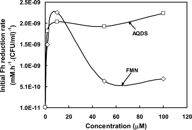
Initial Fh reduction rate by Paenibacillus sp. 300A in the presence of various FMN and AQDS concentrations.
Fh reduction kinetics in the presence of natural ETMs.
Although Paenibacillus sp. 300A showed no detectable reduction of Fh in the absence of ETMs, it was observed to reduce Fh at an initial reduction rate of ∼0.038 × 10⁻⁹ mM · h−1 · (CFU/ml)−1 in the presence of autoclaved 300A sediment slurry (Fig. 3). This Fh reduction was likely due to the ability of Paenibacillus sp. 300A to use ETMs present in 300A sediment slurry. It is hypothesized that in this process, Paenibacillus sp. 300A accepts electrons from the oxidation of lactate and transfers them to oxidized ETMs present in the 300A sediment slurry. The reduced ETMs abiotically transfer electrons to reduce Fh and regenerate oxidized ETMs. Control experiments did not show Fe(II) production, which indicates that the ETMs present in the 300A sediment slurry were not capable of reducing solid-phase Fe(III) oxide abiotically. A similar Fh reduction rate was observed (∼0.03 × 10⁻⁹ mM · h−1 · [CFU/ml]−1 versus no reduction in the absence of sediment extracts) when the experiment was carried out in the presence of 300A sediment extracts (see Fig. S3 in the supplemental material). Paenibacillus sp. 300A was able to use ETMs secreted by S. oneidensis MR-1 to reduce Fh (see Fig. S4). The Fh reduction rate (0.19 × 10⁻⁹ mM · h−1 · [CFU/ml]−1) in the presence of secreted ETMs was comparable to the solid-phase Fe(III) reduction rate of Shewanella species (Table 1). Several Shewanella strains were previously isolated from Hanford 300A (53, 69), which may secrete ETMs and could be used by Paenibacillus sp. 300A for the reduction of insoluble Fe(III) minerals.
Fig 3.

Fh reduction [shown as Fe(II) production] by Paenibacillus sp. 300A in the presence of 300A sediment slurry (slurry plus cells plus Fh). Slurry plus Fh, slurry plus cells, and slurry were used as controls. The error bars represent the standard deviations (n = 3).
U(VI) immobilization kinetics in the presence of bicarbonate buffer.
Paenibacillus sp. 300A was capable of immobilizing U(VI) under anaerobic conditions in bicarbonate-buffered non-growth medium, and the immobilization process was faster than those of other isolates that have been previously isolated from contaminated DOE sites (Fig. 4A; also see Table S1 in the supplemental material). There was relatively little (∼10%) U(VI) immobilization by heat-killed cells. The data show an O2-induced ∼40% remobilization of the anaerobically immobilized U(VI) in bicarbonate-buffered medium (see Table S2), which is consistent with ∼40% of the anaerobically immobilized U(VI) resulting from microbial reduction. The ratio of reduced to adsorbed U(VI) was ∼4:1. Cellulomonas sp. ES6, a strain isolated from Hanford 300A, was capable of immobilizing U(VI), but the immobilization process was relatively slow (see Table S1) even in bicarbonate-buffered medium with a high cell biomass concentration (62). Anaeromyxobacter dehalogenans, isolated from a DOE field research site near Oak Ridge, TN, utilized H2 but not acetate as an electron donor to immobilize U (74). Deinococcus radiodurans R1 is a radiation-resistant isolate studied for U(VI) immobilization, but Deinococcus radiodurans R1 was not able to immobilize U(VI) in the absence of an external ETM (22). Although we observed 51% immobilization of the initial U(VI) concentration (100 μM), this could be due to low initial biomass concentrations and non-growth conditions, since the U(VI) removal process is slow in a bicarbonate-buffered system (62). Our results suggest that Paenibacillus sp. 300A is capable of immobilizing U(VI) through microbial reduction.
Fig 4.

(A) U(VI) immobilization by Paenibacillus sp. 300A in bicarbonate-buffered non-growth medium. U(VI) immobilization lacking cells and immobilization with heat-killed cells were used as abiotic controls. The initial U(VI) concentration used in the immobilization study was 100 μM. The error bars represent the standard deviations (n = 3). (B and C) Transmission electron microscopy (TEM) photomicrographs of unstained thin sections. (D) High-resolution TEM micrograph of U precipitates. (E) Selected-area electron diffraction (SAED) (top) and an energy-dispersive X-ray spectrum of the U particles (bottom).
TEMs of the cells are shown in Fig. 4B to E. Figure 4B shows that uranium deposits were associated primarily with cells. Some uranium was deposited inside the cells (Fig. 4C and D), and, as determined by energy-dispersive X-ray spectroscopy (EDS), the deposited material appeared to be amorphous, nanoparticulate U (Fig. 4D). In addition, TEM images taken at the end of the U(VI) immobilization experiment (72 h) showed that cell membranes were intact and that the cells contained ribosomes, indicating that cells were not lysed at the time of chemical fixation (see Fig. S5A in the supplemental material). TEM images also showed some extracellularly deposited uranium (see Fig. S5B). Whole-cell TEM images were taken as a control to ensure that the cells remained intact while in the bicarbonate or PIPES buffer (see Fig. S5C). We found the uptake of U to be an interesting ability of Paenibacillus sp. 300A, because not many bacterial species are able to tolerate U inside their cells. A more interesting observation is that the cells we imaged look completely healthy, judging from their morphology, with well-pronounced ribosomes and intact membranes (see Fig. S5A). About 80% of the U present in the larger nodules (right underneath the membrane) are composed of amorphous uraninite (∼2.5 nm in diameter), very similar to that on the membranes (Fig. 4E, top).
U(VI) immobilization kinetics in the presence of PIPES buffer.
Paenibacillus sp. 300A completely immobilizes U(VI) under anaerobic conditions in PIPES-buffered non-growth medium (Fig. 5). The abiotic control did not show any immobilization, which confirms that the U(VI) removal was biotic. Heat-killed cells immobilized ∼69% of the initial U within 10 h and remained constant throughout the study period. The immediate U(VI) immobilization was the result of physical adsorption onto cell surfaces (62). We estimated that ∼31% of the U(VI) immobilization was through reduction. For this estimate, we assumed that heat-killed and live cells have the same U(VI) sorption capacity, which has been experimentally demonstrated (51). However, upon exposure to air, only ∼7% of the anaerobically immobilized U(VI) was remobilized (see Table S2 in the supplemental material). This could be due to the recalcitrance of the reduction product to oxidation by molecular oxygen (O2). The ratio of reduced to adsorbed U(VI) was ∼3:7. The observed immobilization capability was fast compared to those of isolates recently isolated from U(VI)-contaminated DOE sites and studied for U(VI) immobilization (see Table S1). Cellulomonas sp. ES6, isolated from Hanford 300A, was capable of immobilizing U(VI), but the immobilization process was slow [∼95% U(VI) immobilization in 100 h] compared to that of Paenibacillus sp. 300A in PIPES-buffered medium [∼95% U(VI) immobilization in 35 h] (see Table S1).
Fig 5.

U(VI) immobilization by Paenibacillus sp. 300A in PIPES-buffered non-growth medium. U(VI) immobilization without cells and immobilization with heat-killed cells were used as the abiotic controls. The initial U(VI) concentration used in the immobilization study was 100 μM. The error bars represent the standard deviations (n = 3).
U(VI) immobilization kinetics in the presence of 300A sediment slurry.
Paenibacillus sp. 300A was able to immobilize approximately 40% of the initial U(VI) in 100 h in the presence of 300A sediment slurry (Fig. 6). The addition of sediment slurry has been shown to enhance the reduction rate of U(VI), most likely because naturally occurring humic substances in the 300A sediment facilitate electron transfer between cells and U(VI) and prevent the formation of stable calcium-uranyl-carbonate complexes (24, 28). The bioreduction of U(VI) can also be inhibited by humic substances (10). The total U(VI) immobilization (40% at 100 h) was slower in the presence of sediment slurry than in bicarbonate-buffered non-growth medium (51% at 70 h). Although identifying the detailed mechanism was not the goal of this study, the decreased U(VI) immobilization could be due to (i) the redirection of electrons from U(VI) reduction pathways or interrupted electron transport to U(VI) because of the presence of Fe(III) in the slurry as a competitive electron acceptor, (ii) U(IV)-humic complex formation preventing the precipitation of reduced U(IV), (iii) preferential use of humic substances as an electron acceptor, or (iv) decreased bioavailability as a result of humic-U(VI) complex formation (10). U(VI) immobilization in soil or sediments is usually slow because of Fe(III) oxide content and the adsorption behavior of the solids (30). The reduction of Fe(III) (Fig. 3) in the presence of 300A sediment slurry shows the presence of metal-reducing activity; the slowed U(VI) immobilization may be due to the enzymatic reduction activity of Paenibacillus sp. 300A being reduced because of U(VI) sorbed into micropores in the sediment slurry being inaccessible (30). Furthermore, the continued U(VI) immobilization over time, compared to that of the control, reveals that Paenibacillus sp. 300A is capable of immobilizing U(VI) in the Hanford 300A subsurface sedimentary environment. Similar U(VI) immobilization experiments were carried out in the presence of 300A sediment slurry amended with Fh. U(VI) adsorbed to sediment slurry amended with Fh within 2 h (see Fig. S6 in the supplemental material), and because of this the U(VI) was less bioavailable and microorganisms were not able to further enhance immobilization through reduction (4, 30).
Fig 6.

U(VI) immobilization by Paenibacillus sp. 300A in the presence of 300A sediment slurry. U(VI) immobilization lacking cells was used as the abiotic control. The initial U(VI) concentration used in the immobilization study was 100 μM. The error bars represent the standard deviations (n = 3).
Conclusions.
This study demonstrates that Paenibacillus sp. 300A isolated from Hanford 300A subsurface sediment biofilms is capable of reducing Fe(III) and immobilizing U(VI) efficiently and considerably faster than recently studied indigenous bacteria isolated from uranium-contaminated sediments. Paenibacillus sp. 300A is capable of scavenging ETMs from the surrounding environment without the metabolic and energetic burden of producing and excreting them, and it is capable of using these ETMs to reduce solid-phase iron oxides. Paenibacillus sp. 300A is capable of immobilizing U(VI) through both adsorption and reduction.
Supplementary Material
ACKNOWLEDGMENTS
This research was supported by the U.S. DOE Office of Biological and Environmental Research under the Subsurface Biogeochemistry Research (SBR) Program (grant DE-FG92-08ER64560) and the DOE-BER SBR Program's Scientific Focus Area (SFA) at the Pacific Northwest National Laboratory (PNNL). We are also grateful to the Franceschi Microscopy and Imaging Center of Washington State University for the use of their facilities and for staff assistance.
A portion of the research was performed in the William R. Wiley Environmental Molecular Sciences Laboratory, a national scientific user facility sponsored by the DOE's Office of Biological and Environmental Research and located at PNNL. PNNL is operated by Battelle for the DOE under contract DE-AC05-76RL01830.
Footnotes
Published ahead of print 7 September 2012
Supplemental material for this article may be found at http://aem.asm.org/.
REFERENCES
- 1. Ahmed B, et al. 2012. Immobilization of U(VI) from oxic groundwater by Hanford 300 Area sediments and effects of Columbia River water. Water Res. 46: 3989– 3998 [DOI] [PubMed] [Google Scholar]
- 2. Anderson RT, et al. 2003. Stimulating the in situ activity of Geobacter species to remove uranium from the groundwater of a uranium-contaminated aquifer. Appl. Environ. Microbiol. 69: 5884– 5891 [DOI] [PMC free article] [PubMed] [Google Scholar]
- 3. Ash C, Priest FG, Collins MD. 1993. Molecular identification of rRNA group 3 bacilli (Ash, Farrow, Wallbanks and Collins) using a PCR probe test. Antonie Van Leeuwenhoek 64: 253– 260 [DOI] [PubMed] [Google Scholar]
- 4. Barnett MO, Jardine PM, Brooks SC. 2002. U(VI) adsorption to heterogeneous subsurface media: application of a surface complexation model. Environ. Sci. Technol. 36: 937– 942 [DOI] [PubMed] [Google Scholar]
- 5. Behrends T, Van Cappellen P. 2005. Competition between enzymatic and abiotic reduction of uranium(VI) under iron reducing conditions. Chem. Geol. 220: 315– 327 [Google Scholar]
- 6. Beyenal H, et al. 2004. Uranium immobilization by sulfate-reducing biofilms. Environ. Sci. Technol. 38: 2067– 2074 [DOI] [PubMed] [Google Scholar]
- 7. Boonchayaanant B, Nayak D, Du X, Criddle CS. 2009. Uranium reduction and resistance to reoxidation under iron-reducing and sulfate-reducing conditions. Water Res. 43: 4652– 4664 [DOI] [PubMed] [Google Scholar]
- 8. Brina R, Miller AG. 1992. Direct detection of trace levels of uranium by laser-induced kinetic phosphorimetry. Anal. Chem. 64: 1413– 1418 [Google Scholar]
- 9. Brown M, Balkwill D. 2009. Antibiotic resistance in bacteria isolated from the deep terrestrial subsurface. Microb. Ecol. 57: 484– 493 [DOI] [PubMed] [Google Scholar]
- 10. Burgos WD, et al. 2007. Soil humic acid decreases biological uranium(VI) reduction by Shewanella putrefaciens CN32. Environ. Eng. Sci. 24: 755– 761 [Google Scholar]
- 11. Cao B, Ahmed B, Beyenal H. 2010. Immobilization of uranium in groundwater using biofilms. In Shah V. (ed), Emerging environmental technologies, vol II Springer, Berlin, Germany [Google Scholar]
- 12. Cao B, et al. 2011. Contribution of extracellular polymeric substances from Shewanella sp. HRCR-1 biofilms to U(VI) immobilization. Environ. Sci. Technol. 45: 5483– 5490 [DOI] [PubMed] [Google Scholar]
- 13. Reference deleted.
- 14. Chien CC, Han CT. 2009. Tellurite resistance and reduction by a Paenibacillus sp. isolated from heavy metal-contaminated sediments. Environ. Toxicol. Chem. 28: 1627– 1632 [DOI] [PubMed] [Google Scholar]
- 15. Cologgi DL, Lampa-Pastirk S, Speers AM, Kelly SD, Reguera G. 2011. Extracellular reduction of uranium via Geobacter conductive pili as a protective cellular mechanism. Proc. Natl. Acad. Sci. U. S. A. 108: 15248– 15252 [DOI] [PMC free article] [PubMed] [Google Scholar]
- 16. Costerton JW, et al. 1987. Bacterial biofilms in nature and disease. Annu. Rev. Microbiol. 41: 435– 464 [DOI] [PubMed] [Google Scholar]
- 17. Costerton JW, Lewandowski Z, Caldwell DE, Korber DR, Lappinscott HM. 1995. Microbial biofilms. Annu. Rev. Microbiol. 49: 711– 745 [DOI] [PubMed] [Google Scholar]
- 18. Decho AW. 2000. Microbial biofilms in intertidal systems: an overview. Cont. Shelf Res. 20: 1257– 1273 [Google Scholar]
- 19. Decho AW. 2010. Overview of biopolymer-induced mineralization: what goes on in biofilms? Ecol. Eng. 36: 137– 144 [Google Scholar]
- 20. Felsenstein J. 1985. Confidence limits on phylogenies: an approach using the bootstrap. Evolution 39: 783– 791 [DOI] [PubMed] [Google Scholar]
- 21. Fredrickson JK. 2007. Hanford 300 Area subsurface as microbial habitat. PNNL-SA-54843 Pacific Northwest National Laboratory, Richland, WA [Google Scholar]
- 22. Fredrickson JK, Kostandarithes HM, Li SW, Plymale AE, Daly MJ. 2000. Reduction of Fe(III), Cr(VI), U(VI), and Tc(VII) by Deinococcus radiodurans R1. Appl. Environ. Microbiol. 66: 2006– 2011 [DOI] [PMC free article] [PubMed] [Google Scholar]
- 23. Fredrickson JK, Kota S, Kukkadapu RK, Liu C, Zachara JM. 2003. Influence of electron donor/acceptor concentrations on hydrous ferric oxide (HFO) bioreduction. Biodegradation 14: 91– 103 [DOI] [PubMed] [Google Scholar]
- 24. Fredrickson JK, et al. 2000. Reduction of U(VI) in goethite (alpha-FeOOH) suspensions by a dissimilatory metal-reducing bacterium. Geochim. Cosmochim. Acta 64: 3085– 3098 [Google Scholar]
- 25. Fredrickson JK, et al. 2002. Influence of Mn oxides on the reduction of uranium(VI) by the metal-reducing bacterium Shewanella putrefaciens. Geochim. Cosmochim. Acta 66: 3247– 3262 [Google Scholar]
- 26. Gerlach R, et al. 2011. Influence of carbon sources and electron shuttles on ferric iron reduction by Cellulomonas sp. strain ES6. Biodegradation 22: 983– 995 [DOI] [PubMed] [Google Scholar]
- 27. Gorby YA, Lovley DR. 1992. Enzymatic uranium precipitation. Environ. Sci. Technol. 26: 205– 207 [Google Scholar]
- 28. Gu BH, et al. 2005. Natural humics impact uranium bioreduction and oxidation. Environ. Sci. Technol. 39: 5268– 5275 [DOI] [PubMed] [Google Scholar]
- 29. Istok JD, et al. 2004. In situ bioreduction of technetium and uranium in a nitrate-contaminated aquifer. Environ. Sci. Technol. 38: 468– 475 [DOI] [PubMed] [Google Scholar]
- 30. Jeon BH, et al. 2004. Microbial reduction of U(VI) at the solid-water interface. Environ. Sci. Technol. 38: 5649– 5655 [DOI] [PubMed] [Google Scholar]
- 31. Jukes TH, Cantor CR. 1969. Evolution of protein molecules. Academic Press, New York, NY: [Google Scholar]
- 32. Kieft T, et al. 1999. Dissimilatory reduction of Fe(III) and other electron acceptors by a Thermus isolate. Appl. Environ. Microbiol. 65: 1214– 1221 [DOI] [PMC free article] [PubMed] [Google Scholar]
- 33. Kostka JE, Dalton DD, Skelton H, Dollhopf S, Stucki JW. 2002. Growth of iron(III)-reducing bacteria on clay minerals as the sole electron acceptor and comparison of growth yields on a variety of oxidized iron forms. Appl. Environ. Microbiol. 68: 6256– 6262 [DOI] [PMC free article] [PubMed] [Google Scholar]
- 34. Krawczyk-Barsch E, Grossmann K, Arnold T, Hofmann S, Wobus A. 2008. Influence of uranium (VI) on the metabolic activity of stable multispecies biofilms studied by oxygen microsensors and fluorescence microscopy. Geochim. Cosmochim. Acta 72: 5251– 5265 [Google Scholar]
- 35. Lewandowski Z, Beyenal H. 2007. Fundamentals of biofilm research. CRC Press, Taylor & Francis Group, Boca Raton, FL: [Google Scholar]
- 36. Liger E, Charlet L, Van Cappellen P. 1999. Surface catalysis of uranium(VI) reduction by iron(II). Geochim. Cosmochim. Acta 63: 2939– 2955 [Google Scholar]
- 37. Liu CX, Gorby YA, Zachara JM, Fredrickson JK, Brown CF. 2002. Reduction kinetics of Fe(III), Co(III), U(VI) Cr(VI) and Tc(VII) in cultures of dissimilatory metal-reducing bacteria. Biotechnol. Bioeng. 80: 637– 649 [DOI] [PubMed] [Google Scholar]
- 38. Lovley DR. 1993. Dissimilatory metal reduction. Annu. Rev. Microbiol. 47: 263– 290 [DOI] [PubMed] [Google Scholar]
- 39. Lovley DR. 1997. Microbial Fe(III) reduction in subsurface environments. FEMS Microbiol. Rev. 20: 305– 313 [Google Scholar]
- 40. Lovley DR, et al. 1998. Humic substances as a mediator for microbially catalyzed metal reduction. Acta Hydrochim. Hydrobiol. 26: 152– 157 [Google Scholar]
- 41. Lovley DR, Phillips EJP. 1992. Bioremediation of uranium contamination with enzymatic uranium reduction. Environ. Sci. Technol. 26: 2228– 2234 [Google Scholar]
- 42. Lovley DR, Phillips EJP. 1986. Organic-matter mineralization with reduction of ferric iron in sediments. Appl. Environ. Microbiol. 51: 683– 689 [DOI] [PMC free article] [PubMed] [Google Scholar]
- 43. Lovley DR, Phillips EJP. 1987. Rapid assay for microbially reducible ferric iron in aquatic sediments Appl. Environ. Microbiol. 53: 1536– 1540 [DOI] [PMC free article] [PubMed] [Google Scholar]
- 44. Lovley DR, Phillips EJP, Gorby YA, Landa ER. 1991. Microbial reduction of uranium. Nature 350: 413– 416 [Google Scholar]
- 45. Reference deleted.
- 46. Marsili E, et al. 2008. Shewanella secretes flavins that mediate extracellular electron transfer. Proc. Natl. Acad. Sci. U. S. A. 105: 3968– 3973 [DOI] [PMC free article] [PubMed] [Google Scholar]
- 47. Nevin KP, Lovley DR. 2000. Lack of production of electron-shuttling compounds or solubilization of Fe(III) during reduction of insoluble Fe(III) oxide by Geobacter metallireducens. Appl. Environ. Microbiol. 66: 2248– 2251 [DOI] [PMC free article] [PubMed] [Google Scholar]
- 48. N′Guessan AL, Vrionis HA, Resch CT, Long PE, Lovley DR. 2008. Sustained removal of uranium from contaminated groundwater following stimulation of dissimilatory metal reduction. Environ. Sci. Technol. 42: 2999– 3004 [DOI] [PubMed] [Google Scholar]
- 49. Nguyen HD, et al. 2012. Microscale geochemical gradients in Hanford 300 Area sediment biofilms and influence of uranium. Water Res. 46: 227– 234 [DOI] [PubMed] [Google Scholar]
- 50. Palmisano A, Hazen T. 2003. Bioremediation of metals and radionuclides: what it is and how it works, 2nd ed LBNL-42595 Lawrence Berkeley National Laboratory, Berkeley, CA: [Google Scholar]
- 51. Panak PJ, Knopp R, Booth CH, Nitsche H. 2002. Spectroscopic studies on the interaction of U(VI) with Bacillus sphaericus. Radiochim. Acta 90: 779– 783 [Google Scholar]
- 52. Petrie L, North NN, Dollhopf SL, Balkwill DL, Kostka JE. 2003. Enumeration and characterization of iron(III)-reducing microbial communities from acidic subsurface sediments contaminated with uranium(VI). Appl. Environ. Microbiol. 69: 7467– 7479 [DOI] [PMC free article] [PubMed] [Google Scholar]
- 53. Popa R, Mashall MJ, Nguyen H, Tebo BM, Brauer S. 2009. Limitations and benefits of ARISA intra-genomic diversity fingerprinting. J. Microbiol. Methods 78: 111– 118 [DOI] [PubMed] [Google Scholar]
- 54. Reardon CL, et al. 2010. Role of outer-membrane cytochromes MtrC and OmcA in the biomineralization of ferrihydrite by Shewanella oneidensis MR-1. Geobiology 8: 56– 68 [DOI] [PubMed] [Google Scholar]
- 55. Reitz T, Merroun ML, Selenska-Pobell S. 2008. Interactions of Paenibacillus sp. and Sulfolobus acidocaldarius strains with U(VI), p 703–710 In Merkel BJ, Hasche-Berger A. (ed), Uranium, mining and hydrogeology. Springer, Berlin, Germany [Google Scholar]
- 56. Renslow RS, et al. 2010. In situ effective diffusion coefficient profiles in live biofilms using pulsed-field gradient nuclear magnetic resonance. Biotechnol. Bioeng. 106: 928– 937 [DOI] [PMC free article] [PubMed] [Google Scholar]
- 57. Riley RG, Zachara JM, Wobber FJ. 1992. Chemical contaminants on DOE lands and selection of contaminant mixtures for subsurface science research DOE/ER-0547T. Pacific Northwest Laboratory, Richland, WA: [Google Scholar]
- 58. Roden EE, Zachara JM. 1996. Microbial reduction of crystalline iron(III) oxides: influence of oxide surface area and potential for cell growth. Environ. Sci. Technol. 30: 1618– 1628 [Google Scholar]
- 59. Saitou N, Nei M. 1987. The neighbor-joining method: a new method for reconstructing phylogenetic trees. Mol. Biol. Evol. 4: 406– 425 [DOI] [PubMed] [Google Scholar]
- 60. Sani RK, Peyton BM, Smith WA, Apel WA, Petersen JN. 2002. Dissimilatory reduction of Cr(VI), Fe(III), and U(VI) by Cellulomonas isolates. Appl. Microbiol. Biotechnol. 60: 192– 199 [DOI] [PubMed] [Google Scholar]
- 61. Sirota-Madi A, et al. 2010. Genome sequence of the pattern forming Paenibacillus vortex bacterium reveals potential for thriving in complex environments. BMC Genomics 11: 710. [DOI] [PMC free article] [PubMed] [Google Scholar]
- 62. Sivaswamy V, et al. 2011. Multiple mechanisms of uranium immobilization by Cellulomonas sp. strain ES6. Biotechnol. Bioeng. 108:264– 276 [DOI] [PubMed] [Google Scholar]
- 63. Smith WA, Apel WA, Petersen JN, Peyton BM. 2002. Effect of carbon and energy source on bacterial chromate reduction. Bioremediat. J. 6: 205– 215 [Google Scholar]
- 64. Tamura K, et al. 2011. MEGA5: molecular evolutionary genetics analysis using maximum likelihood, evolutionary distance, and maximum parsimony methods. Mol. Biol. Evol. 28: 2731– 2739 [DOI] [PMC free article] [PubMed] [Google Scholar]
- 65. Tebo BM, Obraztsova AY. 1998. Sulfate-reducing bacterium grows with Cr(VI), U(VI), Mn(IV), and Fe(III) as electron acceptors. FEMS Microbiol. Lett. 162: 193– 198 [Google Scholar]
- 66. Tratnyek PG, Macalady DL. 1989. Abiotic reduction of nitro aromatic pesticides in anaerobic laboratory systems. J. Agric. Food. Chem. 37: 248– 254 [Google Scholar]
- 67. Truex MJ, Peyton BM, Valentine NB, Gorby YA. 1997. Kinetics of U(VI) reduction by a dissimilatory Fe(III)-reducing bacterium under non-growth conditions. Biotechnol. Bioeng. 55: 490– 496 [DOI] [PubMed] [Google Scholar]
- 68. Turick CE, Tisa LS, Caccavo F. 2002. Melanin production and use as a soluble electron shuttle for Fe(III) oxide reduction and as a terminal electron acceptor by Shewanella algae BrY. Appl. Environ. Microbiol. 68: 2436– 2444 [DOI] [PMC free article] [PubMed] [Google Scholar]
- 69. Turse JE, Marshall MJ, Fredrickson JK, Lipton MS, Callister SJ. 2010. An empirical strategy for characterizing bacterial proteomes across species in the absence of genomic sequences. PLoS One 5: e13968 doi:10.1371/journal.pone.0013968 [DOI] [PMC free article] [PubMed] [Google Scholar]
- 70. Veeramani H, et al. 2011. Products of abiotic U(VI) reduction by biogenic magnetite and vivianite. Geochim. Cosmochim. Acta 75:2512– 2528 [Google Scholar]
- 71. von Canstein H, Ogawa J, Shimizu S, Lloyd JR. 2008. Secretion of flavins by Shewanella species and their role in extracellular electron transfer. Appl. Environ. Microbiol. 74: 615– 623 [DOI] [PMC free article] [PubMed] [Google Scholar]
- 72. Wen Y, et al. 2011. Identification and analysis of the gene cluster involved in biosynthesis of paenibactin, a catecholate siderophore produced by Paenibacillus elgii B69. Environ. Microbiol. 13: 2726– 2737 [DOI] [PubMed] [Google Scholar]
- 73. Wolf M, Kappler A, Jiang J, Meckenstock RU. 2009. Effects of humic substances and quinones at low concentrations on ferrihydrite reduction by Geobacter metallireducens. Environ. Sci. Technol. 43: 5679– 5685 [DOI] [PubMed] [Google Scholar]
- 74. Wu Q, Sanford RA, Loffler FE. 2006. Uranium(VI) reduction by Anaeromyxobacter dehalogenans strain 2CP-C. Appl. Environ. Microbiol. 72: 3608– 3614 [DOI] [PMC free article] [PubMed] [Google Scholar]
- 75. Zachara JM, et al. 1998. Bacterial reduction of crystalline Fe3+ oxides in single phase suspensions and subsurface materials. Am. Miner. 83: 1426– 1443 [Google Scholar]
- 76. Zachara JM, Kukkadapu RK, Fredrickson JK, Gorby YA, Smith SC. 2002. Biomineralization of poorly crystalline Fe(III) oxides by dissimilatory metal reducing bacteria (DMRB). Geomicrobiol. J. 19: 179– 207 [Google Scholar]
Associated Data
This section collects any data citations, data availability statements, or supplementary materials included in this article.