SUMMARY
In IV Class of Misch classification is usually not possible insert fixtures at the same time of the sinus lift procedure. To solve all these situation the use of several appliances have been proposed. Among those the S.I.S. titan plane is the easiest to use. The S.I.S. plane allows to stabilize implants without primary stability and to fix them to osseous ridge. In IV Class of Misch it brings to a real treatment shortening. After six and twelve months RX assessments show bone levels comparables to literature implant success criteria. This technique seems to reach success in Misch IV Class with simultaneous insertion of dental implants, with a simple procedure and without further traumaticity. Literature on S.I.S. plane is lacking and mostly based on case reports. A further investigation based on a larger number of patients is needed.
Keywords: S.I.S., primary stability, sinus lift, implant stabilization
RIASSUNTO
Nelle IV classi di Misch non è di norma possibile inserire impianti dentali contestualmente alla procedura di rialzo di seno. Per queste situazioni è stato proposto l’uso di diversi presidi per stabilizzare immediatamente impianti provi di stabilità primaria. Tra questi risulta di uso più semplice la placca in titanio S.I.S. La placca S.I.S. consente di stabilizzare tra loro ed alla cresta residua gli impianti dentali inseriti in assenza di stabilità primaria. Nelle classi IV di Misch ciò comporta un notevole accorciamento dell’iter terapeutico. Controlli radiografici a 6 mesi ed a 12 mesi dal carico testimoniano stabilità dei livelli ossei in linea con i criteri di successo della letteratura. La tecnica descritta sembra poter assicurare il successo nei casi di IV Classe di Misch con inserimento contestuale di impianti osseo integrati. La tecnica appare semplice e riproducibile senza essere causa di ulteriore traumatismo. La letteratura ad oggi disponibile su questo tipo di tecniche è numericamente limitata e costituita per la maggior parte da case report. È quindi auspicabile uno studio più approfondito su numeri di maggiore rilevanza.
Introduction
In upper-lateral areas rehabilitations an osseous ridge insufficient for inserting dental implants can be often found out. Such a deficit can be attributable to combined effect of post-extraction bone resorption and of maxillary sinus expanding. Examining different clinical situations that can be found out Misch (1) has identified four classes:
CLASS I
More than 12 mm of bone availability. In these cases is possible to introduce fixture of right dimensions according to the usual techniques.
CLASS II
Bone availability included between 12 and 8 mm. In these cases short fixtures could be introduced but is advisable to start begin sinus lifting using not invasive techniques in order to introduce longer fixtures.
CLASS III
Bone Availability included between 8 and 5 mm. In these cases is necessary to obtain a greater lift of the floor sinus. The more used technique is the one with lateral approach described by Misch and Tatum (2, 3) followed by insertion of fixtures at the same time.
CLASS IV
Less than 5 mm of bone availability. In these cases is expected equally a sinus floor lift but with fixture insertion postponed after graft integration.
In that case the impossibility of obtaining a sufficient primary stability to gain osseointegration requires to postpone fixtures insertion. Primary stability and healing in absence of micromovements are strict conditions for obtaining the osseointegration (4–6). Without these conditions a fibrous connection can occur instead of osseointegration (7–9). In order to reduce treatment lenght and number of surgical stages some authors have proposed to solidarize the fixtures between them to eliminate their mobility. Cranin and Russel in 1993 (10) successfully used a Leibinger splint for stabilizing stability-free fixtures introduced after sinus lifting, obtaining a clinical success. Sontheimer in 2000 (11) and Vollmer in 2002 (12) proposed, to the same aim, the use of mini-plates for osteosynthesis. Patyk et to the in 2000 (13) developed an extrasinusal stabilizer plate made of a resorbable material (polilactate). Engelke in 2002 (14) stabilized single fixtures lacking primary stability with osteosynthesis plates welded to a screwed abutment obtaining osseous regeneration and osseointegration in domestic pigs. Lindorf and Müller-Herzog proposed in 2004 (15) the ASIS technique consisiting in using a block of cortical bone coming from the mandibular ramus like an extrasinusal fixture stabilizer in association with a sinus lift. With the same purpose Lang in 1999 (16) realized a titanium plate, later described, specifically destined to stabilize dental implants. Positive results with the same device thought out from Lang were obtained by Kaps in 1999 (17) and, in Italy, by Morlino in 2000 (18).
Case report
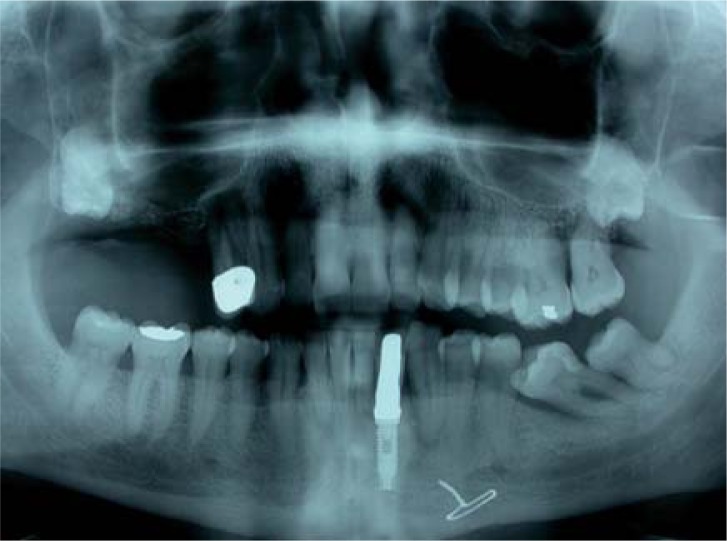
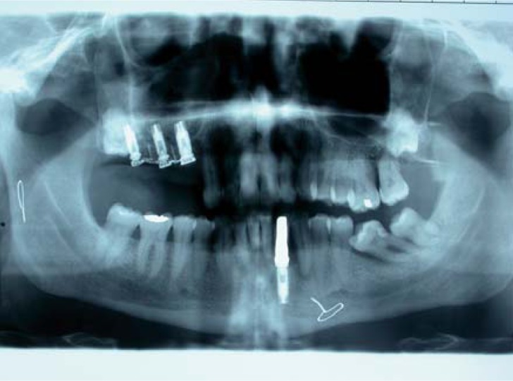
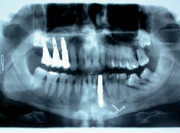
In The January 2006 patient M.U. was examined, masculine sex, years 66, nonsmoker. The medical case history revealed absence of active pathologies. During the examination it was found out the loss of 17, 16, 15 e 36 with atrophy of the edentulous ridge Class IV of Misch, tooth 14 with grade 3 mobility, teeth 37 and 38 mesially tilted with periodontal compromission, teeth 18 and 28 retained, previous mandibular fracture with loss of 31. The patient refuses extractions of 18, 37, 38. Etiological therapy was chosen followed by extraction of 14 and implant supported rehabilitation in zone 16, 15, 14, without extraction of 18, with contemporary maxillary sinus lift DX (Fig. 1).
Figure 1.
Pre-surgical Rx.
Methods
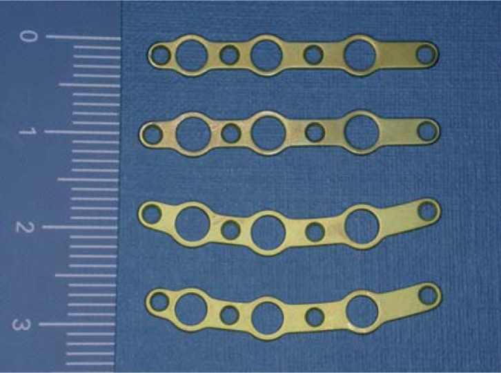
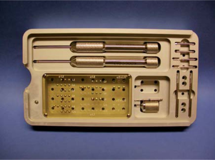
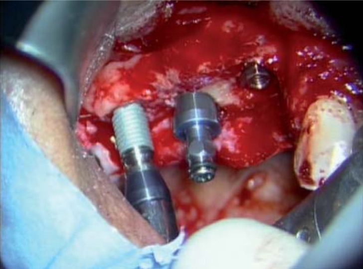
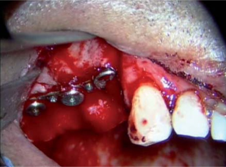
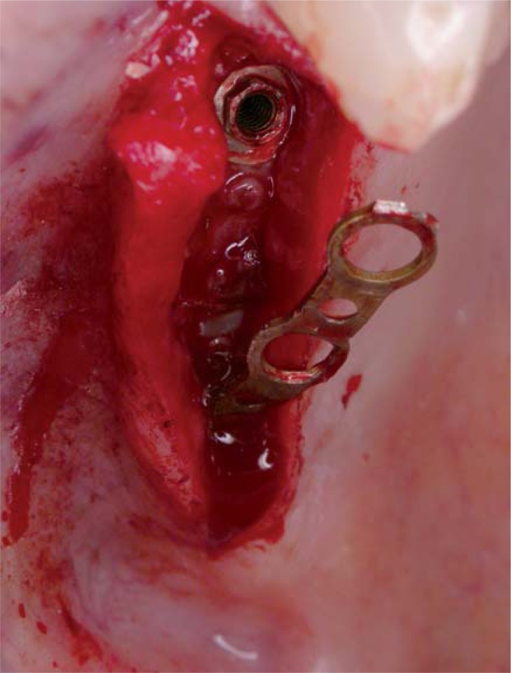
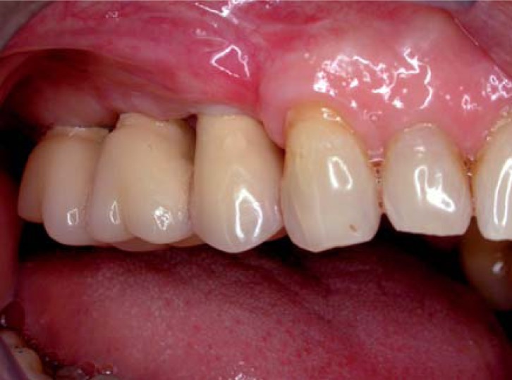
In this IV Class of Misch case, in order to permit fixtures insertion and sinus lift at the same time, it is decided to solidarize the fixtures, lacking primary stability, by means of S.I.S. plate (Sinus Implantat Stabilisator, Mondeal Medical Systems GmbH, Germany - Tuttlingen, distributed in Italy from Geass srl, Italy - Pozzuolo del Friuli) thought out from Lang (16). This device is made of a titanium plates of 0,6 mm thickness (Fig. 2) bringing greater holes, destined to screw the cover screws into the fixture, alternated at smaller holes destined to fix it to the osseous ridge by means of osteosynthesis screws and dedicated tools (Fig. 3) (Syntesis, Geass srl, Italy - Pozzuolo del Friuli). That plate has three presetted holes for the first premolar, the second premolar and the first molar. It is produced in two dimensions, to fit different types of fixture with different types of connections, and in two shapes, straight and curve, to fit different shapes of edentulous ridge. The procedure is carried out 40 day after tooth 14 extraction. The pharmacological therapy is 2 gr of Amoxicilline and Clavulanic Acid. Anesthesia is obtained by blocking the greater palatine, the inferior-orbital nerve and infiltrating the tuberosity region. A crestal incision is made in association with two mesial and distal releasing incisions. After that a buccal window is made with multiblade burs at first than with diamond burs in the final phase. Than the buccal window and the sinus membrane are raised with the usual manual tools, alternating the surgical movements with pauses in order to of to verify the synchronous membrane motion the with the respiratory actions. After obtaining the needed volume and verifying membrane integrity, an initial filling up is managed with heterologous bone (Bio-gen, Bioteck, Arcugnano - Italy) and clot of the patient previously drawn. A 5 mm diameter 13 mm length fixture (Hexa, Geass srl, Italy – Well of the Friuli) is firstly introduced in 14 post extraction socket. After fixing and moulding the S.I.S. plate onto the 14 fixture, two guiding holes are made fitting the plate holes in 15 and 16 position. Than the plate is removed, drilling 15 and 16 holes finished and two 3,75×13 mm fixtures (Hexa, Geass srl, Italy - Pozzuolo del Friuli) are installed. After their insertion the position 15 and 16 fixtures are lacking primary stability (Fig. 4). The three fixtures are now tightly solidarized between them and to the S.I.S. plate by means of the cover screws and to the osseous ridge by osteosynthesis screws (Figs. 5, 6). At the end the fixture-plate complex is totally still and rigidly joined with the ridge. Finally filling is completed up with the same biomaterial and covered with a collagen membrane (Biocollagen, Bioteck, Arcugnano - Italy) and a sutures is performed made of single and mattress stitches (supramyd 4-0, Butterfly Italy S.r.l. - Cavenago). The second procedure carried eight months after the first surgical time out. By means of slightly palatal incision a split-thickness flap is raised and a large band of keratinized gum is moved vestibular. Cover screws are removed as well as osteosynthesis screws and the S.I.S. plate (Fig. 7). After screwing of healing abutments a single stitches suture is performed (supramyd 4-0, Butterfly Italy S.r.l. - Cavenago).When healing is completed a fixed prosthesis is set (Fig. 8).
Figure 2.
Available shapes of S.I.S. plate.
Figure 3.
Tool kit for installing S.I.S. plate.
Figure 4.
Fixtures in 14, 15 and 16 positions.
Figure 5.
In situ S.I.S. plate.
Figure 6.
Post-surgical RX.
Figure 7.
S.I.S. plate removal.
Figure 8.
Final prosthesis setting.
Discussion
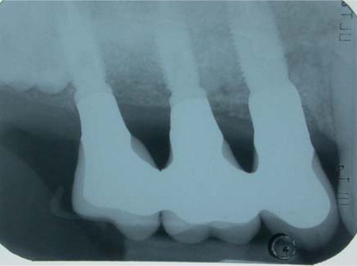
6 (Fig. 9) and 12 (Fig. 10) months after loading radiographic assessments testify stability of the bone levels in line with the literature success criteria. The technology described seems to be able to ensure the success in IV Class of Misch with dental implants insertion at the same time. In comparison with similar techniques it seems to be simpler and more reproducible, less operator related (10–14) and less harmful (15). A potential limit is the S.I.S. plates conformation, that does not permit insertion of the second molar when primary stability is lacking, and does no permit correction of horizontal defects and coronal augmentations. It would be useful to change the shape of the plates in order to permit second molar insertion and insertion of osteosynthesis screws not only along the plate central axis but also in palatal and buccal positions.
Figure 9.
6 months Rx assessment.
Figure 10.
12 months Rx assessment.
Conclusion
The described technique permits obtaining an immediate dental implants stabilization in IV Classes of Misch. This result allows to reduce considerably treatment length. Positive results 12 months after loading, are in line with the literature success criteria and with the few specific literature. This technique is simple and reproducible avoiding further traumas. The literature up till now available about this technique is limited and mostly based on case reports. A further investigation on larger number is needed.
References
- 1.Misch CE. Los Angeles Calif. Apr 19–21, 1990. Subantral augmentation (abstract 16). Ucla Symposium on Implants and the partially edentulous patient. [Google Scholar]
- 2.Misch CE. Maxillary sinus augmentation for endosteal implants: organized alternative treatment plans. Int J Oral Implantol. 1987;4(2):49–58. [PubMed] [Google Scholar]
- 3.Tatum H. Maxillary and sinus implants recontruction. Dent Clin North Am. 1986;30:107–19. [PubMed] [Google Scholar]
- 4.Brånemark PI, Hansson BO, Adell R, Breine U, Lindström J, Hallen O, Öhman A. Osseointegrated implants in the treatment of the edentulous jaw. Experience from a 10-year period. Scandinavian Journal of Plastic Reconstructive Surgery. 1977;16:1–132. [PubMed] [Google Scholar]
- 5.Adell R, Lekholm U, Rockler B, Brånemark PI. A 15-year study of osseointegrated implants in the treatment of the edentulous jaw. International Journal of Oral Surgery. 1981;10:387–416. doi: 10.1016/s0300-9785(81)80077-4. [DOI] [PubMed] [Google Scholar]
- 6.Lioubavina-Hack N, Lang NP, Karring T. Significance of primary stability for osseointegration of dental implants. Clin Oral Impl Res. 2006;17:244–250. doi: 10.1111/j.1600-0501.2005.01201.x. [DOI] [PubMed] [Google Scholar]
- 7.Isidor F. Loss of osseointegration caused by occlusal load of oral implants. A clinical and radiographic study in monkeys. Clinical Oral Implants Research. 1996;7:143–152. doi: 10.1034/j.1600-0501.1996.070208.x. [DOI] [PubMed] [Google Scholar]
- 8.Isidor F. Histological evaluation of peri-implant bone at implants subjected to occlusal overload or plaque accumulation. Clinical Oral Implants Research. 1997;8:1–9. doi: 10.1111/j.1600-0501.1997.tb00001.x. [DOI] [PubMed] [Google Scholar]
- 9.Brunski JB. The influence of force, motion, and related quantities on the response of bone to implants. In: Fitzgerald R Jr, editor. Non Cemented Total Hip Arthroplasty. New York: Raven Press; 1988. p. 43. [Google Scholar]
- 10.Cranin AN, Russell D, et al. Immediate implantation into the posterior maxilla after antroplasty: the Cranin-Russell Operation. J Oral Implantol. 1993;19(2):143–50. [PubMed] [Google Scholar]
- 11.Sontheimer M. One-stage sinus lifting using implant stabilizators. A report on everyday practice. Dent Implantol. 2000;4:2oai-94–303. [Google Scholar]
- 12.Vollmer R, Vollmer M, et al. Sinus elevation and single-stage surgical implant placement with a titanium osteosynthesis bar. Pract Proced Aesthet Dent. 2002;14(4):307–11. [PubMed] [Google Scholar]
- 13.Patyk AJ, Nadalini A, Duesmann O, Merten HA. Development and properties of resorbable implant stabilizer concerning sinus lift operation. Int Poster J Dent Oral Med. 2000;2(2) Poster Abstracts. [Google Scholar]
- 14.Engelke W, Stahr S, et al. Enhancement of primary stability of dental implants using cortical satellite implants. Implant Dent. 2002;11(1):52–7. doi: 10.1097/00008505-200201000-00014. [DOI] [PubMed] [Google Scholar]
- 15.Lindorf HH, Müller-Herzog R. Der autologe Sinus-Implantat-Stabilisator (ASIS) ZMK - Zeitschrift für Zahnheilkunde, Management und Kultur. 4/2004.
- 16.Lang M. Einphasiger sinuslift auch bei extrem geringer knochenhöhe möglich. DZW Spezial. 1999;2:26–27. [Google Scholar]
- 17.Kaps M. Mit einer schiene werder neueWege für der sinuslift möglich. DZW Spezial. 1999;5:24. [Google Scholar]
- 18.Morlino M, Tia M, Pescarandolo S. Tecnica di stabilizzazione impiantare extrasinusale nel rialzo bilaterale di seno mascellare. Quint Int Ita. 2000:5/6. [Google Scholar]


