Abstract
Background
The interaction of adenosine diphosphate with its P2Y1 and P2Y12 receptors on platelets is important for platelet function. However, nothing is known about adenosine diphosphate and its function in human megakaryocytes.
Design and Methods
We studied the role of adenosine diphosphate and P2Y receptors on proplatelet formation by human megakaryocytes in culture.
Results
Megakaryocytes expressed all the known eight subtypes of P2Y receptors, and constitutively released adenosine diphosphate. Proplatelet formation was inhibited by the adenosine diphosphate scavengers apyrase and CP/CPK by 60-70% and by the P2Y12 inhibitors cangrelor and 2-MeSAMP by 50-60%, but was not inhibited by the P2Y1 inhibitor MRS 2179. However, the active metabolites of the anti-P2Y12 drugs, clopidogrel and prasugrel, did not inhibit proplatelet formation. Since cangrelor and 2-MeSAMP also interact with P2Y13, we hypothesized that P2Y13, rather than P2Y12 is involved in adenosine diphosphate-regulated proplatelet formation. The specific P2Y13 inhibitor MRS 2211 inhibited proplatelet formation in a concentration-dependent manner. Megakaryocytes from a patient with severe congenital P2Y12 deficiency showed normal proplatelet formation, which was inhibited by apyrase, cangrelor or MRS 2211 by 50-60%. The platelet count of patients with congenital delta-storage pool deficiency, who lack secretable adenosine diphosphate, was significantly lower than that of patients with other platelet function disorders, confirming the important role of secretable adenosine diphosphate in platelet formation.
Conclusions
This is the first demonstration that adenosine diphosphate released by megakaryocytes regulates their function by interacting with P2Y13. The clinical relevance of this not previously described physiological role of adenosine diphosphate and P2Y13 requires further exploration.
Key words: ADP, proplatelet formation, P2Y13 receptor
Introduction
Megakaryocytes, after their migration from the osteoblastic niche to the vascular niche of the bone marrow, mature and generate platelets by extending long filaments, called proplatelets. Proplatelets protrude through the vascular endothelium into the sinusoid lumen, where platelets, stemming from the proplatelet tips, are released into the bloodstream.1,2 Early during their development, megakaryocytes develop cytoplasmic granules,3 including α-granules, which contain proteins that are essential for hemostasis and tissue repair, and δ-granules, which contain hemostatically active substances, such as the adenine nucleotides adenosine diphosphate (ADP) and adenosine triphosphate (ATP). An essential part of the process of platelet formation is the distribution of organelles and platelet-specific granules into the proplatelet tips and nascent platelets.4 Evidence of proplatelet formation was provided by electron microscopy analysis5 and, more recently, by multiphoton intravital microscopy of intact bone marrow from mice, which also documented the release of platelets into the bloodstream.6 However, many aspects of the mechanisms underlying and controlling proplatelet extension and platelet release have not been delineated, particularly in humans.7 It has been reported that hematopoietic precursors secrete several molecules that regulate the various stages of normal human hematopoiesis in an autocrine and/or paracrine manner.8-10 We recently demonstrated that membrane-bound von Willebrand factor can be detected exclusively in proplatelet-forming megakaryocytes, suggesting that autocrine release of α-granules is associated with proplatelet formation.8 More recently, it has been demonstrated that megakaryocytes express several proteins that are required for vesicular release, such as functional SNARE and related proteins.11 These data further suggest that autocrine-paracrine loops regulate megakaryocyte development and platelet formation.
In the present study, we investigated whether or not ADP and ATP are constitutively released by maturing megakaryocytes and play any role in proplatelet formation. In addition, because purine and pyrimidine nucleotides interact with P2 purinergic receptors on the cell plasma membranes, we aimed at identifying the P2 receptor(s) on megakaryocytes which mediate the effects of ADP and/or ATP. According to their molecular structure, P2 receptors are divided into two subfamilies: G protein-linked or “metabotropic”, termed P2Y, of which eight subtypes have been identified and cloned so far, and ligand-gated ion channels or “ionotropic”, termed P2X, of which seven subtypes have been identified and cloned.12,13 Platelets contain three P2 receptors: P2Y1 and P2Y12, which mediate ADP-induced platelet activation and aggregation,14 and P2X1, which mediates the amplification of platelet aggregation by ATP at high shear.15 We studied the expression of these receptors on megakaryocytes and their involvement in proplatelet formation.
Design and Methods
Materials
Apyrase from potato (grade VII), phosphocreatine (CP), creatine phosphokinase (CPK), 2-methylthioadenosine 5′-monophosphate triethylammonium salt (2-MeSAMP), adenosine 5′-diphosphate (ADP), prostaglandin E1 (PGE1), trypan blue solution (0.4%), propidium iodide and RNAse were from Sigma-Aldrich (Milan, Italy). MRS 2179 and MRS 2211 were from Tocris Bioscience (Missouri, USA). Cangrelor was a kind gift from The Medicines Company (Parsippany, NJ, USA). The active metabolites of clopidogrel (Clopidogrel-AM) and prasugrel (Prasugrel-AM) were kindly provided by Daiichi Sankyo Co., Ltd. (Tokyo, Japan). The following antibodies were used: mouse monoclonal anti–α-tubulin (clone DM1A) and mouse anti–β-actin (clone AC-15) (Sigma-Aldrich, Milan, Italy); anti–human CD41 (FITC) (clone HIP8) (eBioscience, Milan, Italy); mouse monoclonal anti–CD42b (PE) (clone AK2) (Abcam, Cambridge, UK); goat polyclonal anti-CD61 (clone C-20) and goat polyclonal anti–P2Y13 (clone C-18) (Santa Cruz Biotechnology, California, USA); rabbit monoclonal anti–phospho-ERK1/2 (Thr185/Tyr187) (clone AW39) (Millipore, Milan, Italy); mouse monoclonal anti-ERK1/2 (clone 3A7); rabbit monoclonal anti–phospho-Akt (Ser473) and rabbit polyclonal anti–Akt (Cell Signaling Technology, Massachusetts, USA); Alexa Fluor–conjugated antibodies (Invitrogen, Milan, Italy); and horseradish peroxidase-conjugated antibodies (BioRad, Milan, Italy).
Clinical and laboratory assessments
Human cord blood was collected following normal pregnancies and deliveries with informed consent of the parents, in accordance with the ethical committee of the IRCCS Policlinico San Matteo Foundation and the principles of the Declaration of Helsinki. For peripheral blood studies, blood samples were obtained from healthy subjects and a patient with congenital, severe P2Y12 deficiency, 16 who gave their informed consent. The study was approved by the IBS of IRCCS San Matteo Foundation, Pavia, Italy.
Differentiation of megakaryocytes and morphological analysis
CD34+ cells from cord blood and CD45+ cells from peripheral blood samples were separated by immunomagnetic bead selection (Miltenyi Biotec, Bologna, Italy) and cultured, as previously described,8,17 in Stem Span medium supplemented with 10 ng/mL thrombopoietin (TPO), interleukin (IL)-6 and IL-11 at 37°C in a 5% CO2 fully humidified atmosphere. At the end of the culture (13 days for CD34+ cells and 14 days for CD45+ cells), 150x103 cells were collected, cytospun on glass coverslips and stained with a primary antibody against CD61 (1:100) to evaluate megakaryocyte output and maturation.18 Images were acquired using a Olympus BX51 (Olympus, Deutschland GmbH, Hamburg, Germany) through a 60X/1.25 UPlanF1 oil-immersion objective. At least 100 megakaryocytes were evaluated for each specimen.
Proplatelet formation
Megakaryocyte and proplatelet yields were evaluated at the end of the cell culture as previously described.8,17 Briefly, cells were seeded in a 24-well plate and incubated at 37°C in a 5% CO2 fully humidified atmosphere for 16 h. For immunofluorescence evaluation of proplatelet formation, the entire well culture was harvested and then seeded onto glass coverslips, previously coated with poly-L-lysine, and allowed to adhere by centrifugation at 240xg. Cells were then fixed in 4% paraformaldehyde, permeabilized with 0.1% Triton X-100, and stained with anti–α-tubulin antibody (1:700). The coverslips were mounted onto glass slides with ProLong Gold antifade reagent (Invitrogen, Milan, Italy) and images acquired by an Olympus BX51 microscope (Olympus) using a 20X/0.5 UPlanF1 objective. Proplatelets were identified as cells displaying long filamentous structures, ending with plateletsized tips. The percentage of megakaryocytes bearing proplatelets was determined by analyzing the entire area of the coverslips. In some experiments, before being seeded, cells were harvested and pre-incubated in Stem Span medium with the following substances, at the indicated final concentrations, at 37°C for 15 min: apyrase 1 U/mL, phosphocreatine 5 mM and creatine phosphokinase 40 U/mL (CP/CPK), cangrelor 10 μM, 2-MeSAMP 10 μM, MRS 2179 10 μM, MRS 2211 0.1-10 μM, clopidogrel-AM 10 μM, and prasugrel-AM 10 μM. After pre-incubation, cells were seeded in 24-well plates, without being washed, and left for 16 h before proplatelet formation was analyzed as described above.
Assessment of cell viability
A trypan blue exclusion assay was employed to determine the number of viable cells in cultures. Briefly, cells at different days of culture in the presence or absence of apyrase, CP/CPK, cangrelor, 2-MeSAMP or MRS 2211, were centrifuged and suspended in phosphate-buffered saline (PBS). For evaluation of cell viability, 100 μL of cell suspension were mixed with 100 μL trypan blue and visualized by phase-contrast microscopy (Nikon TMS-F, Tokyo, Japan) using a 20X objective. Viable cells (unstained) and dead cells (blue-stained) were counted and the values expressed as percentages of total cell count. Lactate dehydrogenase, which is a marker of cell lysis, was measured enzymatically in cell culture medium at different days of culture, using the lactate dehydrogenase assay and the ARCHITECT c16000 analyzer (all from Abbott Laboratories, Abbott Park, IL, USA), following standard criteria.19
Retrotranscription and quantitative real-time polymerase chain reaction of P2Y receptors
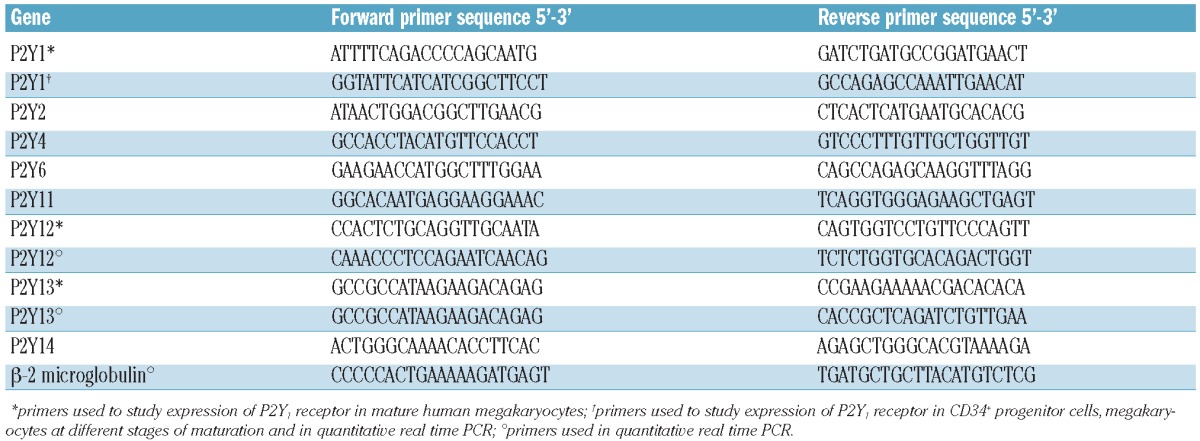
CD34+ cells from cord blood or cord blood derived-CD61+ megakaryocytes at different stages of maturation were separated using immunomagnetic beads (Miltenyi Biotec, Bologna, Italy) and total cellular RNA was extracted as previously described.20 Reverse transcription was performed in a final volume of 20 μL reaction mixture in the presence of: 1 μg RNA, 1x polymerase chain reaction (PCR) buffer, 5 mM MgCl2, 4 mM of each dNTP, 0.625 μM oligo d(T)16, 1.875 μM Random Hexamers, 20 U RNase inhibitor, and 50 U MuLV reverse transcriptase (all from Applera, Monza, Italy). The conditions for the reverse transcription were as follows: 25°C for 10 min, 42°C for 45 min, 99°C for 5 min. The reverse transcribed samples were diluted up to 50 μL with ddH2O. The primers used are listed in Table 1. The amplification reaction was performed in 25 μL using an Applied Biosystems GeneAmp 9700 thermocycler; 20 μL PCR products were electrophoresed in 1% agarose gel or 20% polyacrylamide gel stained with ethidium bromide. For quantitative real-time PCR, reverse transcribed samples were diluted up to 50 μL with ddH2O and 5 μL of the resulting cDNA were amplified in duplicate in a 20 μL reaction mixture with 200 nM of each specific primer and MESA GREEN qPCR MasterMix Plus for SYBR assay no ROX (Eurogentec, Milan, Italy) at 1x final concentration. The amplification reaction was performed in a Rotorgene 6000 (Corbett Life Science, Sydney, Australia) as follows: 95°C for 5 min, followed by 35 cycles at 95°C for 10 s, 60°C for 15 s, 72°C for 20 s. The Rotorgene 6000 Series Software 1.7 was used for the comparative concentration analysis. β-2 microglobulin gene expression was used for the normalization of the samples.
Table 1.
Primers used for RT-PCR in this study.
Preparation of platelet lysates from washed human platelets for western immunoblotting analysis
Peripheral blood from healthy subjects was collected in citric acid/citrate/dextrose (ACD) as anticoagulant (blood:ACD, 6:1, v/v) in the presence of apyrase 0.2 U/mL and PGE1 1 μM. Platelet-rich plasma was prepared by centrifuging blood samples at 200xg for 10 min. The platelet-rich plasma was then aspirated, centrifuged at 1200xg for 10 min and washed with PIPES buffer (20mM PIPES and 136 mM NaCl, pH 6.5). Washed platelets were then lysed in sodium dodecyl sulphate (SDS) buffer (Tris 0.5M, pH 6.8, 2% SDS, 10% glycerol), in the presence of a 2% protease inhibitor cocktail (Sigma), on ice for 30 min. Lysates were clarified by centrifugation at 15700xg at 4°C for 15 min.
Western immunoblotting
Cellular expression of P2Y13
In order to evaluate the cellular expression of P2Y13 protein, megakaryocytes at days 7, 10 and 13 of culture were lysed with Hepes-glycerol lysis buffer (Hepes 50 mM, NaCl 150 mM, 10% glycerol, 1% Triton X-100, MgCl2 1.5 mM, EGTA 1 mM, NaF 10 mM, PMSF 1 mM, Na3VO4 1 mM, 1 μg/mL leupeptin, 1 μg/mL aprotinin). Lysis was performed on ice for 30 min and the lysates clarified by centrifugation at 15700xg at 4°C for 15 min. Protein concentration was measured by the bicinchoninic acid assay (Pierce, Milan, Italy). Samples containing equal amounts of proteins were subjected to SDS-polyacrylamide gel electrophoresis (SDS-PAGE) and then immunoblotted with antibodies against P2Y13 (1:500), CD61 (1:500) or β-actin (1:5000), following the conditions recommended by the manufacturers.
P2Y13 signaling
Megakaryocytes at day 13 of culture were preincubated with or without MRS 2211 10 μM for 15 min and lysed at the end of incubation with Hepes-glycerol lysis buffer. Total cell lysates were probed with antibodies against phospho-Akt (1:1000), Akt (1:1000), phospho-ERK1/2 (1:1000), ERK1/2 (1:1000), CD61 (1:500) or β-actin (1:5000), following the conditions recommended by the manufacturers.
Immunoreactive bands were detected by horseradish peroxidase-labeled secondary antibodies using an enhanced chemiluminescence reagent (Millipore, Milan, Italy). At least three different experiments were performed for each assay.
Flow cytometry analysis of megakaryocyte differentiation and ploidy
For the megakaryocyte differentiation analysis, 200x103 cells were collected on days 7, 10 and 13 of culture and centrifuged at 250xg for 7 min. Cells were then resuspended in PBS and stained with a FITC-conjugated antibody against human CD41 and a PE-conjugated antibody against human CD42b, at room temperature, in the dark for 30 min. To analyze DNA content, megakaryocytes at day 13 of culture were incubated with or without apyrase, CP/CPK, cangrelor, 2-MeSAMP or MRS 2211, as described above. At the end of the treatment, cells were harvested and fixed with cold ethanol 70% and frozen at -20°C overnight. Subsequently, cells were stained with an antibody against CD41 in the dark with 50 μg/mL propidium iodide supplemented with 100 μg/mL RNAse at room temperature for 30 min. After incubation, cells were analyzed immediately by two-color flow cytometry (FACSCalibur flow cytometer; BD Biosciences, Milan, Italy). At least 20,000 events were collected. Data were analyzed offline using FCS Express Version 3.0 software (DeNovo Software).
Measurement of adenine nucleotides in the supernatants of megakaryocyte cultures
The concentrations of ADP and ATP were measured by the luciferine/luciferase technique,21 in the supernatant of the cell culture medium. Samples were prepared by collecting specimens of the conditioned medium at different days of culture and centrifuging them at 17800xg for 10 min; aliquots of the supernatant were stored at -80°C until assay.
Whole blood platelet counts in thienopyridine-treated patients
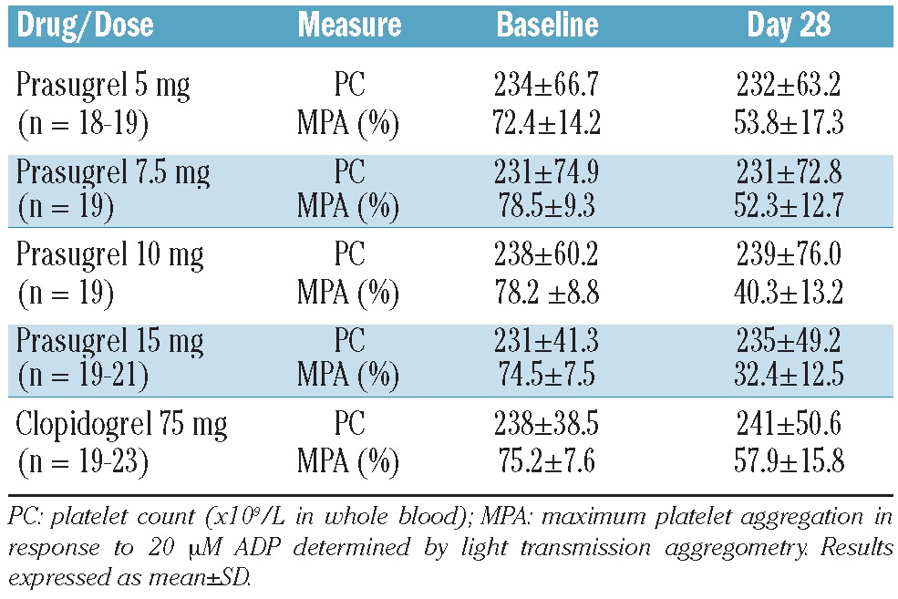
We retrospectively examined circulating platelet counts in a comparative clinical pharmacology study of prasugrel and clopidogrel. 22 During the course of the study, platelet counts were collected as part of routine hematology determinations at baseline (prior to thienopyridine administration) and 7 and 28 days after once daily dosing with the study drugs. Light transmission aggregometry was also performed in the study to determine the aggregation response to 20 μM ADP and any attenuation of this response by the study drugs.
Statistics
Values are expressed as mean ± SD or median and range, when appropriate. Data were analyzed using ANOVA, followed by the post-hoc Bonferroni's t-test. The χ2 test was used to analyze differences in the prevalence of thrombocytopenia among patients with platelet function disorders. P values <0.05 were considered statistically significant. All experiments were independently replicated at least three times, unless specified otherwise.
Results
Adenosine diphosphate is constitutively released by differentiating megakaryocytes in culture and influences proplatelet formation
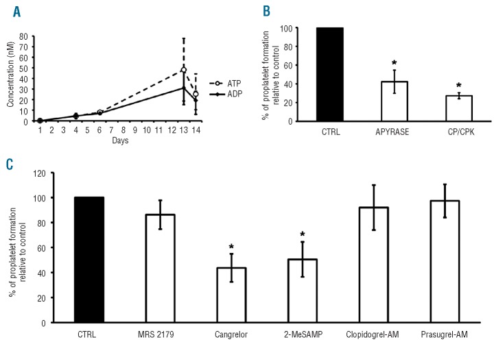
We detected the presence of ADP and ATP in the culture medium of megakaryocytes, at concentrations that increased with time (Figure 1A). This was paralleled by an increase in the percentage of CD41+CD42b+ cells (detected by flow cytometry analysis), from 67±10% on day 6 of culture to 87±10% on day 13. Cell viability, measured by the trypan blue dye exclusion assay, also increased with time (70±5% on day 6 and 89±5% on day 13), while the concentration of lactate dehydrogenase (a marker of cell lysis) in the culture medium did not change significantly (33±5 U/L on day 6 and 37±6 U/L on day 13, P=NS). The percentage of megakaryocytes producing proplatelets was 10±3% (mean ± SD, n=7 separate experiments).
Figure 1.
Role of ADP in megakaryocyte differentiation and platelet release. (A) ADP and ATP were constitutively released into the conditioned medium during megakaryocyte differentiation in culture (means±SD, n=5 separate experiments. (B) On day 13 of maturation, cord blood derived-megakaryocytes were seeded for 16 h in the presence or absence of the ADP scavengers apyrase (1 U/mL) or CP/CPK (5 mM/40 U/mL), and proplatelet formation was quantified (mean±SD, n=5 separate experiments. (C) Megakaryocytes on day 13 of culture were incubated for 16 h with the P2Y1 inhibitor MRS 2179 (10 μM), the P2Y12 inhibitors cangrelor (10 μM) and 2-MeSAMP (10 μM), or the active metabolites of clopidogrel (clopidogrel-AM, 10 mM) or prasugrel (prasugrel-AM, 10 μM) and proplatelet formation was quantified (mean±SD, n=5 separate experiments). *P<0.05 compared to untreated control (CTRL), MRS 2179, clopidogrel-AM and prasugrel-AM treated samples (ANOVA and Bonferroni's t-test as a post-hoc test).
The addition of the ADP scavengers apyrase 1 U/mL, which hydrolyses ATP and ADP, or CP 5 mM plus CPK 40 U/mL (CP/CPK), which transform ADP into ATP, to cell cultures on day 13 inhibited proplatelet formation by about 60-70% (4.6±1% and 2.1±0.1% respectively, mean ± SD, n=5 separate experiments, P<0.05) (Figure 1B), but did not affect the ploidy of cells (data not shown) or their viability (88±5% with apyrase, and 91±7% with CP/CPK). Collectively these data suggest that maturing megakaryocytes constitutively release adenine nucleotides and that released ADP plays a role in proplatelet formation.
Effects of inhibitors of the adenosine diphosphate receptors P2Y1 and P2Y12 on proplatelet formation
Several inhibitors of P2Y receptors were tested for their effects on proplatelet formation by megakaryocytes in culture (expressed as the % of megakaryocytes producing proplatelets): none of them affected cell viability or ploidy (data not shown). Proplatelet formation was not inhibited by the P2Y1 inhibitor MRS 2179 (10 μM) (8.7±2, mean ± SD, n=5 separate experiments), while it was inhibited by about 50-60% by the P2Y12 inhibitors cangrelor (10 μM) (3.9±0.9%, mean ± SD, n=5 separate experiments, P<0.05) and 2-MeSAMP (10 μM) (5±1%, mean ± SD, n=5 separate experiments, P<0.05). Proplatelet structure was not affected by the tested inhibitors (Online Supplementary Figure S1). In contrast, other P2Y12 inhibitors, such as clopidogrel-AM (10 μM) and prasugrel-AM (10 μM) did not affect proplatelet formation (10.5±1.7% and 10.1 ± 1.6%, respectively, mean ± SD, n=5 separate experiments) (Figure 1C). Moreover, the analysis of the results of a clinical pharmacology study that included the oral administration of clopidogrel and prasugrel revealed that, at doses that effectively inhibited ADP/P2Y12 mediated platelet aggregation, neither drug affected circulating platelet counts (Table 2). Notably, platelet counts were stable throughout the 28-day treatment period and at all levels of P2Y12/platelet inhibition. We, therefore, hypothesized that the observed effects of cangrelor and 2-MeSAMP on proplatelet formation were mediated by a P2Y receptor different from P2Y12, with which these inhibitors interact.
Table 2.
Circulating platelet counts at baseline and during the administration of the thienopyridine drugs clopidogrel and prasugrel to patients with stable cardiovascular diseases.
Human megakaryocytes in culture express all the known P2Y mRNA
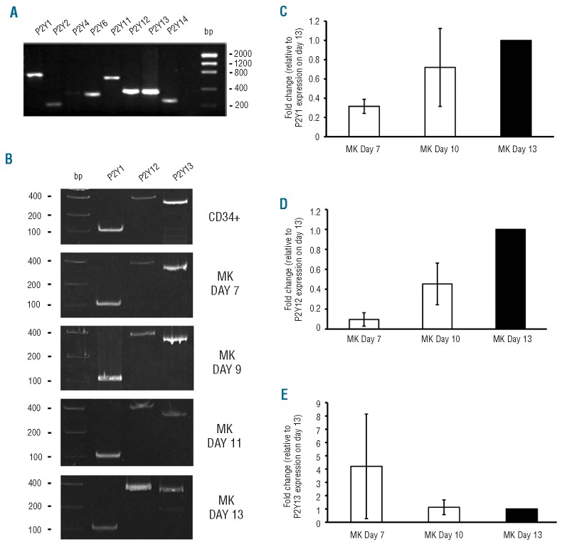
We first examined what types of P2Y receptors are expressed by megakaryocytes and found that mRNA of all the known P2Y receptors (P2Y1, P2Y2, P2Y4, P2Y6, P2Y11, P2Y12, P2Y13, and P2Y14) were expressed by megakaryocytes (Figure 2A). Of the P2Y receptors, P2Y13 is the only known P2Y receptor, other than P2Y1 and P2Y12, that recognizes ADP and its stable analogs as agonists. 13 Time-course analysis demonstrated that P2Y1, P2Y12 and P2Y13 were expressed in CD34+ progenitor cells as well as in all stages of megakaryocyte development (Figure 2B). Quantitative RT-PCR of the three receptors revealed that both P2Y1 and P2Y12 expression increased during megakaryocyte differentiation (Figure 2C-D), while P2Y13 expression decreased (Figure 2E).
Figure 2.
Expression of P2Y mRNA in cultured human megakaryocytes. RNA was extracted from CD34+ progenitor cells or CD61+ megakaryocytes (MK). The amplification products were resolved on 1% agarose gel or 20% polyacrylamide gel and visualized by ethidium bromide staining. (A) Expression of P2Y mRNA in mature human megakaryocytes (representative of 3 experiments). (B) Timecourse analysis of expression of P2Y1, P2Y12 and P2Y13 mRNA in CD34+ progenitor cells and megakaryocytes at different stages of maturation (representative of 3 different experiments). (CD-E) Fold changes in P2Y1, P2Y12 and P2Y13 mRNA expression on days 7, 10 and 13 of megakaryocyte differentiation. For each receptor, mRNA levels were normalized to their expression level on day 13 of culture. Bars represent mean±SD of at least three different experiments.
Human megakarycoytes in culture express the P2Y13 receptor for adenosine diphosphate
Western blot analysis of megakaryocyte lysates on days 7, 10 and 13 of culture showed that P2Y13 protein was present, and that its expression increased with time (Figure 3A). In contrast, no reactive bands were detected in lysates from circulating platelets (Figure 3B).
Figure 3.
Analysis of P2Y13 protein expression in human megakaryocytes in culture and in human platelets. CD61+ human megakaryocytes (MK) or washed human platelets were lysed and subjected to western blot analysis. (A) P2Y13 receptor expression in human megakaryocytes on different days of culture. (B) Expression of CD61 and P2Y13 in human megakaryocytes on day 13 of culture and in washed human platelets. Samples were re-probed with anti–β-actin to ensure equal loading (results are representative of 3 different experiments).
MRS 2211, a specific inhibitor of P2Y13 inhibits proplatelet formation by megakaryocytes in culture
Based on the data of RT-PCR and western blot analysis, we considered P2Y13 as the most likely candidate receptor for ADP on megakaryocytes which mediates the effects of ADP on proplatelet formation. This hypothesis is corroborated by reports in the medical literature demonstrating that both the “P2Y12” inhibitors that inhibited proplatelet formation in our study, cangrelor and 2-MeSAMP, also interact with P2Y13, inhibiting its function.23-25
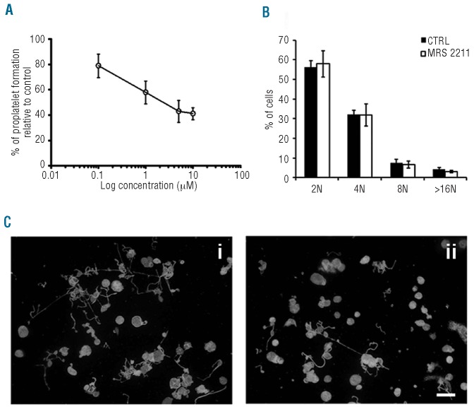
To test our hypothesis, we used the specific P2Y13 inhibitor MRS 2211,26 which inhibited proplatelet formation by megakaryocytes in culture in a concentration-dependent manner; maximal inhibition of approximately 50% was achieved at a concentration of 10 mM (3.4±0.7%, mean ± SD, n=5 separate experiments, P<0.05) (Figure 4A). Like cangrelor and 2-MeSAMP, MRS 2211 had no effects on ploidy (Figure 4B), morphology (Figure 4C) and viability (90±6% versus 89±5%) of megakaryocytes.
Figure 4.
Effect of MRS 2211, a specific P2Y13 inhibitor, on human megakaryocytes in culture. (A) Concentration-dependent inhibition of proplatelet formation by normal megakaryocytes in culture by the specific P2Y13 antagonist MRS 2211 (0.1-10 μM) (mean±SD, n=5 separate experiments). (B) Effects of MRS 2211 (10 μM, 16 h incubation) on megakaryocyte ploidy. Data are expressed as mean±SD of three experiments. (C) Immunoflorescence staining of proplatelet-forming megakaryocytes, after their incubation with: (i) vehicle (control) or (ii) MRS 2211 (10 μM) for 16 h in culture (α-tubulin in green, Hoechst 33258 in blue, scale bars=40 μm) (images were acquired by an Olympus BX51, magnification 20X).
MRS 2211 inhibits Akt and ERK1/2 phosphorylation in human mature megakaryocytes
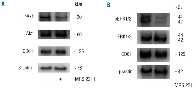
P2Y13 receptor is coupled to PI3K/Akt/GSK327 and ERK1/228 signaling, both of which are involved in the regulation of proplatelet formation.29-31 In order to explore these biochemical pathways, megakaryocytes at day 13 of culture were incubated or not with the P2Y13 receptor antagonist MRS 2211 (10 μM). Western blots indicated that both Akt (Figure 5A) and ERK1/2 (Figure 5B) were highly phosphorylated during proplatelet formation and that their phosphorylation decreased in the presence of MRS 2211.
Figure 5.
Akt and ERK1/2 phosphorylation by proplatelet-forming megakaryocytes in culture is inhibited by MRS 2211. Western blot analysis of (A) Akt and (B) ERK1/2 phosphorylation in human megakaryocytes on day 13 of culture in the presence (+) or absence (−) of the specific P2Y13 inhibitor MRS 2211 (10 μM). Samples were also probed with anti-Akt, anti-ERK1/2, anti-CD61 and anti–β-actin antibodies (representative of 3 experiments).
Proplatelet formation by megakaryocytes from a patient with severe, congenital P2Y12 deficiency
Megakaryocytes were differentiated from the peripheral blood progenitor cells of a previously described patient with severe P2Y12 deficiency16 and ten healthy subjects. Proplatelet formation by the patient's megakaryocytes (8±3%, n=4 separate experiments) was comparable to that observed in healthy subjects (11±4%). Apyrase (2.4%, n=1 experiment), cangrelor (3.7±0.5%, mean ± SD, n=4 separate experiments, P<0.05) and MRS 2211 (3.1±0.6%, mean ± SD, n=4 separate experiments, P<0.05) inhibited proplatelet formation by the patient's megakaryocytes by about 50% compared to their effects on megakaryocytes from control subjects (Figure 6). MRS 2211 had no effects on patient's cell viability or morphology (data not shown).
Figure 6.
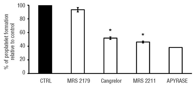
Inhibition of proplatelet formation by megakaryocytes from a patient with congenital, severe P2Y12 deficiency. The effects of the indicated P2Y inhibitors and of apyrase on proplatelet formation by megakaryocytes from a patient with congenital severe P2Y12 deficiency were tested under the same experimental conditions as those used for normal megakaryocytes. Means±SD, n=4 separate experiments, except for apyrase (n=1). *P<0.05 compared to untreated control (CTRL) and MRS 2179 treated samples (ANOVA and Bonferroni's t-test as a post-hoc test).
Platelet count in patients with delta-storage pool deficiency
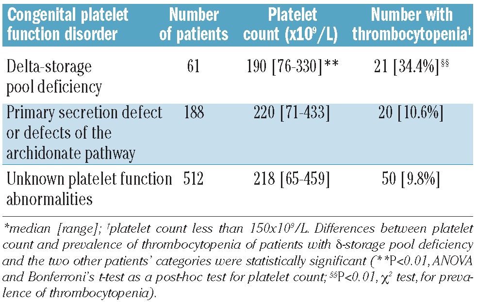
If constitutively released ADP plays a role in proplatelet formation by megakaryocytes, its deficiency should result in decreased platelet production and low platelet counts in circulating blood. It is, therefore, reasonable to assume that patients whose megakaryocytes/platelet lineage is characterized by decreased nucleotide content in storage granules, such as patients with δ-storage pool deficiency (δ-SPD),32 might have lower platelet counts, compared to control subjects. We, therefore, retrospectively analyzed the platelet count in 61 patients who were diagnosed with δ-SPD at the Hemophilia and Thrombosis Center in Milan between 1988 and 2010. These patients' median platelet count was 190x109/L (range, 76x109/L-330x109/L); 21 patients (34.4%) had mild thrombocytopenia (platelet count <150x109/L) (Table 3). These results were significantly different from those observed in two control groups of patients who underwent the same laboratory screening for platelet function disorders, in the same Institution during the same period: (i) patients with abnormalities of platelet secretion of adenine nucleotides not due to deficiency of platelet granules (“primary secretion defects” and “abnormalities of the arachidonate pathway”); and (ii) patients with no known abnormalities of platelet function (Table 3).
Table 3.
Platelet count and prevalence of thrombocytopenia in historical patients with bleeding diatheses, who underwent platelet function testing between 1988 and 2010, according to their diagnosis.
Discussion
Our study demonstrates that, during the in vitro development and maturation of megakaryocytes from blood progenitors, the concentration of the adenine nucleotides ADP and ATP in the culture medium increases, peaking at day 13 of culture, when megakaryocytes form proplatelets; the study also shows that ADP contributes to proplatelet formation through its interaction with the P2 receptor P2Y13. Our results suggest that adenine nucleotides are constitutively released by megakaryocytes, because their increase in the culture medium is paralleled by an increase in the percent of viable, maturing megakaryocytes in culture, but not by an increase in lactate dehydrogenase, which is a marker of cell lysis. The observed constitutive release of adenine nucleotides into the megakaryocyte culture medium was time-dependent and paralleled the previously described time-course of formation of δ-granules in developing megakaryocytes.4 This chronological parallelism and the contemporary release of ADP and ATP observed in our study suggest that adenine nucleotides are released by megakaryocyte δ-granules, where they are stored. Based on the results of our study, it could, therefore, be predicted that conditions associated with a decreased content of ADP in δ-granules may be associated with decreased platelet production, and reflected by a low platelet count in the circulation. δ-SPD is a congenital bleeding diathesis, which is associated with impaired platelet function, characterized by a deficiency of dense granules and their constituents (including ATP and ADP) in megakaryocytes and platelets.32 If the results of our study have a clinical counterpart, patients with δ-SPD should have some degree of thrombocytopenia, which contrasts with the current knowledge that platelet count is normal in these patients. However, an ad hoc study focused on the platelet counts of a large series of patients with δ-SPD has never been published. In our study, we reviewed the data of 61 historical patients with δ-SPD who had been diagnosed at the Angelo Bianchi Bonomi Hemophilia and Thrombosis Center in Milan, Italy, between 1984 and 2010, and found that 34.4% of them had mild thrombocytopenia. The mean platelet count was significantly lower and the prevalence of thrombocytopenia in δ-SPD patients was significantly higher than those observed in 188 patients in whom abnormalities of agonist-induced platelet secretion associated with normal δ-granule content (prevalence of thrombocytopenia, 10.6%) or in 512 patients in whom no known abnormalities of platelet function (prevalence of thrombocytopenia 9.8%) had been diagnosed in the same Institution, within the same time frame (Table 2). In addition, upon review of the published literature, we found that, although in a series of 18 patients with heterogeneous types of δ-SPD only one patient had mild thrombocytopenia, 33 the prevalence of mild thrombocytopenia in other studies was 18% (9/51)34 and 42% (10/24),35 demonstrating that mild thrombocytopenia is not an uncommon finding in patients with δ-SPD.
Having identified a role of constitutively released ADP in proplatelet formation, we tried to identify the ADP receptor on megakaryocytes that mediates this effect. Adenine nucleotides interact with purinergic P2 receptors, of which two subfamilies have been identified: G protein-linked or “metabotropic”, termed P2Y, and ligand-gated ion channels or “ionotropic”, termed P2X.12,13 ADP only interacts with certain types of P2Y receptors.12,13 In our experiments, we initially focused our attention on P2Y1 and P2Y12, which are the only P2Y receptors present on the platelet membrane and bind ADP,12-14 and tested the in vitro effects of inhibitors of these receptors. We found that the specific P2Y1 receptor inhibitor MRS 2179 did not significantly affect proplatelet formation, while the P2Y12 receptor inhibitors cangrelor and 2-MeSAMP inhibited proplatelet formation to a similar degree (about 50-60%) as the ADP scavengers apyrase and CP/CPK did, suggesting that P2Y12 is responsible for the observed effects of released ADP. However, several lines of evidence were in disagreement with this conclusion: (i) patients with severe P2Y12 deficiency have normal platelet counts;16 (ii) thrombocytopenia has never been identified as a side effect of the P2Y12-inhibitory thienopyridine drugs ticlopidine, clopidogrel and prasugrel, with the exception of in those patients, mostly treated with ticlopidine, who experienced thrombotic thrombocytopenic purpura, which causes intravascular consumption of platelets, or myelosuppression and which also lowered the leukocyte and red blood cell counts;36 (iii) we reviewed the records of a phase 1b clinical trial, in which platelet counts were recorded before and after the administration of clopidogrel and prasugrel, and found that neither drug affected platelet counts; and (iv) when we tested the in vitro effect of the active metabolites of clopidogrel and prasugrel, under the same experimental conditions that we used to study the effects of cangrelor and 2-MeSAMP, we found that neither inhibited proplatelet formation. We, therefore, hypothesized that the observed effect of cangrelor and 2-MeSAMP was mediated by their inhibition of a receptor distinct from P2Y12. First of all, we found that mature megakaryocytes express the mRNA of all known P2Y receptors. Previous studies showed that stem cells and progenitor cells express several P2 receptors,37 suggesting that the receptors play a role in cell differentiation and proliferation. Among the P2 receptors, besides P2Y1 and P2Y12, only P2Y13 binds ADP.12,13 P2Y13 mRNA decreased with time during megakaryocyte maturation, while P2Y13 protein peaked at day 13, when megakaryocytes started producing proplatelets, and disappeared in circulating platelets. In order to test whether P2Y13 was responsible for the observed effect of released ADP on proplatelet formation, we used the specific P2Y13 inhibitor MRS 2211,26 and found that it did indeed inhibit proplatelet formation by about 60%. MRS 2211 also inhibited the phosphorylation of Akt and ERK1/2, which are coupled with P2Y13 signaling.27,28 In addition, a review of the published literature revealed that both cangrelor and 2-MeSAMP inhibit not only P2Y12, but also P2Y13.23-25 Evidence for the involvement of P2Y13 does, therefore, seem compelling, considering that, of the six P2Y inhibitors that we tested, only the three that antagonize P2Y13 (cangrelor, 2-MeSAMP and MRS2211) inhibited proplatelet formation, despite being structurally very different from each other. The involvement of P2Y13, and not of P2Y12, in proplatelet formation was also confirmed by the finding that the formation of proplatelets by megakaryocytes from a patient with congenital severe deficiency of P2Y12 was normal and was inhibited by cangrelor and MRS 2211 to a similar degree to that of megakaryocytes from normal subjects. The clinical efficacy of cangrelor in patients with acute coronary syndromes has been tested in randomized clinical trials:38-40 while its effects on in vivo platelet formation and count have not been reported, the short duration of infusion that has been investigated would not be expected to change platelet counts.
To the best of our knowledge, this is the first demonstration that ADP regulates proplatelet formation and that P2Y13 is expressed by maturing megakaryocytes and mediates the observed effects of released ADP. The results of our study open new perspectives in the evaluation of novel signals that trigger proplatelet formation by mature megakaryocytes. Autocrine components, together with environmental factors, seem to play an important role in the regulation of platelet production.
Acknowledgments
the authors thank Cesare Perotti for suppling human cord blood, Riccardo Albertini for LDH measurements and Gianluca Viarengo for assistance with fluorocytometry.
Funding: this paper was supported by grants from the Cariplo Foundation (2006.0596/10.8485), the Regione Lombardia - Project SAL-45, Almamater Foundation (Pavia) and the Regione Lombardia- Progetto di cooperazione scientifica e tecnologica internazionale to AB.
Footnotes
The online version of this article has a Supplementary Appendix.
Authorship and Disclosures: The information provided by the authors about contributions from persons listed as authors and in acknowledgments is available with the full text of this paper at www.haematologica.org.
Financial and other disclosures provided by the authors using the ICMJE (www.icmje.org) Uniform Format for Disclosure of Competing Interests are also available at www.haematologica.org.
Alessandra Balduini, MD, Biotechnology Laboratories, Department of Molecular Medicine, IRCCS San Matteo Foundation, Università degli Studi di Pavia, Via Forlanini 6, 27100 Pavia, Italy. E-mail:alessandra.balduini@unipv.it
References
- 1.Avecilla ST, Hattori K, Heissig B, Tejada R, Liao F, Shido K, et al. Chemokine-mediated interaction of hematopoietic progenitors with the bone marrow vascular niche is required for thrombopoiesis. Nat Med. 2004;10(1):64-71 [DOI] [PubMed] [Google Scholar]
- 2.Italiano JE, Jr, Lecine P, Shivdasani RA, Hartwig JH. Blood platelets are assembled principally at the ends of proplatelet processes produced by differentiated megakaryocytes. J Cell Biol. 1999;147(6):1299-312 [DOI] [PMC free article] [PubMed] [Google Scholar]
- 3.Youssefian T, Cramer EM. Megakaryocyte dense granule components are sorted in multivesicular bodies Blood. 2000;95(12):4004-7 [PubMed] [Google Scholar]
- 4.Richardson JL, Shivdasani RA, Boers C, Hartwig JH, Italiano JE., Jr. Mechanisms of organelle transport and capture along proplatelets during platelet production. Blood. 2005;106(13):4066-75 [DOI] [PMC free article] [PubMed] [Google Scholar]
- 5.Becker RP, De Bruyn PP. The transmural passage of blood cells into myeloid sinusoids and the entry of platelets into the sinusoidal circulation; a scanning electron microscopic investigation. Am J Anat. 1976;145(2):183-205 [DOI] [PubMed] [Google Scholar]
- 6.Junt T, Schulze H, Chen Z, Massberg S, Goerge T, Krueger A, et al. Dynamic visualization of thrombopoiesis within bone marrow. Science. 2007;317(5845):1767-70 [DOI] [PubMed] [Google Scholar]
- 7.Thon JN, Montalvo A, Patel-Hett S, Devine MT, Richardson JL, Ehrlicher A, et al. Cytoskeletal mechanics of proplatelet maturation and platelet release. J Cell Biol. 2010;191(4):861-74 [DOI] [PMC free article] [PubMed] [Google Scholar]
- 8.Balduini A, Pallotta I, Malara A, Lova P, Pecci A, Viarengo G, et al. Adhesive receptors, extracellular proteins and myosin IIA orchestrate proplatelet formation by human megakaryocytes. J Thromb Haemost. 2008;6(11):1900-7 [DOI] [PubMed] [Google Scholar]
- 9.Möhle R, Green D, Moore MA, Nachman RL, Rafii S. Constitutive production and thrombin-induced release of vascular growth factor by human megakaryocytes and plateles. Proc Natl Acad Sci USA. 1997;94(2):663-8 [DOI] [PMC free article] [PubMed] [Google Scholar]
- 10.Casella I, Feccia T, Chelucci C, Samoggia P, Castelli G, Guerriero R, et al. Autocrineparacrine VEGF loops potentiate the maturation of megakaryocytic precursors through Flt1 receptor. Blood. 2003;101(4):1316-23 [DOI] [PubMed] [Google Scholar]
- 11.Thompson CJ, Schilling T, Howard MR, Genever PG. SNARE-dependent glutamate release in megakaryocytes. Exp Hematol. 2010;38(6):504-15 [DOI] [PMC free article] [PubMed] [Google Scholar]
- 12.Boeynaems J-M, Communi D, Suarez Gonzales N, Robaye B. Overview of the P2 receptors. Sem Thromb Hemost. 2005;31(2):139-49 [DOI] [PubMed] [Google Scholar]
- 13.von Kügelen I. Pharmacological profiles of cloned mammalian P2Y-receptor subtypes. Pharmacol Ther. 2006;110(3):415-32 [DOI] [PubMed] [Google Scholar]
- 14.Cattaneo M, Gachet C. ADP receptors and clinical bleeding disorders. Arterioscler Thromb Vasc Biol. 1999;19(10):2281-5 [DOI] [PubMed] [Google Scholar]
- 15.Hechler B, Lenain N, Marchese P, Vial C, Heim V, Freund M, et al. A role of the fast ATP-gated P2X1 cation channel in thrombosis of small arteries in vivo. J Exp Med. 2003;198(4):661-7 [DOI] [PMC free article] [PubMed] [Google Scholar]
- 16.Cattaneo M, Lecchi A, Randi AM, McGregor JL, Mannucci PM. Identification of a new congenital defect of platelet function characterized by severe impairment of platelet responses to adenosine diphosphate. Blood. 1992;80(11):2787-96 [PubMed] [Google Scholar]
- 17.Pecci A, Malara A, Badalucco S, Bozzi V, Torti M, Balduini CL, et al. Megakaryocytes of patients with MYH9-related thrombocytopenia present an altered proplatelet formation. Thromb Haemost. 2009;102(1):90-6 [DOI] [PubMed] [Google Scholar]
- 18.Williams N, Levine RF. The origin, development and regulation of megakaryocytes. Br J Haematol. 1982;52(2):173-80 [DOI] [PubMed] [Google Scholar]
- 19.Kaplan LA, Pesce AJ. Clinical Chemistry: Theory, Analysis and Correlation. St. Louis, Missouri: Mosby Ed.; 1996 [Google Scholar]
- 20.Malara A, Gruppi C, Rebuzzini P, Visai L, Perotti C, Moratti R, et al. Megakaryocytematrix interaction within bone marrow: new roles for fibronectin and factor XIII-A. Blood. 2011;117(8):2476-83 [DOI] [PubMed] [Google Scholar]
- 21.Dangelmaier CA, Holmsen H. Platelet dense granule and lysosome content. In: Harker LA, Zimmerman TS, eds. Methods in Hematology: Measurement of Platelet Function. Edinburgh, Scotland: Churchill Livingstone; 1983:92-114 [Google Scholar]
- 22.Jernberg T, Payne CD, Winters KJ, Darstein C, Brandt JT, Jakubowski JA, et al. Prasugrel achieves greater inhibition of platelet aggregation and a lower rate of non-responders compared with clopidogrel in aspirin-treated patients with stable coronary artery disese. Eur Heart J. 2006;27(10):1166-73 [DOI] [PubMed] [Google Scholar]
- 23.Marteau F, Le Poul E, Communi D, Communi D, Labouret C, Savi P, et al. Pharmacological characterization of the human P2Y13 receptor. Mol Pharmacol. 2003;64(1):104-12 [DOI] [PubMed] [Google Scholar]
- 24.Fumagalli M, Trincavelli L, Lecca D, Martini C, Ciana P, Abbracchio MP. Cloning, pharmacological characterization and distribution of the rat G-protein-coupled P2Y13 receptor. Biochem Pharmacol. 2004;68(1):113-24 [DOI] [PubMed] [Google Scholar]
- 25.Wang L, Olivecrona G, Götberg M, Olsson ML, Winzell MS, Erlinge D. ADP acting on P2Y13 receptors is a negative feedback pathway for ATP release from human red blood cells. Circ Res. 2005;96(2):189-96 [DOI] [PubMed] [Google Scholar]
- 26.Kim YC, Lee JS, Sak K, Marteau F, Mamedova L, Boeynaems JM, et al. Synthesis of pyridoxal phosphate derivatives with antagonist activity at the P2Y13 receptor. Biochem Pharmacol. 2005;70(2):266-74 [DOI] [PMC free article] [PubMed] [Google Scholar]
- 27.Ortega F, Pérez-Sen R, Miras-Portugal MT. Gi-coupled P2Y-ADP receptor mediates GSK-3 phosphorylation and beta-catenin nuclear translocation in granule neurons. J Neuorchem. 2008;104(1):62-73 [DOI] [PubMed] [Google Scholar]
- 28.Ortega F, Pérez-Sen R, Delicado EG, Miras-Portugal MT. Erk1/2 activation is involved in the neuroprotecitve action of P2Y13 and P2X7 receptors against glutamate excitotoxicity in cerebellar granule neurons. Neuropharmacology. 2011;61(8):1210-21 [DOI] [PubMed] [Google Scholar]
- 29.Ono M, Matsubara M, Shibano T, Ikeda Y, Murata M. GSK-3 negatively regulates megakaryocyte differentiation and platelet production from primary human bone marrow cells in vitro. Platelets. 2011;22(3):196-203 [DOI] [PubMed] [Google Scholar]
- 30.Jiang F, Jia Y, Cohen I. Fibronectin-and protein kinase C-mediated activation of ERK/MAPK are essential for proplateletlike formation. Blood. 2002;99(10):3579-84 [DOI] [PubMed] [Google Scholar]
- 31.Mazharian A, Watson SP, Séverin S. Critical role for ERK1/2 in bone marrow and fetal liver-derived primary megakaryocyte differentiation, motility, and proplatelet formation. Exp Hematol. 2009;37(10):1238-49 [DOI] [PMC free article] [PubMed] [Google Scholar]
- 32.Cattaneo M. Inherited platelet-based bleeding disorders. J Thromb Haemost. 2003;1(7):1628-36 [DOI] [PubMed] [Google Scholar]
- 33.Weiss HJ, Witte LD, Kaplan KL, Lages BA, Chernoff A, Nossel HL, et al. Heterogeneity in storage pool deficiency: studies on granule-bound substances in 18 patients including variants deficient in alpha-granules, platelet factor 4, beta-thromboglobulin, and platelet-derived growth factor. Blood. 1979;54(6):1296-319 [PubMed] [Google Scholar]
- 34.Nieuwenhuis HK, Akkerman JW, Sixma JJ. Patients with a prolonged bleeding time and normal aggregation tests may have storage pool deficiency: studies on one hundred six patients. Blood. 1987;70(3):620-3 [PubMed] [Google Scholar]
- 35.Pujol-Moix N, Hernández A, Escolar G, Español I, Martínez-Brotóns F, Mateo J. Platelet ultrastructural morphometry for diagnosis of partial delta-storage pool disease in patients with mild platelet dysfunction and/or thrombocytopenia of unknown origin. A study of 24 cases. Haematologica. 2000;85(6):619-26 [PubMed] [Google Scholar]
- 36.Cattaneo M. The platelet P2Y12 receptor for adenosine diphosphate: congenital and druginduced defects. Blood. 2011;117(7):1202-12 [DOI] [PubMed] [Google Scholar]
- 37.Wang L, Jacobsen SE, Bengtsson A, Erlinge D. P2 receptor mRNA expression profiles in human lymphocytes, monocytes and CD34+ stem and progenitor cells. BMC Immunol. 2004;5:16. [DOI] [PMC free article] [PubMed] [Google Scholar]
- 38.Bhatt DL, Lincoff AM, Gibson CM, Stone GW, McNulty S, Montalescot G, et al. Intravenous platelet blockade with cangrelor during PCI. N Engl J Med. 2009;361(24):2330-41 [DOI] [PubMed] [Google Scholar]
- 39.Harrington RA, Stone GW, McNulty S, White HD, Lincoff AM, Gibson CM, et al. Platelet inhibition with cangrelor in patients undergoing PCI. N Engl J Med. 2009;361(24):2318-29 [DOI] [PubMed] [Google Scholar]
- 40.Angiolillo DJ, Firstenberg MS, Price MJ, Tummala PE, Hutyra M, Welsby IJ, et al. ; BRIDGE Investigators. Bridging antiplatelet therapy with cangrelor in patients undergoing cardiac surgery: a randomized controlled trial. JAMA. 2012;307(3):265-74 [DOI] [PMC free article] [PubMed] [Google Scholar]


