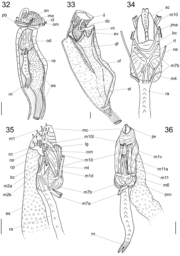
Figures 32–36.
Calliostoma tupinamba anatomy. 32 Head and hemocel, ventral view, scale bar = 5 mm 33 Buccal cavity and esophagus opened longitudinally, ventral-inner view, odontophore removed 34 Odontophore, dorsal view 35–36 Buccal mass and central nervous system, right and ventral views. Scales bar = 2 mm.

