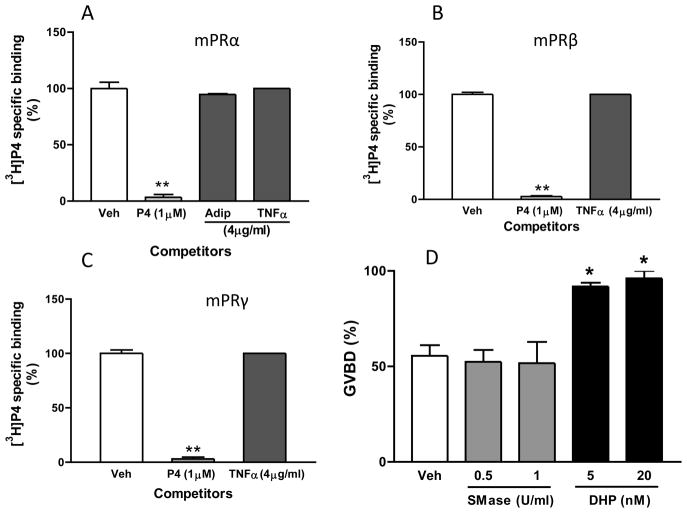
Figure 2.
The proposed role of mPRs in sphingolipid signaling is not supported by empirical data showing a lack of TNFα binding to mPRs (A–C) and the ineffectiveness of exogenous neutral sphingomyelinase in inducing meiotic maturation of zebrafish oocytes (D). A–C, Single point competition with 1μM progesterone (P4), 4μg/ml TNFα or 4μg/ml adiponectin (adip) for [3H]-progesterone binding to plasma membranes of MDA-MB-231 breast cancer cells over expressing mPRα (A), mPRβ (B), or mPRγ (C). Data represent mean disintegrations per minute (DPM)/50μg protein ± SEM, n=3; **, P < 0.0001 compared with vehicle (veh) control by one-way ANOVA and Dunnett’s multiple comparison test. D, Comparison of the effects of exogenous B. cerus spingomyelinase (SMase, 0.5 and 1.0 U/ml) and 17,20β-dihydroxy-4-pregene-3-one (DHP, 20 and 50 nM) on percent GVBD of denuded zebrafish oocytes. Large, fully grown zebrafish oocytes (diameter> 550μm) were denuded and the oocytes (30–40 oocytes/1 ml Leibovitz L-15 medium/well, 4 wells/treatment) were incubated with the various treatments for 3 hrs and scored at the end of the incubation period for germinal vesicle breakdown (GVBD) as described previously [91]. Data represent mean % oocytes completing GVBD ± SEM, n=4; *, P < 0.05 compared with vehicle (veh) control by one-way ANOVA and Dunnett’s multiple comparison test.

