
Fig. 4.
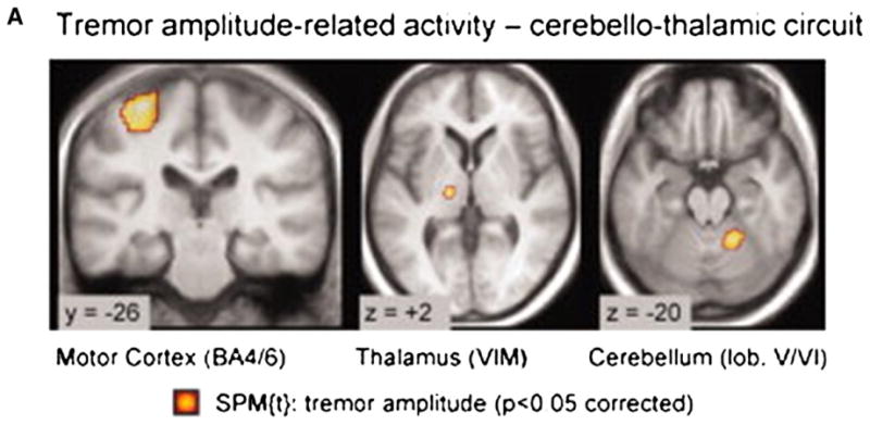
Tremor-related cerebral activity in tremor-dominant Parkinson disease (PD). Cerebral regions where activity cofluctuated with left sided tremor amplitude (Statistical Parametric Mapping [SPM] t-contrast on the electromyographic (EMG) amplitude regressor in 19 tremor-dominant patients). Activity was localized to the motor cortex (MC), ventral intermediate nucleus of the thalamus, and cerebellum. They found motor cortex activity correlated with tremor amplitude. Conversely, GPi activity correlated with changes in tremor amplitude.
From Helmich et al. (2011) Fig. 1A, p272.