

Abstract
Proximal screw pullout is a well-recognized problem in anterior scoliosis surgery, with a rate of pseudarthrosis or screw pullout ranging from 15 to 30%. To prevent screw pullout at the top of the construct, the authors have devised the concept of a claw for the top instrumented vertebra. The claw consists of a classic anterior vertebral body screw inserted parallel to the inferior end-plate and in the posterior portion of the vertebral body 8 mm in front of the spine canal. After rib desarticulation, a laminar hook of a small size is inserted over the superior aspect of the pedicle of the same vertebra. The rod is then inserted into the two side openings of the screw and the hook. Compression across the hook and the screw is then performed, making a claw construct. This concept can also be extended in the case of early revision for a proximal screw pullout, where it is possible to revise the instrumentation with an offset connector linking the rod to the superior portion of the pedicle where the suprapedicule hook has been inserted. We report two cases where a suprapedicle claw was successfully used in anterior scoliosis correction of a right thoracic curve. Such a concept may represent the solution to proximal screw pullout in anterior scoliosis correction.
Keywords: Anterior spine surgery, Anterior vertebral body screw, Scoliosis, Screw pullout
Introduction
Recently, anterior spine fusion for correction of adolescent idiopathic scoliosis has gained popularity [3, 4]. The advantages of anterior spine fusion over posterior fusion could be shorter fusion [3, 4], better correction of hypokyphosis and possible better cosmetic outcome.
However, the drawback of such an approach seems to be the high rate of pseudarthrosis with implant breakage and screw pullout [3, 4]. Because of the smaller size of the proximal vertebrae, smaller size screws are inserted in these vertebrae. As the proximal thoracic part of the curve is stiffer than the lower part, a smaller screw and increased curve stiffness explain why the proximal screw pullout is mostly observed at the top of the instrumentation and more rarely at the bottom. Some authors have inserted washers on the opposite cortex to prevent screw pullout [1]. Recently, a biomechanical study showed that the addition of pullout resistance nuts increased the resistance of the screw by 2-fold [8]. The placement of a nut on the opposite vertebral cortex requires further dissection, larger approaches and a longer screw to engage the nut or washer.
We therefore thought of a claw construct that would be easier to insert than a controlateral washer and that would prevent screw pullout at the proximal vertebra level.
Materials and methods
Description of the suprapedicle claw construct (Figs 1, 2, 3)
Fig. 1.

Schematic representation of the claw construct on the saw bone. The vertebral screw is inserted at the inferior aspect of the vertebral body 8 mm in front of the spine canal and parallel to the inferior endplate. The laminar hook inserted in an offset configuration sits on the superior aspect of the pedicle
Fig. 2.

Superior aspect of the canal intrusion of the laminar hook over the pedicle: this corresponds to the blade thickness and is 2 mm
Fig. 3.

Lateral aspect of the spine after the claw construct has been made on the rod
A standard 5 or 6 mm pedicle screw from the AO Universal spine system (Synthes, Paoli, Pa., USA) is inserted at the lower aspect of the top vertebral body to be instrumented. The screw starting point is 5 mm above the inferior end-plate and 8 mm in front of the spine canal. The screw is inserted in a coronal or mildly oblique fashion away from the spine canal. The screw is bicortical and its side opening is turned posteriorly. A side opening laminar hook of the smallest size is then inserted above the pedicle. Its side opening faces anteriorly to match the side opening of the screw below. The offset configuration of the two side-opening hook and screw allow perfect alignment of the two implants. The rod will then be inserted into the two side openings and the claw compressed over the rod.
Case report (Figs 4, 5)
Fig. 4.

Case number 1: 48° King II right thoracic curve: the midsacral line passes through the disc T12–L1
Fig. 5.

Postoperative correction with the suprapedicle claw construct at the level of T6
A 14-year-old girl with adolescent idiopathic scoliosis presented with a progressive 48° right thoracic curve. After informed consent, the patient was brought to the OR. The patient was positioned in a strict lateral decubitus position and a thoracotomy through the bed of the 6th rib was then carried out. The 6th rib was completely removed. After complete discectomy screws were inserted from T6 to T12 through the same thoracotomy. In T6 and T7, a 5 mm screw was inserted. In the lower vertebrae, 6 mm screws were inserted. At the top of the instrumentation, the claw construct was performed at T6. The 6th rib head articulating with the 6th vertebral body was disarticulated and separated from the pedicle and the transverse process. To achieve such separation a combination of sharp dissection with the Cobb elevator and a rongeur was necessary, as the costovertebral ligaments represent a difficult structure to separate. The epidural space above the pedicle was then visualized. The 5th intercostal nerve root was not seen as it lies far above. The suprapedicle hook could then be easily inserted. Bone graft was then packed into all the disc spaces. A 6-mm titanium rod was then inserted proximally into the claw. The claw was tightened and the rod was then cantilevered into the other screws to correct the deformity. Blood loss for the whole procedure was 600 cc and there was no bleeding from the epidural space and at the time of hook insertion. The pleura was then closed over the rod and the thoracotomy closed in a standard fashion over a chest tube. Throughout the procedure, spinal cord monitoring remained perfectly normal. At 1 year follow-up there was no loss of correction, the fusion appeared solid, and the patient reported no complaints (Fig. 5).
Variation of the suprapedicle claw construct in the case of revision for proximal screw pullout
This concept is best illustrated by the following case. An 11-year-old female with juvenile onset idiopathic scoliosis was initially treated with a brace beginning at 6 years of age. Despite bracing, the thoracic curve progressed to a magnitude of 52° (Fig. 6). Bending films showed the curve to reduce down to 33°. Preoperative bone density measurement to assess for osteoporosis and ability of the thoracic spine to maintain anterior hardware revealed 0.832 g/cm, which was within 1 SD of age matched controls. She underwent anterior fusion and instrumentation from T6 to T12 to restore normal thoracic sagittal balance and to prevent crankshaft. An anterior interbody graft was placed at T11–12 to maintain sagittal profile at the thoracolumbar junction. Rib autograft was used for bone grafting. Fluoroscopy was used intraoperatively to confirm the bicortical fixation of all screws and adequate instrumentation (Fig. 7). The patient did well, but on the 6th postoperative day standing films revealed proximal screw pullout (Fig. 7). The patient was sent home with a brace. Discussion prior to discharge included a scheduled return to the operating room for revision. She returned to the hospital 10 days later and underwent removal of the screw from T6 with placement of a structural allograft placed eccentrically on the concavity of the interspace of T6–7. A supra-pedicular hook was placed and attached to the previously placed rod, thanks to an offset lateral connector attaching the suprapedicle hook to the rod. The patient was discharged on postoperative day 5 after the chest tube had been removed.
Fig. 6.

Eleven-year-old patient with progressive 53° right thoracic scoliosis
Fig. 7.

The intraoperative fluoroscopy picture shows satisfactory correction and positioning of the screws, which are all bicortical. The standing film on day 6 shows proximal screw pullout at the level of the 6th vertebra
Follow-up X-rays at 3 months revealed maintained postoperative correction (Fig. 8).
Fig. 8.
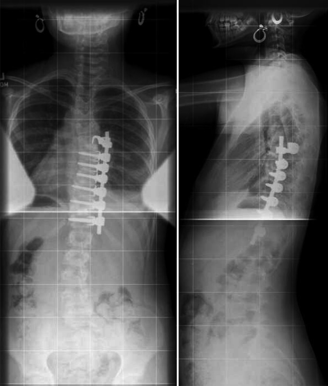
Revision with a suprapedicle claw is made, restoring the initial correction. At 6 months follow-up, the correction is stable
Discussion
Proximal screw pullout or coronal fracture of the proximal vertebral body has remained one of the problems of anterior scoliosis surgery. To address this problem, different systems have been used in the past. Staples were initially used in the Dwyer and Zielke instrumentation [10]. However, toggling and pullout of the proximal screw was observed with this system. Other systems such as the Kaneda system have used two rods and therefore two screws per vertebra, in an attempt to prevent screw pullout and increase stability [6]. Such systems are, however, indicated for thoracolumbar or lumbar curve and cannot be applied to the upper thoracic vertebrae, which are too small to accommodate two screws. The other original idea from the surgeons who devised the AOUSS was to used washers on the opposite side of the vertebral body [1]. Lieberman [8] has shown in vitro that adding an opposite nut augmented the pull out strength 2-fold. It is, however, our opinion that the addition of a washer or nut on the opposite side requires additional dissection and is difficult. It also renders necessary the use of a longer screw, which can theoretically be dangerous for the aorta, which lies in the concavity of the curve. The concept we have developed can be criticized for the following reasons. One may be afraid to invade the spine canal with the hook blade, leading to spinal cord compression. The imprint of the hook blade in the canal is only 2 mm (Fig. 2). If one reads the reports on pedicle screw fixation in the spine, it is accepted that a protrusion of a screw into the spine canal of less than 4 mm does not lead to neurologic complications [5, 9, 11]. Clinical experience in previous posterior scoliosis surgery has shown that hooks were safe and that their intrusion in the spine canal led only exceptionally to neurologic complications. Another concern could be an injury to the segmental nerve root, as it lies above our construct and could be compressed by our hook. We recognize this as a potential problem, yet we did not encounter it in our two cases. As the hook is in a supralaminar position, it lies away from the above exiting nerve root. Bleeding due to injury of the epidural veins is a theoretical problem that we did not encounter during our case. Such bleeding is often encountered during posterior surgery at the time of pedicle or laminar hook insertion and usually stops once the hook is inserted. We therefore think that such bleeding should not theoretically be dangerous. One could fear to injure the Adamkieviecz artery during the claw procedure. Most commonly, the artery arises from T9–T12 in 75% of cases and in 10% from L1–L2 [7]. The artery arises classically in 80% on the left side [7], then courses through the foramen, under the pedicle and not above. From a purely theoretical perspective, injury of the Adamkieviecz artery with our construct would be extremely unlikely if the claw construct is used at T8 or above. At any rate, the blade of the hook sits above the pedicle and remains far from the Adamkievicz artery. The risk of injury to this artery is therefore theoretically extremely small. Another concern would be the failure mode of the claw construct and its potential risks to the spine canal. We have tested this failure mode in the laboratory in a pullout experiment on 36 vertebrae. The pullout strength of our construct was increased 2-fold compared with a single screw and the failure mode of the vertebra was never judged dangerous for the neural structure [2].
Conclusions
From all these considerations and our cases, we believe that the concept of a suprapedicular claw construct at the top of the instrumented thoracic vertebra is valid and will prevent the often observed proximal screw pullout.
References
- 1.Aebi M, Thalgott JS, Webb JK. AOASIF principles in spine surgery. Scoliosis anterior correction and stabilisation. Berlin Heidelberg New York: Springer; 1998. [Google Scholar]
- 2.Alobaid A, Arlet V, Busato A, Steffen T (2005) Pullout strength of the suprapedicle claw construct. A biomechanical study. Eur Spine J (in press) [DOI] [PMC free article] [PubMed]
- 3.Betz RR, Shufflebarger H. Anterior versus posterior instrumentation for the correction of thoracic idiopathic scoliosis. Spine. 2001;26:1095–1100. doi: 10.1097/00007632-200105010-00023. [DOI] [PubMed] [Google Scholar]
- 4.Bitan FD, Neuwirth MG, Kuflik PL, Casden A, Bloom N, Siddiqui S. The use of short and rigid anterior instrumentation in the treatment of idiopathic thoracolumbar scoliosis: a retrospective review of 24 cases. Spine. 2002;27:1553–1557. doi: 10.1097/00007632-200207150-00014. [DOI] [PubMed] [Google Scholar]
- 5.GertzbeinSpine 199015112326693 [Google Scholar]
- 6.Kaneda K, Shono Y, Satoh S, Abumi K. New anterior instrumentation for the management of thoracolumbar and lumbar scoliosis. Application of the Kaneda two-rod system. Spine. 1996;21:1250–1261. doi: 10.1097/00007632-199605150-00021. [DOI] [PubMed] [Google Scholar]
- 7.Lazorthes G, Gouaze A, Zadeh JO, et al. Arterial vascularization of the spinal cord. Recent studies of the anastomotic substitution pathways. J Neurosurg. 1971;35:253–262. doi: 10.3171/jns.1971.35.3.0253. [DOI] [PubMed] [Google Scholar]
- 8.Lieberman IH, Khazim R, Woodside T. Anterior vertebral body screw pullout testing. A comparison of Zielke, Kaneda, Universal spine system, and Universal spine system with pullout-resistant nut. Spine. 1998;23:908–910. doi: 10.1097/00007632-199804150-00012. [DOI] [PubMed] [Google Scholar]
- 9.Liljenqvist UR, Halm HF, Link TM. Pedicle screw instrumentation of the thoracic spine in idiopathic scoliosis. Spine. 1997;22:2239–2245. doi: 10.1097/00007632-199710010-00008. [DOI] [PubMed] [Google Scholar]
- 10.Moe JH, Purcell GA, Bradford DS. Zielke instrumentation (VDS) for the correction of spinal curvature. Analysis of results in 66 patients. Clin Orthop. 1983;180:133–153. [PubMed] [Google Scholar]
- 11.Papin P, Arlet V, Marchesi D, Rosenblatt B, Aebi M. Unusual presentation of spinal cord compression related to misplaced pedicle screws in thoracic scoliosis. Eur Spine J. 1999;8:156–159. doi: 10.1007/s005860050147. [DOI] [PMC free article] [PubMed] [Google Scholar]