Abstract
Background: The strength of abdominal muscle and back extensors or their balances are commonly mentioned as major indicators of potential low back pain (LBP). Former studies on anthropometrics in terms of trunk muscle strength seemed to lack precision in methodology. Furthermore, the extension-flexion ratio, which is a good parameter of trunk muscle balance, was not as much studied as simple maximum torques in this area of study. Objectives: To investigate relationship between trunk muscle strength and lumbar lordosis, sacral angle in patients who did not show significant abnormal findings on their simple lateral radiograph. Methods: Thirty-one subjects were participated and their mean age was 35. Lumbar simple lateral radiograph was taken and lordotic angle was obtained by altered Cobb’s method. Sacral angle was also examined on the same film. The relationship between these angles and muscle strength (isometric maximum torques and ratios of them) was investigated by the correlation analysis. Results: None of the isometric maximum torques was related to sacral angle or lordotic angle. However, the ratio of extension to flexion was significantly related to the lordotic angle (Pearson’s correlation coefficient=0.491, p<0.01). Other ratios were not related to any of the angles. Conclusions: An imbalance in trunk muscle strength can influence significantly lordotic curve of lumbar spine and might be one risk factor for potential low back pain.
Keywords: Lordosis, Sacral angle, Isometric measurement, Abdominal muscle function
Introduction
Low back pain (LBP) has been related with anthropometric, postural, muscular, and mobility characteristics. Numerous etiologic factors have been linked to various conditions including obesity, increased lumbar lordosis, poor abdominal muscle strength, imbalance between flexor and extensor trunk muscle strength, reduced spinal mobility, tight hamstrings, and leg-length inequality [1].
It is commonly accepted that trunk musculature and intra-abdominal pressure produced by muscular activity stabilize spinal structures [6]. Many studies have demonstrated an association between the functional capacity of trunk muscles and LBP. These studies reported patients with LBP have weaker trunk muscles than healthy individuals [2].
Excessive lordosis has been advocated as the major cause of postural pain, facet pain and radiculopathy [12]. Among the causes of LBP, the alteration of lumbar curvature on sagittal plane mostly due to aberration of posture plays a great role on LBP [8]. It is widely accepted by chiropractors and physicians that abdominal and back musculatures affect pelvic inclination and lumbar lordosis [4, 15]. Because the abdominal muscles originate from the crest and symphysis of the pubis and inserts on the xyphoid process and cartilages of five to seven ribs anatomically, it can tilt the pelvis posteriorly, hence change the curvature of lumbar spine [4, 5].
Recently, for the investigation of relationship among this curvature alteration and muscular function, several attempts [4, 15, 17] were made to measure lordotic curve and abdominal muscle strength objectively. These methods, however, were not very successful to obtain satisfactory precision. B-200 isostation whose reliability is well stated in many literatures [3, 9, 13] was used to measure abdominal muscle strength. Its accuracy and ability to digitalize the data are also great virtues for the examiner. Radiograph study which also has good reproducibility was adopted to measure lumbosacral angle and lordotic angle.
In the present study, we hypothesized that imbalance of trunk muscles due to weakness of abdominal muscles, can increase the lordotic curvature of lumbar spine, which can be an important factor of LBP.
Methods
Subjects
This study consisted of 11 males and 20 females patients who have visited East-West Spine Center, Kyunghee Medical Center, from May to September 2001. The mean age of subjects was 35 years (range 13–61 years). They visited the Center as outpatients for managing LBP.
Subjects experienced LBP in the region of the back between L1 and the gluteal folds within the previous 3 months. We excluded those who had acute back pain less than 1 week from the study. Complete neurologic examination was done to subjects. Among these, subjects with acute radicular signs or symptoms were also excluded. In addition, if they had any scoliosis of greater than 15° or severe straightening of lumbar spine was also excluded. All of the subjects took simple radiologic examination on low back in the first visit. Trunk muscle strength was measured by the Isostation B-200 (Isotechnologies Inc., NC, USA) on the same day.
The measurement of trunk muscle strength
The B-200 test was originally performed on three categories; range of motion, isometric maximum torque, and dynamic test. Only isometric maximum torque, however, was adopted for this study.
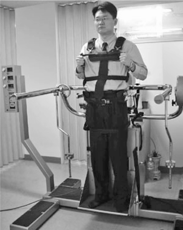
Subjects were placed in an upright neutral position with maximum resistance and mechanical locks placed on all the three axes (Fig. 1). They were then asked to perform maximal isometric contraction in the order of lateral flexion, flexion-extension, and then rotation. The same tests were repeated twice in all directions to enhance the accuracy. Due to possible problems of maladaptation to the device, we selected the higher value according to the manual of the B-200. The ratios of extension to flexion (E/F), right to left lateral flexion (R/L LF), and right to left rotation(R/L Rot) were calculated, respectively, to assess trunk muscle balances by the built-in software.
Fig. 1.

Subject secured in B-200
Measurement of lordosis and sacral angle
After radiographic films were developed, sacral angle and lumbar lordosis were measured. Most of the cases were examined by goniometer on the horizontal light box manually. The same examiner measured the angles twice and the mean value was accepted.
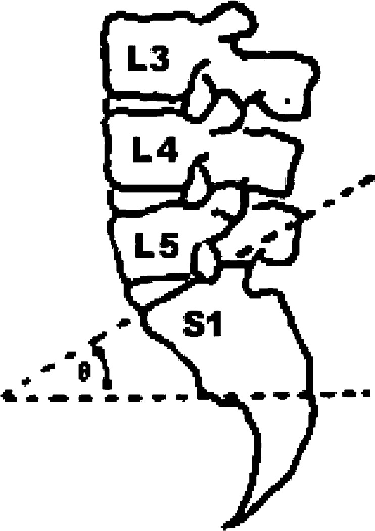
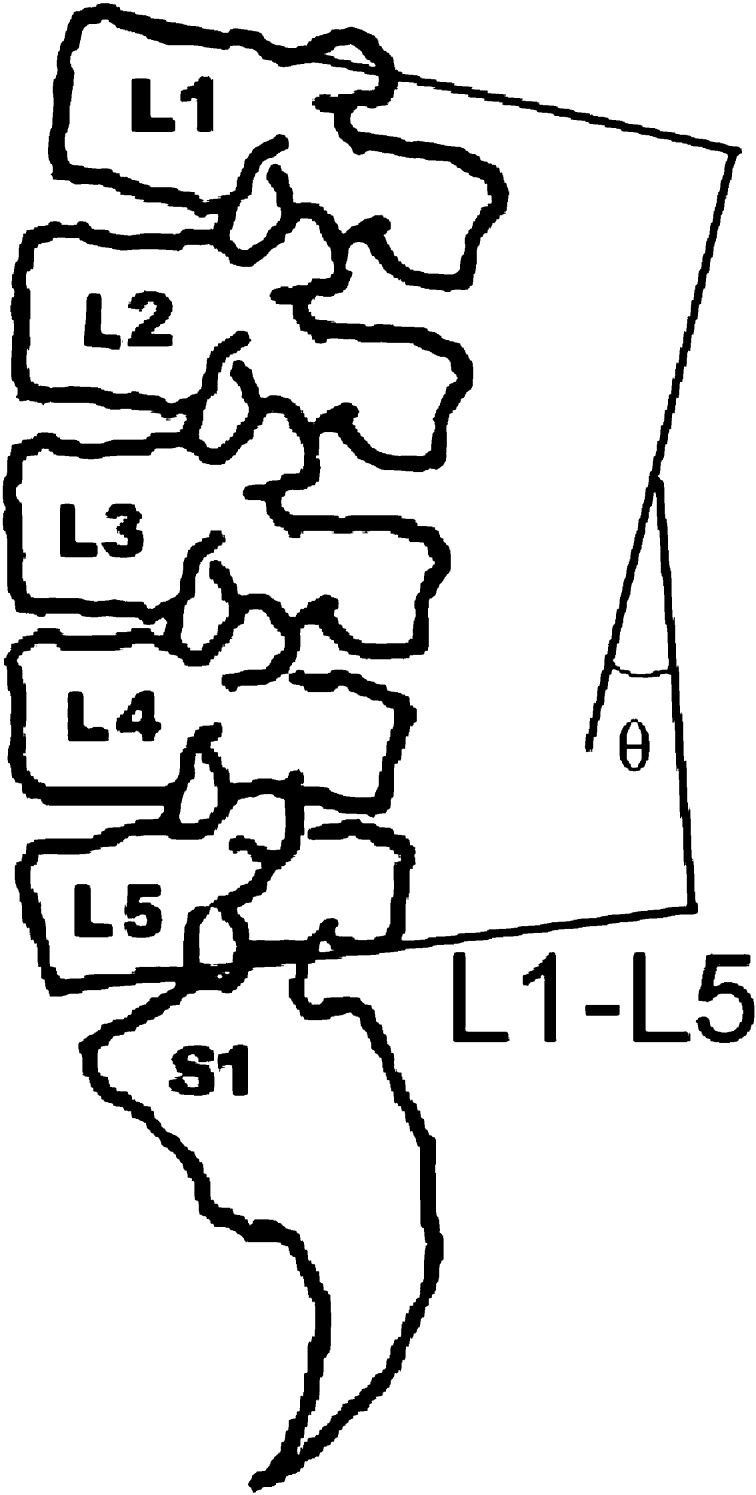
Sacral angle was defined as an angle between horizontal line parallel to bottom end of the film and superior endplate of sacrum (Fig. 2). To measure lordotic angle, a permutation of the method of Cobb was applied. Lordotic angle was obtained between inferior endplate of L5 and superior endplate of L1 in the lateral view following Polly’s method [11] (Fig. 3).
Fig. 2.
Measurement of lumbosacral angle
Fig. 3.
Measurement of lumbar lordosis
The subjects stood upright while the radiographs were taken with theirs arms folded across the chest. The X-ray beam was centered on L3. Siemens OPTITOP was used for simple radiograph and 7×17 inch films (Fuji, Japan) were used.
All of the selected films were radiologically normal and showed no significant abnormal findings in their readings by radiologists. Some of the radiographs were viewed in the network monitor and the angles were directly measured by computerized goniometer of the PACS (Picture Archiving and Communication System) software.
Statistical analysis
The Pearson’s correlation coefficients between lumbar lordosis, sacral angle and ratios of maximum torques, muscle strength were calculated by the SPSS for Windows 8.0. In the case that the coefficient is between 0.5 and 0.8, it is regarded that there is a moderate correlation with each other [7].
Results
The subjects’ characteristics were shown in Table 1. Two cases that had sacral angle less than 10° due to severe straightening were excluded. One more case that had scoliosis whose Cobb’s angle was over 15° was also excluded from the analysis. As a result, thirty-one subjects were used in the analysis.
Table 1.
Sex and age distribution of the subjects
| Male | Female | |
|---|---|---|
| No. of subjects | 11 | 20 |
| Age | 30.55±8.72 | 37.00±13.86 |
All values are mean ± SD
The isometric maximum torque values were shown by gender (Table 2). In lateral flexions and rotations, the right side was slightly bigger than the left side.
Table 2.
Isometric maximum strength
| Right rotation | Left rotation | Flexion | Extension | Right lateral flexion | Left lateral flexion | |
|---|---|---|---|---|---|---|
| Male | 50.60±16.56 | 50.10±13.60 | 79.11±26.08 | 99.56±32.46 | 101.42±31.35 | 98.87±30.51 |
| Female | 29.75±7.32 | 26.45±6.37 | 45.78±11.20 | 60.46±16.59 | 58.07±13.03 | 55.94±17.52 |
| Total | 37.15±15.11 | 34.84±14.82 | 57.61±23.85 | 74.33±29.79 | 73.45±29.66 | 71.17±30.67 |
All values are mean ± SD
The lumbar lordosis obtained was 27.19 (8.53 and lumbosacral angle was 31.77 (6.02, respectively.
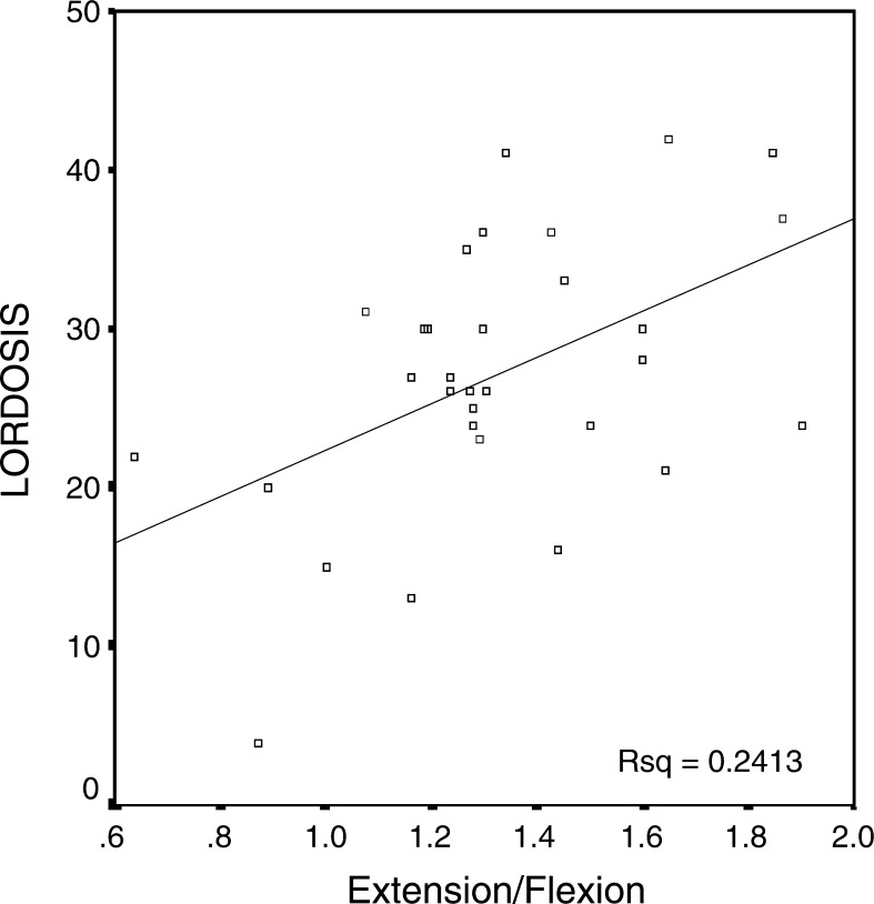
Lumbar flexor and extensor muscle strength were not significantly related with lordotic angle in both males and females. Only the ratio of extension to flexion showed a significant but very moderate relationship with lordotic angle (R=0.491, p<0.01), but not with sacral angle (Table 3). Its scatter pattern was shown in a graph (Fig. 4). The R/L ratio of rotation or lateral flexion did not show any significant correlation with sacral angle or lordotic angle. The extension maximum torque was significantly related with sacral angle only in men (p<0.01). Maximum torques except extension in men were not correlated with sacral angle.
Table 3.
Correlations between lordosis and maximum torques or their ratios
| Mean±SD (ft-lb) | Correlation with lordosis* | Correlation with lumbosacral angle* | |
|---|---|---|---|
| Flexion | |||
| M | 79.11±26.08 | 0.128 | 0.196 |
| F | 45.78±11.20 | -0.127 | -0.156 |
| Extension | |||
| M | 99.56±32.46 | 0.502 | 0.735** |
| F | 60.46±16.59 | 0.128 | -0.159 |
| E/F | 1.32±0.28 | 0.491** | 0.126 |
| R/L Rot | 1.11±0.30 | -0.278 | 0.060 |
| R/L LF | 1.06±0.19 | 0.080 | 0.015 |
*Values are Pearson’s correlation coefficients; **Statistically significant relationship (p<0.01)
All values are mean ± SD
Fig. 4.
The scatter plot of lordosis and the ratio extension of flexion
Discussion
The importance of sagittal contour and maintenance of appropriate lumbar lordosis has received increasing attention over the last few years [4, 6, 11, 13, 15, 17]. Although the effects of hyperlordosis or hypolordosis are not yet well established, loss of lumbar lordosis can have significant adverse consequences [11, 15]. Investigators have claimed that anthropometric characteristics such as increased lumbar lordosis and diminished abdominal muscle force by them can increase the risk of chronic LBP [11]. This would be very important for physicians or physical therapists. Although, there are controversies whether excessive lordosis can cause chronic LBP, many therapists apply strengthening technique of abdominal muscle to the hyperlordotic LBP patients [5].
The lumbosacral angle was calculated as the angle from the horizontal line to the superior aspect of the sacrum [8, 12]. Although there are some disputes about physiological range, this angle should measure at about 30° and determines the degree of the superincumbent lordosis of lumbar spine. In this study, both lordotic angle and sacral angle were slightly smaller compared findings from former studies [3, 8, 10, 11]. Lumbosacral joint is an important determinant to lumbar lordosis and is unstable because it is an inflexion point in spinal curvature [10]. In addition, because the sacrum is firmly attached to the pelvis, the lumbosacral angle also implies pelvic tilt [12]. In this study, lumbosacral angle and lordotic angle showed significant correlation (r=0.544).
The manner of determining of the lumbosacral angle has been quite inaccurate and subjective as merely visual observation was used mostly. To minimize this inaccuracy, radiograph was taken and the lumbosacral angle was measured twice.
The measurement of lordosis was well reported by Polly et al. [11]. Originally, the method of Cobb used in this study was proposed as a measure of coronal plane deformity not as a measure of sagittal contour. However, the reliability and reproducibility of this method are well established in both of the planes. We selected the method that measures angle from superior aspect of L1 to inferior aspect of L5 that reflects lordosis best [11].
Isometric strength was selected and evaluated by isostation B-200 to measure the function of abdominal and back muscles. Isometric testing is the most established method of assessing lumbar strength and therefore, is the most well defined methodology [3]. To avoid contaminating the result by using body and jerk to the machine during the test, subjects were asked to increase their force slowly and hold the contraction for 5 s.
Many literatures [11, 12, 16, 17] suggest that lumbar lordosis and abdominal muscle function are related to each other. The weakness of abdominal muscle permits an anterior pelvic tilt and a lordotic posture [16, 17]. On the other hand, abdominal muscle can tilt pelvis posteriorly and reduce lordosis concurrently. Former studies [4, 15, 17], however, could not prove these hypotheses with sufficient accuracy. Walker et al. [15] measured abdominal performance by leg-lowering test and lordosis by curve tracing flexible ruler. They could not find any relationship between lordosis, abdominal muscle performance and pelvic tilt. Even though the reliability of leg-lowering test is well stated by Kendall et al. [5], this test lacks precision to perform analysis. Similarly, Heino et al. [4] examined the relationship between hip extension range of motion and pelvic tilt, lordosis, abdominal muscle force. No relationship was found among these clinical variables. Youdas et al. [17] had proved that abdominal muscle force was associated with angle of pelvic inclination in women but not in men. Standing lumbar lordosis was associated with abdominal muscle length in women.
We also failed to show relativity between lordosis or lumbosacral angle and flexion or extension in both sexes except extension in men. This can be interpreted that the major extensor, erector spinae, can increase lumbosacral angle functionally. However, because the number of cases in men was not big enough (n=11) and cases in women were far from this tendency, one should be very careful to make any conclusions from these results. Further studies may be needed for more investigation.
We considered the ratio of extension to flexion, instead, as more specific indicator of imbalance of back muscles as Lee et al. [6] stated in his study. The E/F ratio may provide a method for intra-individual assessment of the trunk muscle, and it is a commonly used parameter in evaluating trunk muscle balance. Normal values for this ratio have varied study to study, but 1.3 is the most commonly cited [6]. In the present study, we obtained the mean of 1.32, which is similar to the one from former study [6].
The majority of studies have shown a right to left ratio for lateral flexion and rotation of 1.0, but some normative studies revealed asymmetries possibly due to hand dominance [3]. Here, 1.11 for rotation ratio and 1.06 for lateral flexion both superiority to the right side.
We need to mention about the controversial relationship between LBP and E/F ratio, which implies trunk muscle balance even though it was not investigated in this study. Lee et al. [6] demonstrated that patients with LBP have lower E/F ratios than does the normal population, and stated extensor strength was reduced more than flexor strength in patients with LBP. On the other hand, Tsuji et al. [14] suggested that the loss of lumbar lordosis might occur to compensate the increasing facet joint pressure in an unknown pathology. However, considerable references elucidated weakness of abdominal muscle is the key factor of non-organic LBP [2, 5, 10]. We suggest many other factors must be taken into consideration on concluding that hyper or hypolordosis is directly associated with LBP.
Conclusion
In this anthropometric study, lumbar lordosis is moderately correlated with the ratio of maximum torque of extension to flexion. This suggests that the imbalance between trunk muscles can cause excessive lordosis, possibly a major reason for chronic LBP. Other parameters such as flexion, extension, ratios of right to left lateral flexion and rotation revealed no significant correlation. On the other hand, sacral angle was only statistically correlated with extension in men.
In this cross-sectional study, we had certain limitations that the group size was rather small, the intra-examiner and inter-examiner validation of manual measurement of lordotic and sacral angles were lacked, as well as the investigation of relationship between pain stage and E/F ratio was also something to be desired. Nevertheless, we consider this study has obvious virtues that the E/F ratio was revealed to be a more useful parameter than the maximum torque or L/R ratios in explaining imbalance of trunk muscles which can develop into potential LBP.
Further studies with more subjects combined with assessment of LBP or activities of daily life will be needed.
Acknowledgments
This experiment complies with the current law of South Korea inclusive ethics approval. Supported by Research Fund from KyungHee University.
Footnotes
Supported by Reasearch Fund from KyungHee University
References
- 1.Bayramoglu M, Akman MN, Kilinc S, Cetin N, Yavuz N, Ozker R. Isokinetic measurement of trunk muscle strength in women with chronic low back pain. Am J Phys Med Rehabil. 2001;80(9):650–655. doi: 10.1097/00002060-200109000-00004. [DOI] [PubMed] [Google Scholar]
- 2.Beimborn DS, Morrissey MC. A review of the literature related to trunk muscle performance. Spine. 1988;13(6):655–660. [PubMed] [Google Scholar]
- 3.Gomez T, Beach G, Cooke C, Hrudey W, Goyert P. Normative database for trunk range of motion, strength, velocity, and endurance with the isostation B-200 lumbar dynamometer. Spine. 1991;16(1):15–21. doi: 10.1097/00007632-199101000-00003. [DOI] [PubMed] [Google Scholar]
- 4.Heino JG, Godges JJ, Carter CL. Relationship between hip extension range of motion and postural alignment. J Orthop Sports Phys Ther. 1990;12:243–247. doi: 10.2519/jospt.1990.12.6.243. [DOI] [PubMed] [Google Scholar]
- 5.Kendall FP, McCreary, Provance PG. Muscles testing and function. 4. Baltimore: Williams & Wilkins; 1993. pp. 133–134. [Google Scholar]
- 6.Lee JH, Yuichi Hoshino, Kozo Nakamura, Yusei Kariya, Kazuo Saita, Kuniomi Ito Trunk muscle weakness as a risk factor for low back pain. Spine. 1999;24(1):54–57. doi: 10.1097/00007632-199901010-00013. [DOI] [PubMed] [Google Scholar]
- 7.Morton RF, Hebel JR. A study guide to Epidemiology and Biostastics. 3. Gaithersburg: Aspen Publication; 1989. p. 94. [Google Scholar]
- 8.Na YM, Kang SW, Bae HS, Kang MJ, Park JS, Moon JH. The analysis of spinal curvature in low back pain patients. J Korean Acad Rehab Med. 1996;20(3):669–674. [Google Scholar]
- 9.Newton M, Waddell G. Trunk strength testing with iso-machines Part 1. Spine. 1993;18(7):801–811. doi: 10.1097/00007632-199306000-00001. [DOI] [PubMed] [Google Scholar]
- 10.Noh YH, Keum DH. A clinical observation for the lumbosacral angle changes between initial and follow up treatment in low back pain patients. J Oriental Rehab Med. 2000;10(1):11–21. [Google Scholar]
- 11.Polly DW, Kilkelly FX, McHale KA, Asplund LM. Measurement of lumbar lordosis. Spine. 1996;21(13):1530–1536. doi: 10.1097/00007632-199607010-00008. [DOI] [PubMed] [Google Scholar]
- 12.Rene Cailliet (1995) Low back pain syndrome, 5th edn. F. A. Davis Company, Philadelphia, pp23–25, 100–102
- 13.Stuart M. The influence of lordosis on axial trunk torque and trunk muscle myoelectric activity. Spine. 1992;17(10):1187–1193. doi: 10.1097/00007632-199210000-00010. [DOI] [PubMed] [Google Scholar]
- 14.Tsuji T, Matsuyama Y, Sato K, Hasegawa Y, Yimin Y, Iwata H. Epidemiology of low back pain in the elderly: correlation with lumbar lordosis. J Orthop Sci. 2001;6(4):307–311. doi: 10.1007/s007760100023. [DOI] [PubMed] [Google Scholar]
- 15.Walker ML, Rothstein Finucane JM. SD, Lamb RL. Relationship between lumbar lordosis, pelvic tilt, and abdominal muscle performance. Phys Ther. 1987;4:512–516. doi: 10.1093/ptj/67.4.512. [DOI] [PubMed] [Google Scholar]
- 16.Youdas JW, Garrett TR, Egan KS, Therneau TM. Lumbar lordosis and pelvic inclination in adults with chronic low back pain. Phys Ther. 2000;80(3):261–275. [PubMed] [Google Scholar]
- 17.Youdas JW, Garrett TR, Harmsen S. Lumbar lordosis and pelvic inclination of asymptomatic adults. Phys Ther. 1996;76:1066–1081. doi: 10.1093/ptj/76.10.1066. [DOI] [PubMed] [Google Scholar]


