Abstract
The use of concentrated platelet-rich plasma (cPRP) as a source of growth factors is reported to be beneficial for an enhanced osteogenesis in spine surgery. Today both bovine and autologous thrombin is used for activating the platelets and thus releasing the growth factors. In order to prevent transmission of organisms and the development of antibodies, autologous thrombin seems to be the logical choice. However, the preparation of autologous thrombin is cumbersome and consumes a part of the cPRP. In order to overcome this problem, a commercial autologous thrombin kit (activAT, Dideco, a Sorin Group company, Italy) has been developed which produces autologous thrombin out of platelet-poor plasma. A possible disadvantage of this kit could be the rest fraction of 1.18% of ethanol in the platelet gel. In a pig model, the influence of different ethanol concentrations on the ischiadic nerve was studied. The study consisted out of four groups; a control group (n=6), a group with platelet gel 0% ethanol (n=6), a group with platelet gel 1.18% ethanol (n=6) and a group with platelet gel 3.8% ethanol (n=7). In all the groups, the ischiadic nerve was dissected and the myelin sheet opened after which the wound was closed (control group) or one of the three therapies was applied. After 12 h, the animal was sacrificed and the ischiadic nerve was submitted for histological examination. Myelin sheaths appeared normal in all cases. No axonal swelling was observed. No statistically significant differences were observed for neutrophilic and eosinophilic infiltration nor for collagen necrosis between groups. Platelet gel prepared by the use of a commercial autologous thrombin kit and containing 1.18% of ethanol can be safely used on nerves.
Keywords: Platelet gel, Autologous thrombin, Ethanol, Nerves, Spine
Introduction
Concentrated platelet-rich plasma (cPRP) activated with thrombin is used for enhancing osteogenesis in orthopaedic surgery [6]. The enhanced osteogenesis is explained by the acute and slow release of growth factors by activated platelets [7, 8]. Either autologous or bovine thrombin can be used for activating the platelets. Using bovine thrombin gives a consistent thrombin activity but may lead to the development of antibodies to bovine thrombin [11, 14]. The choice for autologous thrombin seems very attractive since there will be no exposure to bovine proteins. Unfortunately, the preparation of autologous thrombin is cumbersome and it is difficult to anticipate the obtained thrombin activity.
Recently, a commercial autologous thrombin kit has become available. The kit produces thrombin, with a consistent thrombin activity, out of platelet poor plasma (PPP). However, the production process requires the use of ethanol. The ethanol content in the thrombin serum is 13 volume percent. This content will be further diluted by mixing 1 mL of the thrombin serum with 10 mL cPRP to approximately 1.18 volume percent.
Since platelet gel has been described as beneficial for spinal fusion where also nerves will be exposed to the platelet gel, this study wants to evaluate the influence of platelet gel containing 1.18 % ethanol on nerve tissue.
Materials and methods
After approval of the Ethical Committee for animal experiments (ECP 04/20), fourteen pigs (Line 36, Rattlerow Seghers, Buggenhout, Belgium) with an average weight of 26.5±1.7 kg were used for the study. Since in each pig both ischiadic nerves were used, a total of 28 experiments were performed. All ischiadic nerves (n=28) were randomised in four groups. Each group consisted of 7 experiments. All animals received care in accordance with institutional guidelines and national laws [1].
Description of the activAT device
The activAT device (Dideco, a Sorin Group Company, Mirandola, Italy) produces autologous thrombin with a minimum thrombin activity of 50 IU/mL. The thrombin is produced by bringing platelet poor plasma into a syringe filled with negatively charged glass beads, thus offering the negative surface charge required to initiate the formation of thrombin from prothrombin. A thrombin reagent (containing 2.4 mL ethanol and 0.0219 g CaCl2 dihydrate) is added to the chamber and isolates prothrombin from plasma and, in the presence of calcium, converts prothrombin to active thrombin.
Preparation of the cPRP
After induction, the vena jugularis interna was dissected and approximately 450 mL of blood was withdrawn in a blood bag containing 63 mL of CPD anticoagulant solution (Baxter, Brussels, Belgium). Subsequently, a Suprema autotransfusion cell separator (Dideco, a Sorin Group Company, Mirandola, Italy) separated the blood by centrifugation in concentrated red blood cells (RBC), platelet poor plasma (PPP) and concentrated platelet rich plasma (cPRP). Cell counts were performed on the different blood products and the processed volumes were recorded. The PPP and RBC were immediately retransfused to the animal, while the cPRP was stored at room temperature. In groups 3 and 4, 12 mL of PPP was withdrawn before retransfusion for preparing thrombin.
Surgery
In each animal, both ischiadic nerves were used and each nerve was randomised. First the right side was instrumented and after closure of the wound the left side was prepared. After dissection of the ischiadic nerve, the myelin shaft was opened and platelet gel applied on the nerve. Twelve hours after application, the ischiadic nerve was excised and submitted for histological examination, after which the animal was sacrificed.
Description of groups and preparation of autologous thrombin
Group 1 served as a control group and after opening the myelin sheet no therapy was applied to the nerve.
In group 2, cPRP was used in combination with autologous thrombin. Thrombin was prepared by mixing 10 mL cPRP with 0.33 mL of 10% CaCl2 in a glass syringe. After the mixture became solid, the supernatant, rich in thrombin, was aspirated. The platelet gel was made by mixing 10 mL cPRP with 2.5 mL thrombin. The gel was applied on the ischiadic nerve.
In group 3, cPRP was used in combination with autologous thrombin prepared with the activAT device. Twelve millilitre of PPP was aspirated followed by 0.5 mL of air, after which the thrombin reagent was added. After thorough mixing, the mixture was injected in the process syringe containing the gel beads. After 60 min the thrombin was pushed out of the process syringe into a small syringe. Ten millilitre of cPRP was then mixed with 1 mL of the thrombin. Since previous investigations demonstrated rather low thrombin activity in pig blood, 0.33 mL of 10% CaCl2 was added, when no gelling took place after 5 min.
In group 4, cPRP was used in combination with autologous thrombin prepared with the activAT device. Additional ethanol was added in order to obtain a final ethanol content of 3.8% instead of 1.3% in the platelet gel. To obtain this value, 2 mL of thrombin instead of 1 mL was added to the 10 mL cPRP and after mixing, 0.2 mL of 100% ethanol (VWR International, Leuven, Belgium) was added. If no gelling occurred after 5 min, 0.33 mL of 10% CaCl2 was added.
Histopathological examination
The samples for histopathological examination were immediately fixed in phosphate buffered formalin. After fixation for 48 h, the samples were dehydrated and paraffin wax embedded. Four micrometer thick sections were cut and stained with haematoxylin and eosin. Ten micrometer sections were cut and stained with Luxol fast blue for visualisation of the myelin sheaths. Five sections of each nerve were examined.
The histological scoring system, used for the evaluation, was arbitrarily chosen by one senior pathologist on the basis of the range of lesions observed in all of the sections. Subsequently, another senior pathologist reading the sections blindly applied the scoring system. This histological scoring system had not been used previously.
Statistics
A Kruskal–Wallis One Way Analysis on Ranks (Sigmastat, SPSS Inc, Erkrath, Germany) was used to analyse the difference between the four groups. A Dunnett test was performed to compare the control group versus the 3 treatment groups. The difference was considered significant when P value was ≤0.05.
Results
Out of the 28 experiments, three were not used in the final analysis. In those three experiments the ethanol content in the thrombin syringe was lower than the desired 3.8% due to a dilution error.
The characteristics of the withdrawn blood and processed blood components are presented in Table 1.
Table 1.
Characteristics of withdrawn and processed blood products
| Average±SD | |
|---|---|
| Blood withdrawn [mL] | 471±16 |
| Blood processed with Suprema machine [mL] | 360±25 |
| Initial hematocrit [%] | 25.4±1.8 |
| Initial platelet count [x 1000/mm3] | 287±91 |
| Hematocrit PRP [%] | 8.5±3.6 |
| Hematocrit PPP [%] | 0.1±0.0 |
| Hematocrit RBC [%] | 68.8±2.4 |
| Platelet count PRP [×1000/mm3] | 1610±728 |
| Platelet count PPP [×1000/mm3] | 53±41 |
| Platelet count RBC [×1000/mm3] | 66±29 |
| Increase platelet count above baseline [×baseline] | 5.6±1.5 |
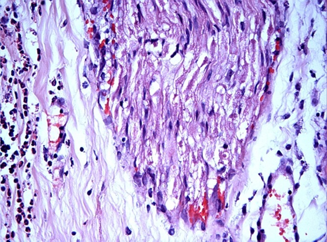
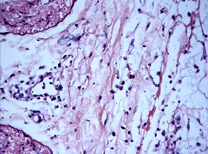
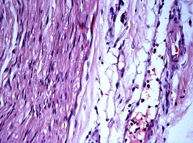
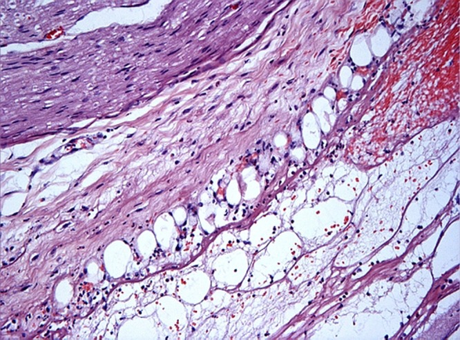
In all the groups, there were some hemorrhages in the surrounding adipose tissue and in the epineurium on one side of the nerve. Infiltrating neutrophilic granulocytes and occasional eosinophilic granulocytes, lymphocytes and macrophages were observed in all groups. The scores for the infiltrating neutrophilic granulocytes and eosinophilic granulocytes are summarised in Table 2. In some sections, small foci of collagen necrosis were observed in the epineurium, in association with larger neutrophilic infiltrates. Scoring of collagen necrosis is summarised in Table 3. Myelin sheaths appeared normal in all cases. No axonal swelling was observed. Some representative histopathological findings are shown in Figs. 1–4.
Fig. 2.

Control group. Control sciatic nerve. Nerve fascicle (centre). Note the infiltrating neutrophylic granulocytes between the dense collagen bundles of the epineurium (left). H&E staining. Magification 400×
Fig. 3.

AKTIVAT (group 3). AKTIVAT treated sciatic nerve. Two nerve fascicles (left). Scattered neutrophylic granulocytes are present between the dense collagen bundles of the epineurium (middle and right). H&E staining. Magnification 400×
Table 2.
Neurophilic and eosinophylic granulocyte infiltration
| Neutrophilic granulocytes infiltration | |||||||
|---|---|---|---|---|---|---|---|
| Experiment | 1 | 2 | 3 | 4 | 5 | 6 | 7 |
| Group 1 | 1 | 2 | 1 | 1 | 2 | 1 | |
| Group 2 | 2 | 1 | 2 | 1 | 2 | 1 | |
| Group 3 | 2 | 2 | 2 | 1 | 2 | 2 | |
| Group 4 | 2 | 2 | 2 | 2 | 2 | 1 | 2 |
| Eosinophilic granulocyte infiltration | |||||||
| Experiment | 1 | 2 | 3 | 4 | 5 | 6 | 7 |
| Group 1 | 0 | 0 | 0 | 1 | 1 | 0 | |
| Group 2 | 1 | 0 | 0 | 0 | 1 | 0 | |
| Group 3 | 1 | 0 | 1 | 1 | 1 | 1 | |
| Group 4 | 1 | 1 | 1 | 1 | 1 | 0 | 1 |
1: Few cells scattered in the epineurium
2: Many cells in the epineurium, extending between the nerve fascicles, occasionally forming aggregates
3: Numerous cells forming aggregates between the nerve fascicles
4: Cells diffusely present in the epineurium and passing through the perineurium
5: Many cells infiltrating the endoneurium
Table 3.
Collagen necrosis
| Collagen necrosis score | |||||||
|---|---|---|---|---|---|---|---|
| Experiment | 1 | 2 | 3 | 4 | 5 | 6 | 7 |
| Group 1 | 1 | 0 | 0 | 0 | 0 | 0 | |
| Group 2 | 0 | 0 | 0 | 0 | 0 | 0 | |
| Group 3 | 1 | 1 | 0 | 0 | 0 | 0 | |
| Group 4 | 1 | 1 | 1 | 0 | 0 | 0 | 0 |
1: small solitary focus of collagen necrosis in epineurium
2: large solitary or multiple small foci of collagen necrosis in epineurium
3: coalescing foci of collagen necrosis in epineurium between nerve fascicles
4: collagen necrosis involving large portions of epineurium and extending to perineurium
Fig. 1.

Control group. Control sciatic nerve. Nerve fascicle (left) and perineurium (right). Note the presence of some neutrophylic granulocytes in the interstitium near a postcapillary venule and adhesion of neutrophylic granulocytes to the endothelium of the venule. H&E staining. Magnification 400×
Fig. 4.

3.8% ethanol group (group 4). 3.80% treated sciatic nerve. Nerve fascicle (upper left corner). Fibrin and haemorrhage in epineurium and surrounding adipose tissue. Scattered neutrophylic granulocytes are present in and around the haemorrhage. H&E staining. Magnification 200×
No statistically significant differences were observed for neutrophilic and eosinophilic infiltration nor for collagen necrosis between groups.
Discussion
Platelet gel as a source for autologous growth factors [3, 5, 10] release, is considered as a tool for enhancement of bone generation. However, in order to release these growth factors, platelets need to be activated with thrombin. Most centres in the USA use bovine thrombin instead of autologous thrombin. By using bovine thrombin, one has a very consistent thrombin activity but also runs the risk for antibodies generation against the bovine thrombin [11, 14]. This can lead to serious adverse reactions when the patient is exposed for a second time to bovine thrombin. Unfortunately, it is difficult to exclude this possibility since most commercial fibrin glues contain bovine thrombin and the majority of patients do not know if they were previously exposed. The majority of European centres prefer to use autologous thrombin. However, independent of the technique used for the preparation of the thrombin, the final thrombin activity is difficult to predict. This is an important issue because if the thrombin activity is too low, one could still be forced to use bovine thrombin to obtain a consistent cPRP gel. Only scarce information is available in the literature on what should be the minimum thrombin activity to obtain a good cPRP gel. Yoshida et al. [13] stated that the optimal thrombin activity necessary for obtaining a good adhesive strength of cryofibrinogen prepared from a patients own autologous plasma to be used as fibrin glue is 50 IU/mL. The thrombin activity claimed by the manufacturer is 50 IU/mL, which should be sufficient based on the available information. The reason why we were not able to achieve a good cPRP gel after mixing the activAT thrombin with 10 mL of cPRP, can be explained by the tendency to hypercoagulability in pigs [4] as shown by lower plasminogen and plasmin levels and a shortened PTT [2, 4]. This finding is combined with a prolonged thrombin time due to enhanced formation of fibrin degradation products (anti-thrombin VI).
Another disadvantage of today’s techniques for preparing autologous thrombin is that the traditional technique uses a portion of the cPRP and thus reduces the amount available for production of the cPRP gel. In order to overcome these problems, a commercial thrombin kit is now available, which claims a minimum thrombin activity and uses PPP for the production of the autologous thrombin. The activAT autologous thrombin kit (Dideco, a Sorin Group company, Mirandola, Italy), tested in our study, has an ethanol content of 13 volume percent. After mixing with 10 mL cPRP, the final ethanol content in the cPRP gel would be 1.18%. No information is currently available in the literature on the effects of this ethanol concentration on the nerve. Topical exposure of absolute alcohol for 30 s on the sciatic nerve, shows 4 h after the exposure splitting of the myelin throughout the entire sheath, with swollen edematous axons and fragmented and widely separated neurofilaments; followed by the classical pattern described for Wallerian degeneration [12]. Exposure of diluted ethanol (10–50%) on the sciatic nerve showed two distinct populations of myelinated fibres. A central core of fibres appeared to be normal. These were sharply demarcated at about half the radius of the nerve from a rim of altered fibres. In this outer zone, the fibres showed myelin sheaths that were more lightly stained and about twice as thick as the sheaths of the more centrally placed normal fibres. This outer zone became smaller with lower ethanol concentrations [9]. Since the damage to the nerve is almost immediate, our evaluation period of 12 h seems to be a good observation window. In our study, we notice none of the histological changes described above. Our results reveal no structural damage at the myelin sheets or axons caused by using 1.18% ethanol concentration or a three times higher concentration. Although we used a pig model for our study we believe that the results can be extrapolated to humans since the histology of the sciatic nerve in the pig is in no way different from the histology of the sciatic nerve in other animal species.
Based on our observations, we conclude that it is now possible to produce a reproducible and consistent cPRP gel by using patient derived blood products only. The major advantage of this technique is the avoidance of allergic reactions and the transmission of infectious diseases without jeopardising the quality of the final cPRP gel.
Conclusion
cPRP gel with an ethanol content up to 3.8 volume percent can be safely used on nerve tissue and thus for spinal fusion operations.
References
- 1.Guide for the Care and Use of Laboratory Animals (1996) National Academy of Sciences. ISBN: 0-309-05377-3
- 2.Höhle P (2000) Zur Übertragbarkeit tierexperimenteller endovaskulärer Studien: Unterschiede der Gerinnungs- und Fibrinolyse-Systeme bei häufig verwendeten Tierspezies im Vergleich zum Menschen. Ph.D. thesis Rheinisch-Westfälischen Technischen Hochschule Aachen
- 3.Kevy S, Jacobson M. Comparison of methods for point of care preparation of autologous platelet gel. JECT. 2004;36:28–35. [PubMed] [Google Scholar]
- 4.Kostering H, Mast W, Kaethner T, Nebendahl K, Holtz W. Blood coagulation studies in domestic pigs (Hanover breed) and minipigs (Goettingen breed) Lab Anim. 1983;17(4):346–349. doi: 10.1258/002367783781062262. [DOI] [PubMed] [Google Scholar]
- 5.Landesberg R, Roy M, Glickman R. Quantification of growth factor levels using a simplified method of platelet rich plasma gel preparation. J Oral Maxillofac Surg. 2000;58:297–300. doi: 10.1016/S0278-2391(00)90058-2. [DOI] [PubMed] [Google Scholar]
- 6.Lowery G, Kulkarni S, Pennisi A. Use of autologous growth factors in lumbar spinal fusion. Bone. 1999;25:47S–50S. doi: 10.1016/S8756-3282(99)00132-5. [DOI] [PubMed] [Google Scholar]
- 7.Marx R, Carlson E, Eichstaedt R, Schimmele S, Strauss J, Georgeff K. Platelet rich plasma Growth factor enhancement for bone grafts. Oral Surg Oral Med Oral Pathol Oral Radiol Endod. 1998;85:638–646. doi: 10.1016/S1079-2104(98)90029-4. [DOI] [PubMed] [Google Scholar]
- 8.Slater M, Patava J, Kingham K, Mason R. Involvement of platelets in stimulating osteogenic activity. J Orthop Res. 1995;13:655–663. doi: 10.1002/jor.1100130504. [DOI] [PubMed] [Google Scholar]
- 9.Taylor J, Woolsey R. Dilute ethyl alcohol effects on the sciatic nerve of the mouse. Arch Phys Med Rehabil. 1976;57(5):233–237. [PubMed] [Google Scholar]
- 10.Weibrich G, Buch R, Kleis W, Hafner G, Hitzler W, Wagner W. Quantification of thrombocyte growth factors in platelet concentrates produced by discontinuous cell separation. Growth Factors. 2002;20(2):93–97. doi: 10.1080/08977190290031950. [DOI] [PubMed] [Google Scholar]
- 11.Winterbottom Antigenic responses to bovine thrombin exposure during. 2001;surgery:a. [Google Scholar]
- 12.Woolsey R, Taylor J, Nagel J. Acute effects of topical ethyl alcohol on the sciatic nerve of the mouse. Arch Phys Med Rehabil. 1972;53:410–414. [PubMed] [Google Scholar]
- 13.Yoshida H, Hirozane K, Kamiya A. Adhesive strength of autologous fibrin glue. Biol Pharm Bull. 2000;23(3):313–317. doi: 10.1248/bpb.23.313. [DOI] [PubMed] [Google Scholar]
- 14.Zehnder J, Leung L. Development of antibodies to thrombin and factor V with recurrent bleeding in a patient exposed to topical bovine thrombin. Blood. 1990;76:2011–2016. [PubMed] [Google Scholar]


