Abstract
Recently, the presence of a high-intensity zone (HIZ) within the posterior annulus seen on T2-weighted MRI has aroused great interest and even controversy among many investigators, particularly on whether the HIZ was closely associated with a concordant pain response on awake discography. The study attempted to interpret the correlation between the presence of the HIZ on MRI and awake discography, as well as its characteristic pathology. Fifty two patients with low back pain without disc herniation underwent MRI and discography successively. Each disc with HIZ was correlated for an association between the presence of a HIZ and the grading of annular disruption and a concordant pain response. Eleven specimens of lumbar intervertebral discs which contain HIZ in the posterior annulus from 11 patients with discogenic low back pain were harvested for histologic examination to interpret the histologic basis of a nociceptive response during posterior lumbar interbody fusion (PLIF). The study found that in all of 142 discograms in 52 patients, 17 presented HIZ. All 17 discs with HIZ showed painful reproduction and abnormal morphology with annular tears extending either well into or through the outer third of the annulus fibrosus. The consecutive sagittal slices through the HIZ lesion showed that a notable histologic feature of the formation of vascularized granulation tissue in the outer region of the annulus fibrosus. The current study suggests that the HIZ of the lumbar disc on MRI in the patient with low back pain could be considered as a reliable marker of painful outer anular disruption.
Keywords: Discogenic low back pain, High-intensity zone, Lumbar intervertebral disc, Discography
Introduction
Recently, there has been interest in the presence of an area of high intensity zone (HIZ) within the posterior annulus seen on T2-weighted, sagittally oriented MRI sequence [1]. Some investigators [8, 16] believed that the presence of a HIZ was closely associated with a concordant pain response on awake discography, but others[2, 9, 11] disagreed. The pathology of HIZ has not been described clearly up to now. Yu et al. [19] thought that it represents the collections of mucoid fluid within the fissure of the annulus. Lam et al. [9] considered it as a reflection of the edge neovasculization of posterior annulus or a healing annular tear. Whether the presence of a HIZ is a reliable marker of painful disc, and whether a defined nociception histology in HIZ lesion can be identified has not been clarified.
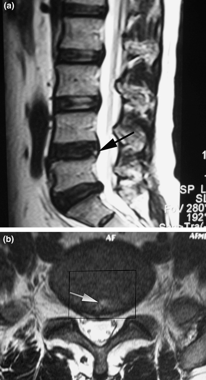
According to the original study by Aprill and Bogduk[1], the HIZ was localized in midline within the posterior annulus (Fig. 1). This makes it easy for us to obtain the disc specimen that contains HIZ for histologic study during posterior lumbar interbody fusion for the patient with discogenic low back pain. Our goal is to investigate the histology of HIZ and its clinical significance.
Fig. 1.

a Sagittal T2-weighted magnetic resonance image (MRI) showing a high-intensity zone (arrow) within the posterior annulus at L4-L5. b Axial T2-weighted magnetic resonance image (MRI) shows a high-intensity zone (arrow) within the posterior annulus at L4-L5. The rectangle indicates range of disc excision (PLIF procedure) that is used for histological examination.
Materials and Methods
From April 2000 to August 2003, fifty two consecutive patients with low back pain without radiculopathy underwent MRI and awake discography (age range, 18–62 years; mean age, 38.8 years; 39 men, 13 women). All the patients had already undergone normal physical therapy and nonsteroidal anti-inflammatory medication for more than 6 months without relief of their symptoms. MRI was taken using Siemens 1.5 T Magnetom (Siemens Corp., New York, NY). Spin echo T1-weighted (repetition time[TR]< 800) proton density (1,000< TR< 2,000; echo time [TE] < 30) and T2-weighted (TR=2000; 50<TE<100) sagittal and axial 4-mm thick MR images were generated. Subsequently, each patient underwent awake discography. The original plan was to conduct the discography on each patient for the discs L3-L4, L4-L5, and L5-S1. In practice, it was not possible to test all the patients at each of these discs. Some patients were also tested for L2-L3 discs when the discography was negative at the three lower levels. A total of 142 discograms were obtained. A positive concordant pain response was defined if the patient had reproduction of his or her usual pain response pattern on injection of the contrast. A negative discogram would be considered if the pain produced did not correspond to the usual symptoms and referred pain patterns. CT was performed after each discogram. The Dallas Discogram Scale for annular disruption was used to classify each CT discogram to identify the depth of posterior annular disruption [14].
According to the Aprill and Bogduk standard, there were 17 HIZ discs in 17 of the 52 patients. Out of them, 11 were localized in L4-L5 discs and 6 in L5-S1 discs. Using Nonparametric correlation test, each HIZ disc was correlated for an association between the presence of an HIZ and the grade of annular disruption and a concordant pain response.
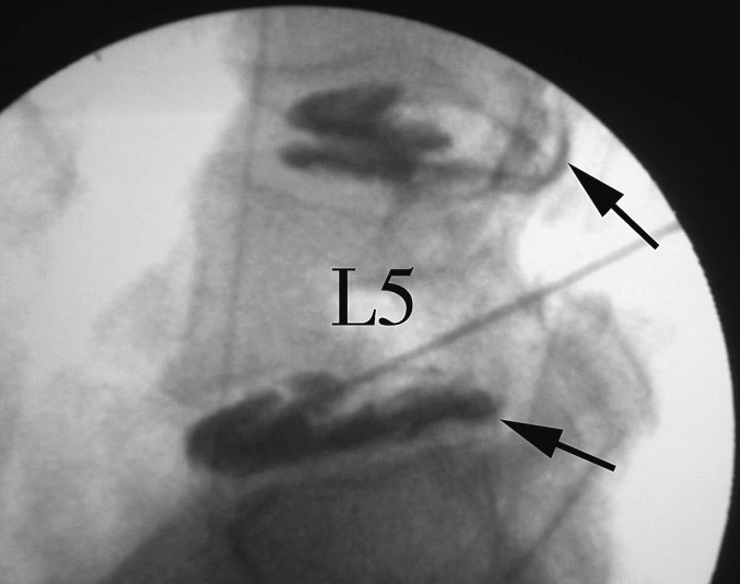
Of the 17 patients with HIZ discs, 11 patients underwent posterior interbody fusion. Eleven biopsy specimens of the posterior midline of L4 to S1 intervertebral discs that contained HIZs on MRI sequences were excised en bloc during posterior interbody fusion. All the 11 painful discs with HIZs were proved by preoperative discography (Fig. 2). To assess the pathological features precisely, all the specimens of discs should contain the nucleus pulposus, the annulus fibrosus, and the posterior longitudinal ligament in continuity.
Fig. 2.

Lateral discography film (same patient as Fig. 1) taken during injection showed anular tears (arrow) at L4-L5 and L5-S1. Discography produced concordant pain at two discs.
The specimens were fixed in 10% neutral formalin, embedded in paraffin, slit sagittally and sliced into serial sections 5 μm thick. Serial sections were stained with haematoxylin and eosin.
Results
All the 17 HIZ discs presented grade 2 or 3 annular disruption and the concordant pain response (Table 1), as seen on the discograms. The HIZ was not found in any histologically normal disc. In all the 142 discograms, we noticed a significant correlation between the presence of a HIZ and the grading of annular disruption (Table 1; R=0.464, P=0.0001).
Table 1.
Correlation between presence of a HIZ and grade of anular disruption
| Grading | With HIZ | Without HIZ | Total |
|---|---|---|---|
| 0 | 0 | 36 | 36 |
| 1 | 0 | 57 | 57 |
| 2 | 2 | 8 | 10 |
| 3 | 15 | 24 | 39 |
| Total | 17 | 125 | 142 |
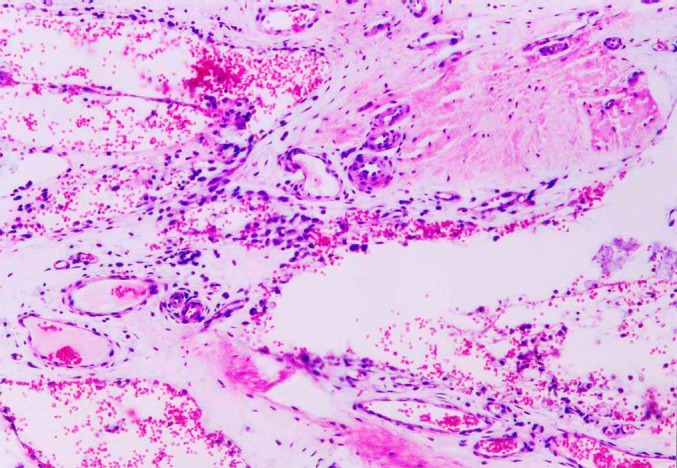
The histologic study of the consecutive sagittal slices of the HIZ lesions showed that the normal lamellar structure was replaced by a disorganized, vascularized granulation tissue that consisted of small round cells, fibroblasts, and newly formed blood vessels around the tears extended from the nucleus pulposus to the outer region of the annulus fibrosus (Fig. 3). Blood proliferation and inflammatory cell infiltration were seen extending along the margins of the tear into the middle and inner annulus, and sometimes the tear was bridged with granulation tissue. Ingrowth of vascularized tissue was sometimes observed to end abruptly at the junction between the inner third of the annulus and the nucleus pulposus. The lamellar structures of annulus fibrosus contiguous to the HIZ sites were arrayed relatively regularly, then became disrupted and confused. The structures of annulus fibrosus distant from the HIZ sites were relatively normal. The distinction between the nucleus pulposus and the inner annulus fibrosus was lost. In the nucleus pulposus, the fibrosis and the cell density of the matrix were markedly increased, and the round chondrocytes were transformed into oval fibroblasts.
Fig. 3.

The sagittal slice in the HIZ lesion showed the ingrowth of the vascularized granulation tissue. (HE×200)
Discussion
The high signal contained within the annulus of a disc, separated from the nucleus pulposus, on lumbar spine MRI was termed as the HIZ by Aprill and Bogduk [1] in 1992. They correlated the finding with computed tomography discography and found that it had a positive predictive value of 86% for a severely disrupted, painful disc on awake discography. This appeared to be a significant breakthrough in the diagnosis of chronic low back pain. Evidence has since been reported which supported [8, 16] or questioned [2, 9, 11] the original postulate of Aprill and Bogduk. Schellhas et al. [16] confirmed the association with pain on discography. They found that 87 of 100 discs with HIZs proved concordantly painful on discography. All the 87 painful discs exhibited abnormal morphology with annular tears extending either well into or through the outer third of the annulus fibrosus. Sixty-five out of 67 discs without HIZs were associated with nonconcordant pain or no pain. Lam et al. [9] found that there was a significant correlation between HIZ and an exact or similar pain reproduction. They believed that the HIZ in patients with low back pain is likely to represent painful internal disc disruption. Saifuddin et al. [15] found that the clinical significance of the HIZ was limited as its sensitivity was as low as 26.7%. Ricketson et al. [12] found no correlation between the presence of an HIZ and discographically identified annular tears. Our current study indicated that in patients with low back pain, the HIZ seems to be a reliable marker of painful outer annular disruption.
The incidence of HIZs in the asymptomatic population is not clear. Bogduk [2] later pointed out that low intensity zone indicating annular fissures might occur in asymptometic persons; however, these fissures may become painful and assume a higher intensity signal when “activated”. Therefore, the HIZ indicates a clinically painful disc. Carragee et al. [4] observed the prevalence of HIZ in the asymptomatic population. They proposed that the presence of an HIZ does not reliably indicate the presence of a symptomatic internal disc disruption because the prevalence of HIZ in asymptomatic individiduals with degenerative disc disease is too high (25%).
The HIZ will not be regarded as a reliable marker of painful annular disruption in patients with low back pain only until the nociceptive histology of HIZ is clarified. Yu et al. [19] studied the correlation of internal architectural components of lumbar intervertebral discs with MRI findings and cryomicyotomy using fresh cadaveric spines. They correlated the MRI finding to the pathologic classification of annular tears. They classified annular tears into three types: concentric, transverse, and radial. The concentric tear was a crescentic or oval cavity associated with rupture of the short transverse fibers connecting the lamellae in the annulus fibrosus. Transverse tears were a rupture of Sharpey’s fibers near their attachments to the ring apophysis. The radial tear was a fissure extending from the nucleus to the outermost surface of the annulus. Authors reported that fluid or mucoid material filled in each annular tear because the tear had become inflamed, and this inflammation accounts for the bright signal. Ross et al. [13] reported that visualization of radial tears could be enhanced with injection of gadolinium-DPTA, indicating granulation tissue or neovascularization induced by inflammation. The current histological study indicated that the HIZ in the patients with low back pain represented the ingrowth of the vascularized granulation tissue into the tears in the posterior part of the painful disc. In a regular MRI scanning set-up, the sagittal sections are 3–4 mm in thickness. Therefore, the HIZ may not show on T2-weighted MRI sequences, if the size of the granulation tissue is too small or contains too much of mature granulation tissue with little vascularity.
We believe that the annular tear could possibly originate from trauma. Structurally the weak posterior part of annulus fibrosus may be subjected to damage [17], which is consistent with the torn lesions shown on discography. The mechanism of the ingrowth of the granulation tissue into the annular tear is not clear. We postulate that the ingrowth of the vascularized granulation tissue seems to be a healing reaction to the annular injury. Unfortunately, the poor healing seems inevitable in such a circumstance of poor blood supply and high tensile strain. Moreover, the disc tissue is supposed to be antigenic and may induce the immunoreaction and the recruitment of inflammatory cells that further impede healing. The inflammatory granulation tissue can certainly produce some proinflammatory cytokine and mediators such as prostaglandin E2, IL-6, and IL-8 [3] that can sensitize the nociceptors within the painful discs [5, 6, 7, 10]. The discogenic low back pain might occur when the intradiscal pressure increases with the trunk motion such as flexion or extension. Additionally, the mechanism of painful discography could be the same, as the pain reproduction might also occur when the intradiscal pressure sharply increases as the contrast media is injected into the disc.
Although we had clearly described the nociceptive histology of the HIZ in painful discs, we still do not know the true significance of the HIZ because of the absence of a control of asymptomatic individuals. Certainly, it is too difficult to obtain the specimens of the HIZ from asymptomatic individuals due to ethical consideration. Yasuma et al. [18] demonstrated vascular invasion as a sign of aging of the intervertebral disc. In their cadaver study, they noticed capillary invasion in patients over 40 years old. Based on their study, we considered that vascular invasion into the posterior annulus could be the reason that the aging or degenerative lumbar discs of some adults without chronic low back pain presented the HIZs. It differs from the HIZ in painful disc. According to our study, we believe that the HIZ in the lumbar disc in the patient with low back pain could be considered as a reliable marker of painful outer annular disruption.
Footnotes
Supported by a grant for scientific research of great project of clinical new technique from 304th Hospital (to Dr. Peng)
References
- 1.Aprill C, Bogduk N. High-intensity zone: a diagnostic sign of painful lumbar disc on magnetic resonance imaging. Br J Radiol. 1992;65:361–369. doi: 10.1259/0007-1285-65-773-361. [DOI] [PubMed] [Google Scholar]
- 2.Bogduk N. Point of view. Spine. 1998;23:1259–1260. doi: 10.1097/00007632-199806010-00017. [DOI] [Google Scholar]
- 3.Burke JG, Watson RWG, McCormack D, et al. Intervertebral discs which cause low back pain secrete high levels of proinflammatory mediators. J Bone Joint Surg (Br) 2002;84:196–201. doi: 10.1302/0301-620X.84B2.12511. [DOI] [PubMed] [Google Scholar]
- 4.Carragee E, Paragioudakis SJ, Khurana S. Lumbar high-intensity zone and discography in subjects without low back problems. Spine. 2000;25:2987–2992. doi: 10.1097/00007632-200012010-00005. [DOI] [PubMed] [Google Scholar]
- 5.Coppes MH, Marani E, Thomeer RTWM, Groen GJ. Innervation of “ painful ” lumbar discs. Spine. 1997;22:2342–2350. doi: 10.1097/00007632-199710150-00005. [DOI] [PubMed] [Google Scholar]
- 6.Coppes MH, Marani E, Thomeer RTWM, Oudega M, Groen GJ. Innervation of annulus fibrosis in low back pain. Lancet. 1990;336:189–190. doi: 10.1016/0140-6736(90)91723-N. [DOI] [PubMed] [Google Scholar]
- 7.Freemont AJ, Peacock TE, Goupile P, Hoyland JA, O’Brien J, Jayson MIV. Nerve ingrowth into diseased intervertebral disc in chronic back pain. Lancet. 1997;350:178–181. doi: 10.1016/S0140-6736(97)02135-1. [DOI] [PubMed] [Google Scholar]
- 8.Ito M, Incorvaia KM, Yu SF, et al. Predictive sins of discogenic lumbar pain on magnetic resonance imaging with discography correlation. Spine. 1998;23:1252–1260. doi: 10.1097/00007632-199806010-00016. [DOI] [PubMed] [Google Scholar]
- 9.Lam KS, Carlin D, Mulholland RC. Lumbar disc high-intensity zone: the value and significance of provocative discography in the determination of the discogenic pain source. Eur Spine J. 2000;9:36–41. doi: 10.1007/s005860050006. [DOI] [PMC free article] [PubMed] [Google Scholar]
- 10.Peng B, Wu W, Hou X, Li P, Zhang C, Yang Y (2004) The pathogenesis of discogenic low back pain. Combined meeting of leading scientific spine societies. Porto, Portugal, 536
- 11.Rankine JJ, Gill KP, Huthcinson CE, Ross ERS, Williamson JB. The clinical significance of the high-intensity zone on lumbar spine magnetic resonance imaging. Spine. 1999;24:1913–1920. doi: 10.1097/00007632-199909150-00009. [DOI] [PubMed] [Google Scholar]
- 12.Rickeson R, Simmons JW, Hauser BO. The, prolapsed intervertebral disc: The high-intensity zone with discography correlation. Spine. 1996;21:2758–2762. doi: 10.1097/00007632-199612010-00010. [DOI] [PubMed] [Google Scholar]
- 13.Ross JS, Modic MT, Masaryk TJ. Tears of the annulus fibrosus: Assessment with Gd-DTPA-enhanced MR imaging. Am J Roentgenol. 1990;154:159–162. doi: 10.2214/ajr.154.1.2136783. [DOI] [PubMed] [Google Scholar]
- 14.Sachs BL, Vanharanta H, Spivey MA, et al. Dallas discogram description: A new classification of CT/discography in low back disorders. Spine. 1987;12:287–294. doi: 10.1097/00007632-198704000-00018. [DOI] [PubMed] [Google Scholar]
- 15.Saifuddin A, Braithwaite I, White J, Taylor BA, Renton P. The value of lumbar spine magnetic resonance imaging in the demonstration of anular tears. Spine. 1998;23:453–437. doi: 10.1097/00007632-199802150-00009. [DOI] [PubMed] [Google Scholar]
- 16.Schellhas KP, Pollei SR, Gundry CR, Heithoff KB. Lumbar disc high-intensity zone: corrrelation of magnetic resonance imaging and discography. Spine. 1996;21:79–86. doi: 10.1097/00007632-199601010-00018. [DOI] [PubMed] [Google Scholar]
- 17.Tsuji H, Hirano N, Ohshima H, Terahata N, Motoe T. Structural variation of the anterior and posterior annulus fibrosus in the development of human lumbar intervertebral disc: a risk factor for intervertebral disc rupture. Spine. 1993;18:204–210. [PubMed] [Google Scholar]
- 18.Yasuma T, Arai K, Yamauchi Y. The histology of lumbar intervertebral disc herniation: the significance of small blood vessels in the extruded tissue. Spine. 1993;18:1761–1765. doi: 10.1097/00007632-199310000-00008. [DOI] [PubMed] [Google Scholar]
- 19.Yu S, Haughton VM, Setter LA, Wagner M. Comparison of MR and discography in detecting radial tears of the annulus: a postmortem study. Am J Neuroradiol. 1989;110:1077–1081. [PMC free article] [PubMed] [Google Scholar]


