Abstract
The rat L5/6 facet joint, from which low back pain can originate, is multisegmentally innervated from the L1 to L5 dorsal root ganglia (DRG). Sensory fibers from the L1 and L2 DRG are reported to non-segmentally innervate the paravertebral sympathetic trunks, while those from the L3 to L5 DRGs segmentally innervate the L5/6 facet joint. Tumor necrosis factor alpha (TNFα) is a mediator of peripheral and central nervous system inflammatory response and plays a crucial role in injury and its pathophysiology. In the current study, change in TNFα in sensory DRG neurons innervating the L5/6 facet joint following facet joint injury was investigated in rats using a retrograde neurotransport method and immunohistochemistry. Neurons innervating the L5/6 facet joints, retrogradely labeled with fluoro-gold (FG), were distributed throughout DRGs from L1 to L5. Most DRG FG-labeled neurons innervating L5/6 facet joints were immunoreactive (IR) for TNFα before and after injury. In the DRG, glial fibrillary acidic protein (GFAP)-IR satellite cells emerged and surrounded neurons innervating L5/6 facet joints after injury. These satellite cells were also immunoreactive for TNFα. The numbers of activated satellite cells and TNFα-IR satellite cells were significantly higher in L1 and L2 DRG than in L3, L4, and L5 DRG. These data suggest that up-regulation of glial TNFα may be involved in the pathogenesis of facet joint pain.
Keywords: Sensory innervation, Lumbar facet joint, Tumor necrosis factor, Satellite cells, Dorsal root ganglion, Low back pain
Introduction
Many studies have indicated that the lumbar facet joints are a possible source of low back pain [12, 26]. Morphologically, the joint capsule is well innervated, receiving a nerve supply from medial branches of the dorsal rami. Each medial branch segmentally innervates at least two or three facet joints. For example, the human L4/5 facet joint is innervated by the medial branches of the dorsal rami from the L3 and L4 spinal nerves [3, 4, 6]. The L5/6 facet joint is multisegmentally innervated by dorsal root ganglia (DRG) from L1 to L5 and nerve fibers from L1 and L2 DRG pass through the paravertebral sympathetic trunks in rats [14, 15, 22, 23].
Proinflammatory cytokines, such as tumor necrosis factor alpha (TNFα), are known mediators of peripheral inflammatory response, and are also synthesized and released in various nervous diseases [1, 5, 11, 13]. In peripheral nerve injury, TNFα expression is up-regulated in endoneurial macrophages and Schwann cells, resulting in pain in rats [24, 25]. The TNFα is produced at nerve injury sites, DRG, and spinal cord, and this results in painful neuropathy [18, 19, 20]. The TNFα and p55 TNF receptor are up-regulated in glia and neurons in DRG and spinal cord after sciatic nerve injury and result in neuropathic pain in mice [18]. However, TNFα expression in DRG glia and neurons innervating facet joints has not yet been fully investigated.
The aim of this study is to determine if there is any change in TNFα in DRG neurons and satellite cells innervating the L5/6 facet joint after facet joint injury. A previously used rat model of cervical facet injury [17] has been adapted in this study.
The L1 and L2 DRG neurons innervating the L5/6 facet joint through sympathetic trunks and L3, L4, and L5 DRG neurons innervating the L5/6 facet joint via routes other than sympathetic trunks, are discussed separately.
Materials and methods
Retrograde FG labeling
Twenty male Sprague–Dawley (SD) rats weighing 250–300 g were used. The protocols for animal procedures in these experiments followed the 1996 revision of the National Institutes of Health Guidelines for the Care and Use of Laboratory Animals and received approval from the ethics committee of our institution.
Rats were anesthetized with sodium pentobarbital (40 mg/kg, i.p.) and treated aseptically throughout the experiments. A midline dorsal longitudinal incision was made over the lumbar spine. The left L5/6 facet joint capsule was exposed under a microscope. A 26-gauge needle whose tip was filled with two fluoro-gold crystals (FG; Fluorochrome, Denver, CO, USA) was advanced into the facet joint. After delivery of the crystals, the hole was immediately sealed with cyanoacrylate to prevent leakage of the FG. The fascia and skin were then closed.
Five days after the application of FG, when FG had time to reach cell bodies in the DRG [14–17], the rats were anesthetized with sodium pentobarbital (40 mg/kg, i.p.), and perfused transcardially with 0.9% saline, followed by 500 ml of 4% paraformaldehyde in phosphate buffer (0.1 M, pH 7.4). Bilateral DRGs from T13 to L6 levels were resected. The specimens were immersed in the same fixative solution overnight at 4°C. After storing in 0.01 M phosphate buffered saline (PBS) containing 20% sucrose for 20 h at 4°C, each DRG was sectioned at 10 μm thickness on a cryostat, and mounted on poly-l-lysine-coated slides.
Immunohistochemistry for GFAP and TNF
Specimens were treated for 90 min in blocking solution, 0.01 M PBS containing 0.3% Triton X-100 and 3% skim milk, at room temperature. They were processed for GFAP and TNFα immunohistochemistry using rabbit antibody to GFAP (marker for satellite cells; 1:1000; Dako, Carpinteria, CA, USA), and mouse antibody to TNFα (1:100; Endogen, Rockford, IL, USA) for 20 h at 4°C, followed by incubation with goat anti-rabbit Alexa 594 fluorescent antibody conjugate (for GFAP-immunoreactivity 1:400; Molecular Probes Inc., Eugene, OR, USA), and goat anti-mouse Alexa 488 fluorescent antibody conjugate (for TNFα-IR; 1:400).
After each step, the sections were rinsed three times with 0.01 M PBS. The sections were observed with a fluorescence microscope. The numbers of FG-labeled neurons, FG-labeled TNFα-IR neurons, GFAP-IR satellite cells surrounding FG-labeled neurons, and TNFα expression in the GFAP-IR satellite cells were counted by an independent observer, who had not performed surgery or immunohistochemistry and was blinded to whether the specimen was from the facet-injury group or the control group. Evaluation of the counts was similarly blinded.
Microscopic observation
The DRG sections were examined using a fluorescence microscope (Nikon, Japan). The FG-labeled neurons were detected using a UV-1A filter (excitation wavelength 365 nm, emission wavelength 420 nm). Each FG-labeled neuron was then examined to determine whether it was positive for GFAP using a G-1A filter (excitation wavelength 546 nm, emission wavelength 575 nm) and for TNFα using an FITC filter (excitation wavelength 465 nm, emission wavelength 505 nm).
Statistical analysis
The data were compared using an unpaired t-test. A P value of less than 0.05 was considered statistically significant. Data are presented as means ± SEM.
Results
FG-labeled DRG neurons
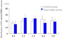
The FG-labeled DRG neurons, in which FG was transported from the facet joint, were present in the left DRG from L1 to L5 (Fig. 1, 2). No labeled neurons were observed in the bilateral T13 or L6 DRG or in the contralateral DRG from L1 to L5. There was no significant difference in number and distribution of FG-labeled neurons between controls and the injured model (P>0.05) (Fig. 1).
Fig. 1.
Distribution of FG-labeled DRG neurons innervating L5/6 facet joints. These neurons were observed from L1 to L5 DRG. Error bars represent standard errors of the means. There is no significant difference in number between each level, and between the control and facet joint injury groups (P>0.05)
Fig. 2.
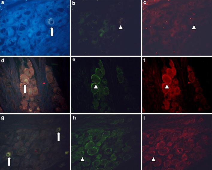
Photomicrographs showing fluorescence of FG-labeled DRG neurons, GFAP-IR satellite cells, and TNFα-IR at the left L3 level. a, d, and g show FG-labeled neurons innervating the L5/6 facet joint (arrows). b, e, and h: cells labeled with red fluorescence are GFAP-IR satellite cells. c, f, and i: cells labeled with green fluorescence are TNFα-IR neurons and satellite cells. a, b, and c are micrographs of the same section harvested from a rat in the control group triple-labeled to show DRG neurons, GFAP-IR (in red) and TNFα-IR neurons and satellite cells (in green). d, e, and f are micrographs of the same triple-labeled section harvested from a rat in the facet joint injury group; and g, h, and i are micrographs of the same triple-labeled section from another rat in the facet joint injury group. In the control group, FG-labeled cells are not surrounded by GFAP-IR satellite cells (b). FG-labeled neurons weakly express TNFα-IR (c; arrowhead). In the facet joint injury group, FG-labeled cells (d and g) are surrounded by GFAP-IR satellite cells (e and h; arrowheads). FG-labeled neurons and the GFAP-IR satellite cells around the FG-labeled neurons weakly express TNFα-IR (f and i; arrows indicate neurons and arrowheads indicate satellite cells)
GFAP-IR satellite cells emerged and expressed TNFα in DRG
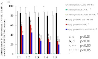
The FG-labeled TNFα-IR neurons were present in the left DRG from L1 to L5. Figure 2 shows FG-labeled TNFα-IR neurons. Most FG-labeled neurons were TNFα-IR. Figure 3 indicates ratios of TNFα-IR neurons to FG-labeled neurons at each level. There were no significant differences in the ratios of FG labeled and TNFα-IR neurons between each level (P>0.05). There were no significant differences in the ratios of FG labeled and TNFα-IR neurons between control (83±10%) and injured groups (80±8%); P>0.05.
Fig. 3.
Distribution of FG-labeled and TNFα-IR cells; GFAP-IR satellite cells around FG-labeled neurons; GFAP- and TNFα-IR satellite cells at each level in both control and facet joint injury groups. The ratio of FG-labeled and TNFα-IR neurons in both groups was not significantly different (P>0.05). The ratio of GFAP-IR satellite cells around FG-labeled neurons (a, c: P<0.01), and GFAP- and TNFα-IR satellite cells around FG-labeled neurons (b, d: P<0.01) in the facet joint injury group was significantly higher than that in the control group. The ratios of GFAP-IR satellite cells to FG-labeled neurons in L1 and L2 DRG were significantly higher than in L3, L4, and L5 DRG (*,***P<0.05). The ratios of GFAP- and TNFα-IR satellite cells to FG-labeled neurons in L1 and L2 DRG were also higher than in L3, L4, and L5 DRG (**,****P<0.05)
GFAP-IR satellite cells were not observed in the control group. However, GFAP-IR satellite cells emerged in the injury group. Some GFAP-IR satellite cells were distributed around FG-labeled neurons (Fig. 2). The ratio of GFAP-IR satellite cells around FG-labeled neurons to total FG-labeled neurons was 46±5%. The ratios of GFAP-IR satellite cells around FG-labeled neurons to total FG-labeled neurons in L3, L4, and L5 DRGs were significantly less than in L1 and L2 DRGs in the injured group (P<0.05) (Fig. 3).
The TNFα immunoreactivity was observed in GFAP-satellite cells. The ratio of TNFα- and GFAP-double stained cells to GFAP-satellite cells around FG-labeled neurons was 35±5% (Fig. 3).
The ratios of TNFα- and GFAP-IR satellite cells around FG-labeled neurons to total FG-labeled neurons in L3, L4, and L5 DRGs were significantly less than that in L1 and L2 DRGs. (P<0.05) (Fig. 3).
Discussion
FG-labeled DRG neurons innervating rat L5/6 facet joints
The rat L5/6 facet joint is innervated by DRG from L1 to L5 by two distinct systems: innervation from corresponding and adjacent segments and from distant segments. In the latter innervation, sensory nerve fibers enter the paravertebral sympathetic trunks and reach L1 or L2 DRG [14, 16, 22, 23]. The present study similarly demonstrates that the rat L5/6 facet joint is innervated by ipsilateral DRG from L1 to L5.
TNFα expressing satellite cells surround sensory DRG neurons innervating the facet joint after facet capsule injury
In the current study, GFAP-IR satellite cells were not observed in the control group. However, GFAP-IR satellite cells emerged around FG-labeled neurons innervating the facet joint after facet joint injury. Some GFAP-IR satellite cells around FG-labeled neurons were co-labeled with TNFα-IR. The GFAP activation and TNFα expression in satellite cells around neurons innervating facet joints were more frequently seen in upper DRG than in lower DRG.
In primary sensory nerves and spinal cord, glial cells such as astrocytes, microglia, Schwann cells, and satellite cells are activated in response to ischemia, traumatic injury, and inflammation [11, 21, 24, 26]. Recently, it has been reported that the glial activation in the DRG and spinal dorsal horn resulting from peripheral nerve injury produces hyperalgesia and allodynia in rats [8, 29]. This activation of glial cells is thought to be involved in the pathogenesis of neuropathic pain. The current data suggest that facet joint capsule injury activates satellite cells in DRG, and may increase facet joint pain intensity.
In peripheral nerve injury, TNFα expression is up-regulated in endoneurial macrophages and Schwann cells, resulting in pain in rats [24, 25]. The TNFα produced at nerve injury sites is axonally transported to DRG neurons and the spinal cord dorsal horn where it correlates with the expression of TNF type 1 and 2 (p55 and p75) receptors, which do not usually exist in the DRG neurons in rats. This may activate central cytokines in the pathogenesis of painful neuropathy [18–20]. The TNFα-IR satellite cells were found to surround neurons expressing TNF p55 receptors in rats [18]. We conclude that TNFα from these satellite cells appears to induce these neurons to synthesize neuropeptides and inflammatory agents [18]. Indeed, TNFα induces substance P (SP) [7], which along with calcitonin gene-related peptide (CGRP), is well known to be associated with neuropathic pain in rats [9]. It has been reported that DRG neurons innervating rat facet joints are immunoreactive for SP and CGRP, and that the ratios of CGRP-IR in L1 and L2 DRG neurons were significantly higher than in L3, L4, and L5 DRG in a rat model of facet joint inflammation [16]. Satellite cells predominantly activated in L1 and L2 DRG compared to L3, L4, and L5 DRG in the current study may be related to the activation of CGRP.
Recently, TNFα has been found to be abundantly expressed in herniated nucleus pulposus in rats [10]. Inhibition of TNFα prevents nucleus pulposus induced thrombus formation, intraneural edema and reduction of nerve conduction velocity in rats [27]. These findings support the suggestion that TNFα is involved in mechanisms of inflammatory pain, neuropathic pain, and low back pain in spinal disorders.
Facet joint pain is a well-recognized clinical problem for some patients. This study is limited because this facet capsule injury model does not correspond exactly to human facet joint pain. Nevertheless, the present study suggests that TNFα in glial cells is related to facet joint pain. These current findings contribute information regarding the relationship between TNFα and facet joint pain.
Footnotes
Seiji Ohtori, Masayuki Miyagi and Tetsuhiro Ishikawa contributed equally to this work.
References
- 1.Bartholdi D, Schwab ME. Expression of pro-inflammatory cytokine and chemokine mRNA upon experimental spinal cord injury in mouse: an in situ hybridization study. Eur J Neurosci. 1997;9:1422–1438. doi: 10.1111/j.1460-9568.1997.tb01497.x. [DOI] [PubMed] [Google Scholar]
- 2.Brandt J, Haibel H, Reddig J, Sieper J, Braun J. Successful short term treatment of severe undifferentiated spondyloarthropathy with the anti-tumor necrosis factor-alpha monoclonal antibody infliximab. J Rheumatol. 2002;29:118–122. [PubMed] [Google Scholar]
- 3.Bogduk N, Long DM. The anatomy of the so-called “articular nerves” and their relationship to facet denervation in the treatment of low back pain. J Neurosurg. 1979;51:172–177. doi: 10.3171/jns.1979.51.2.0172. [DOI] [PubMed] [Google Scholar]
- 4.Bogduk N. The innervation of the lumbar spine. Spine. 1983;8:286–293. doi: 10.1097/00007632-198304000-00009. [DOI] [PubMed] [Google Scholar]
- 5.Botchkina GI, Meistrell ME, Botchkina IL, Tracey KJ. Expression of TNF and TNF receptors (p55 and p75) in the rat brain after focal cerebral ischemia. Mol Med. 1997;3:765–781. [PMC free article] [PubMed] [Google Scholar]
- 6.Cavanaugh JM, el-Bohy A, Hardy WN, Getchell TV, Getchell ML, King AI. Sensory innervation of soft tissues of the lumbar spine in the rat. J Orthop Res. 1989;7:378–388. doi: 10.1002/jor.1100070310. [DOI] [PubMed] [Google Scholar]
- 7.Ding M, Hart RP, Jonakait GM. Tumor necrosis factor-alpha induces substance P in sympathetic ganglia through sequential induction of interleukin-1 and leukemia inhibitory factor. J Neurobiol. 1995;28:445–454. doi: 10.1002/neu.480280405. [DOI] [PubMed] [Google Scholar]
- 8.Fenzi F, Benedetti MD, Moretto G, Rizzuto N. Glial cell and macrophage reactions in rat spinal ganglion after peripheral nerve lesions: an immunocytochemical and morphometric study. Arch Ital Biol. 2001;139:357–365. [PubMed] [Google Scholar]
- 9.Hökfelt T. Neuropeptides in perspective: the last ten years. Neuron. 1991;7:867–879. doi: 10.1016/0896-6273(91)90333-U. [DOI] [PubMed] [Google Scholar]
- 10.Igarashi T, Kikuchi S, Shubayev V, Myers RR. Volvo Award winner in basic science studies: Exogenous tumor necrosis factor-alpha mimics nucleus pulposus-induced neuropathology. Molecular, histologic, and behavioral comparisons in rats. Spine. 2000;25:2975–2980. doi: 10.1097/00007632-200012010-00003. [DOI] [PubMed] [Google Scholar]
- 11.Muñoz-Fernandez MA, Fresno M. The role of tumour necrosis factor, interleukin 6, interferon-gamma and inducible nitric oxide synthase in the development and pathology of the nervous system. Prog Neurobiol. 1998;56:307–34018. doi: 10.1016/S0301-0082(98)00045-8. [DOI] [PubMed] [Google Scholar]
- 12.Mooney V, Robertson J. The facet syndrome. Clin Orthop Res. 1976;115:149–156. [PubMed] [Google Scholar]
- 13.Myers RR, Wagner R, Sorkin LS (1999) Hyperalgesic actions of cytokines on peripheral nerves. Cytokines and Pain, L.R. Watkins and S.F. Maier, Switzerland 133–157
- 14.Ohtori S, Takahashi K, Chiba T, Yamagata M, Sameda H, Moriya H. Substance P and calcitonin gene-related peptide immunoreactive sensory DRG neurons innervating the lumbar facet joints in rats. Auton Neurosci. 2000;86:13–17. doi: 10.1016/S1566-0702(00)00194-6. [DOI] [PubMed] [Google Scholar]
- 15.Ohtori S, Takahashi K, Chiba T, Yamagata M, Sameda H, Moriya H. Phenotypic inflammation switch in rats shown by calcitonin gene-related peptide immunoreactive dorsal root ganglion neurons innervating the lumbar facet joints. Spine. 2001;26:1009–1013. doi: 10.1097/00007632-200105010-00005. [DOI] [PubMed] [Google Scholar]
- 16.Ohtori S, Takahashi K, Moriya H. Inflammatory pain mediated by a phenotypic switch in brain-derived neurotrophic factor-immunoreactive dorsal root ganglion neurons innervating the lumbar facet joints in rats. Neurosci Lett. 2002;323:129–132. doi: 10.1016/s0304-3940(02)00120-9. [DOI] [PubMed] [Google Scholar]
- 17.Ohtori S, Takahashi K, Moriya H. Calcitonin gene-related peptide immunoreactive DRG neurons innervating the cervical facet joints show phenotypic switch in cervical facet injury in rats. Eur Spine J. 2003;12:211–215. doi: 10.1007/s00586-003-0573-4. [DOI] [PMC free article] [PubMed] [Google Scholar]
- 18.Ohtori S, Takahashi K, Moriya H, Myers RR. TNF-alpha and TNF-alpha receptor type 1 upregulation in glia and neurons after peripheral nerve injury: studies in murine DRG and spinal cord. Spine. 2004;29:1082–1088. doi: 10.1097/00007632-200405150-00006. [DOI] [PubMed] [Google Scholar]
- 19.Shubayev VI, Myers RR. Axonal transport of TNF-alpha in painful neuropathy: distribution of ligand tracer and TNF receptors. J Neuroimmunol. 2001;114:48–56. doi: 10.1016/S0165-5728(00)00453-7. [DOI] [PubMed] [Google Scholar]
- 20.Shubayev VI, Myers RR. Anterograde TNF alpha transport from rat dorsal root ganglion to spinal cord and injured sciatic nerve. Neurosci Lett. 2002;320:99–101. doi: 10.1016/S0304-3940(02)00010-1. [DOI] [PubMed] [Google Scholar]
- 21.Stoll G, Jander S. The role of microglia and macrophages in the pathophysiology of the CNS. Prog Neurobiol. 1999;58:233–247. doi: 10.1016/S0301-0082(98)00083-5. [DOI] [PubMed] [Google Scholar]
- 22.Suseki K, Takahashi Y, Takahashi K, Chiba T, Tanaka K, Moriya H. CGRP-immunoreactive nerve fibers projecting to lumbar facet joints through the paravertebral sympathetic trunk in rats. Neurosci Lett. 1996;221:41–44. doi: 10.1016/S0304-3940(96)13282-1. [DOI] [PubMed] [Google Scholar]
- 23.Suseki K, Takahashi Y, Takahashi K, Chiba T, Tanaka K, Morinaga T, Nakamura S, Moriya H. Innervation of the lumbar facet joints. Origin and functions. Spine. 1997;22:477–485. doi: 10.1097/00007632-199703010-00003. [DOI] [PubMed] [Google Scholar]
- 24.Wagner R, Myers RR. Schwann cells produce TNF-alpha: expression in injured and non-injured nerves. Neuroscience. 1996;73:625–629. doi: 10.1016/0306-4522(96)00127-3. [DOI] [PubMed] [Google Scholar]
- 25.Wagner R, Myers RR. Endoneurial injection of TNF-alpha produces neuropathic pain behaviors. Neuroreport. 1996;7:2897–2901. doi: 10.1097/00001756-199611250-00018. [DOI] [PubMed] [Google Scholar]
- 26.Watkins LR, Martin D, Ulrich P, Tracey KJ, Maier SF. Evidence for the involvement of spinal cord glia in subcutaneous formalin induced hyperalgesia in the rat. Pain. 1997;71:225–235. doi: 10.1016/S0304-3959(97)03369-1. [DOI] [PubMed] [Google Scholar]
- 27.Yabuki S, Onda A, Kikuchi S, Myers RR. Prevention of compartment syndrome in dorsal root ganglia caused by exposure to nucleus pulposus. Spine. 2001;26:870–875. doi: 10.1097/00007632-200104150-00008. [DOI] [PubMed] [Google Scholar]
- 28.Yamashita T, Cavanaugh JM, el-Bohy AA, Getchell TV, King AI. Mechanosensitive afferent units in the lumbar facet joint. J Bone Joint Surg (Am) 1990;72:865–870. [PubMed] [Google Scholar]
- 29.Zhou XF, Deng YS, Chie E, Xue Q, Zhong JH, McLachlan EM, Rush RA, Xian CJ. Satellite-cell-derived nerve growth factor and neurotrophin-3 are involved in noradrenergic sprouting in the dorsal root ganglia following peripheral nerve injury in the rat. Eur J Neurosci. 1999;11:1711–1722. doi: 10.1046/j.1460-9568.1999.00589.x. [DOI] [PubMed] [Google Scholar]


