Abstract
Background
Excessive sympathetic activity contributes to the pathogenesis and progression of hypertension. Enhanced cardiac sympathetic afferent reflex (CSAR) is involved in sympathetic activation. This study was designed to determine the roles of angiotensin (Ang)-(1–7) in paraventricular nucleus (PVN) in modulating sympathetic activity and CSAR and its signal pathway in renovascular hypertension.
Methodology/Principal Findings
Renovascular hypertension was induced with two-kidney, one-clip method. Renal sympathetic nerve activity (RSNA) and mean arterial pressure (MAP) were recorded in sinoaortic-denervated and cervical-vagotomized rats with anesthesia. CSAR was evaluated with the RSNA and MAP responses to epicardial application of capsaicin. PVN microinjection of Ang-(1–7) and cAMP analogue db-cAMP caused greater increases in RSNA and MAP, and enhancement in CSAR in hypertensive rats than in sham-operated rats, while Mas receptor antagonist A-779 produced opposite effects. There was no significant difference in the angiotensin-converting enzyme 2 (ACE2) activity and Ang-(1–7) level in the PVN between sham-operated rats and hypertensive rats, but the Mas receptor protein expression in the PVN was increased in hypertensive rats. The effects of Ang-(1–7) were abolished by A-779, adenylyl cyclase inhibitor SQ22536 or protein kinase A (PKA) inhibitor Rp-cAMP. SQ22536 or Rp-cAMP reduced RSNA and MAP in hypertensive rats, and attenuated the CSAR in both sham-operated and hypertensive rats.
Conclusions
Ang-(1–7) in the PVN increases RSNA and MAP and enhances the CSAR, which is mediated by Mas receptors. Endogenous Ang-(1–7) and Mas receptors contribute to the enhanced sympathetic outflow and CSAR in renovascular hypertension. A cAMP-PKA pathway is involved in the effects of Ang-(1–7) in the PVN.
Introduction
Sympathetic activity is enhanced in patients with essential [1] or secondary hypertension [2]–[5] and various hypertensive models [6]–[10]. Excessive sympathetic activity contributes to the pathogenesis of hypertension and progression of organ damage [11]–[13]. Intervention of the sympathetic activation is considered to be an antihypertensive strategy [14]–[16]. Cardiac sympathetic afferent reflex (CSAR) is known as a positive-feedback, sympathoexcitatory cardiovascular reflex [17], [18]. Previous studies in our lab have shown that the CSAR is enhanced in renovascular hypertensive rats [6], [19] and spontaneously hypertensive rats (SHR) [10], which contributes to the sympathetic activation and hypertension [20], [21].
Paraventricular nucleus (PVN) is an important component of the central neurocircuitry of the CSAR [22] and plays a major role in the integration of sympathetic outflow and cardiovascular activity via projections to the intermediolateral column (IML) of the spinal cord and the rostral ventrolateral medulla (RVLM) [23]. Angiotensin (Ang)-(1–7) is known as an important biological active peptide of renin–angiotensin system (RAS) family. Angiotensin-converting enzyme 2 (ACE2) hydrolyzes Ang II or Ang I to Ang-(1–7). Many of Ang-(1–7) effects are primarily mediated by Mas receptors [24] and are selectively blocked by its specific antagonist D-Alanine-Ang-(1–7) (A-779) [25]. Ang-(1–7) immunoreactive staining is present in the PVN [26] including parvocellular and magnocellular subdivisions [27]. The endogenous Ang-(1–7) level in the hypothalamus of rats is comparable to Ang I and Ang II [28]. The Mas receptors are expressed predominantly in the mouse and rat brain and particularly in the forebrain [29]. It has been reported that blockade of endogenous Ang-(1–7) by microinjection of A-779 into the PVN reduces renal sympathetic tone in normal rats [30]. Ang-(1–7) level in hypothalamus is increased in aortic coarctation-induced hypertensive rats [31]. However, it is not known whether Ang-(1–7) in the PVN is involved in the excessive sympathetic activation and the enhanced CSAR in hypertension.
It has been found that activation of Mas receptors by Ang-(1–7) increases intracellular cAMP level and activates protein kinase A (PKA), while inhibition of either adenylyl cyclase (AC) or PKA activity attenuates Ang-(1–7)-induced ERK1/2 activation in glomerular mesangial cells [32]. Ang-(1–7) inhibits vascular growth through the prostacyclin-mediated production of cAMP and activation of cAMP-dependent PKA [33]. These results show that the cAMP-PKA signaling pathway is involved in the activity of Ang-(1–7) and Mas receptors in some peripheral tissues. However, whether cAMP-PKA pathway in the PVN is involved in the CSAR and sympathetic activation and the effects of Ang-(1–7) in hypertension is not understood. The present study was designed to determine whether Ang-(1–7) in the PVN contributed to the enhanced CSAR and sympathetic activation, and whether the cAMP-PKA pathway in the PVN was involved in the effects of Ang-(1–7) in hypertension.
Materials and Methods
Experiments were carried out in male Sprague–Dawley rats. The procedures were approved by the Experimental Animal Care and Use Committee of Nanjing Medical University (No. 20110316) and complied with the Guide for the Care and Use of Laboratory Animals (NIH publication no. 85–23, revised 1996). The rats were kept in a temperature-controlled room on a 12 h–12 h light–dark cycle with free access to standard chow and tap water.
Renovascular hypertensive model
Goldblatt two-kidney one-clip (2K1C) method was used to induce renovascular hypertension in rats as previously reported [6], [20]. Simply, the rat weighing 160–180 g was anesthetized with intraperitoneal administration of sodium pentobarbital (50 mg kg−1). A retroperitoneal flank incision was performed to expose right renal artery. The artery was partly occluded by placing a U-shaped silver clip with an internal diameter of 0.20 mm on the artery to induce renovascular hypertension. Normotensive sham-operated (Sham) rat received similar surgical process except using silver clip. The criterion of hypertension is set as systolic blood pressure (SBP) of tail artery >160 mm Hg in conscious state [19], [20]. Six 2K1C rats were excluded, for their SBP was not high enough to meet the criterion.
SBP measurements in conscious state
Rats were trained by SBP measurement daily for at least 10 days before 2K1C or sham operation to minimize stress-induced SBP fluctuation. The SBP of tail artery was measured weekly in conscious state by using a noninvasive computerized tail-cuff system (NIBP, ADInstruments, Australia) [19], [20]. The rats were warmed for 10–20 min at 28°C before the measurements in order to allow the detection of tail artery pulsations and to achieve the pulse level ready. The SBP was obtained by averaging 10 measurements.
General procedures of acute experiment
Acute experiments were carried out at the end of the 4th week after the 2K1C or sham operation. Rat was intraperitoneally anesthetized with urethane (800 mg kg−1) and α-chloralose (40 mg kg−1). Supplemental doses of anesthesia were used to maintain an appropriate degree of anesthesia that was assessed by the absence of corneal reflex and paw withdrawal response to a noxious pinch. The rat was mechanically ventilated with room air using a rodent ventilator (model 683, Harved Apparatus Inc, USA). The right carotid artery was cannulated and connected with a pressure transducer (MLT0380, ADInstruments, Australia) for continuous recording of arterial blood pressure (ABP), mean arterial pressure (MAP) and heart rate (HR). Bilateral baroreceptor denervation and vagotomy were carried out and identified as previously reported [21], [34].
Renal sympathetic nerve activity (RSNA) recordings
A retroperitoneal incision was made and the left renal sympathetic nerve was isolated. The renal nerve was cut distally to eliminate its afferent activity. The nerve was placed on a pair of silver electrodes and immersed in warm mineral oil. The nerve signals were amplified with a four channel AC/DC differential amplifier (DP-304, Warner Instruments, Hamden, CT, USA) with a high pass filter at 10 Hz and a low pass filter at 3,000 Hz. The RSNA was integrated at a time constant of 100 ms. At the end of each experiment, the background noise was determined after section of the central end of the nerve and was subtracted from the integrated values of the RSNA [35]. The raw and integrated RSNA, ABP, MAP and HR were simultaneously recorded with a PowerLab data acquisition system (8/35, ADInstruments, Australia).
Evaluation of CSAR
A limited left lateral thoracotomy was performed to expose the heart and the pericardium was removed. The CSAR was induced by stimulating cardiac sympathetic afferents with epicardial application of a piece of filter paper (3 mm×3 mm) containing capsaicin (1.0 nmol in 2.0 μl) on the anterior wall of the left ventricle. The filter paper was removed 1 minute later, and the ventricular surface was rinsed three times with 10 ml of normal saline (37°C). The CSAR was evaluated by the RSNA and MAP responses to the epicardial application of capsaicin [34], [35].
PVN microinjection
The rats were placed in a stereotaxic frame (Stoelting, Chicago, USA). The stereotaxic coordinates for the PVN are 1.8 mm caudal from bregma, 0.4 mm lateral to the midline and 7.9 mm ventral to the dorsal surface according to Paxinos & Watson's rat atlas. The bilateral PVN microinjections were completed within 1 min and the microinjection volume was 50 nL for each side of the PVN. At the end of the experiment, 50 nl of Evans Blue dye (2%) was injected into each microinjection site. The microinjection sites were histologically verified with microscope. Rats with microinjection sites outside the PVN were excluded from data analysis.
PVN Sample Preparation
The rat was euthanized with an overdose of pentobarbital. The brain of the rat was quickly removed, frozen in liquid nitrogen and stored at −70°C until being sectioned. Coronal sections of the brain were made with a cryostat microtome (Leica CM1900-1-1, Wetzlar, Hessen, Germany) at the PVN level. The PVN area was punched out with a 15-gauge needle (1.5 mm ID). The punched tissues were subsequently homogenized and centrifuged. The total protein in the homogenate supernatant was extracted and measured by using protein assay kit (BCA; Pierce).
Measurement of Mas receptor protein expression
The Mas receptor protein expression in the PVN was determined with Western blotting method [36]. Briefly, after process of electrophoresis and transmembrane, proteins on nitrocellulose membrane were probed with rabbit polyclonal Mas receptor antibody (1∶200, Alomone Labs, Israel). This was followed by incubation with horseradish peroxidase–conjugated goat anti-rabbit IgG (1∶5000; Immunology Consultants Lab, USA). The bands were visualized by enhanced chemiluminescence using the ECL system (Pierce Chemical). GAPDH (Bioworld Technology Inc., USA) protein was used as a loading control. The total amount of Mas receptors protein is expressed as the percentage of Mas receptors to GAPDH protein.
Measurement of Ang-(1–7) level
The level of Ang-(1–7) in PVN tissue homogenate supernatant was measured using a commercial peptide enzyme immunoassay kit (MyBioSource LLC, USA) following the manufacturer's instructions [37].
Measurement of ACE2 activity
ACE2 activity in the PVN was determined by a fluorimetric ACE2 activity assay kit (Anaspec Inc. Fremont, CA, USA). The assay is based on the use of the ACE2 substrate Mca/Dnp fluorescence resonance energy transfer (FRET) peptide. In the FRET peptide the fluorescence of Mca is quenched by Dnp. Upon cleavage into two separate fragments by the ACE2 enzyme, the fluorescence of Mca is recovered, and can be monitored at excitation/emission = 330 nm/390 nm. Briefly, PVN tissue homogenate supernatant was added to a black flat-bottom 96-well plate containing ACE2 substrate [37], [38]. The change in fluorescence was monitored using a Fluorescence Reader (Gemini EM, Molecular Devices, USA). All fluorescence readings are expressed in relative fluorescence units (RFU) and the ACE2 activity is expressed as amounts of RFU of substrate converted to product per unit time and is normalized for protein content [39], [40].
Chemicals
Ang-(1–7) and A-779 were purchased from Bachem (Bubendorf, Switzerland). Dibutyryl-cAMP (db-cAMP), 9-(tetrahydro-2-furanyl)-9H-purin-6-amine (SQ22536), rp-adenosine-3′,5′-cyclic monophosphothionate (Rp-cAMP) and capsaicin were purchased from Sigma Chemical Co (St. Louis, MO, USA). All the chemicals were dissolved in normal saline.
Experiment protocols
Experiment 1
The PVN microinjection of saline, three doses of Ang-(1–7) (0.03, 0.3 and 3 nmol), Mas receptor antagonist A-779 (3 nmol) and Ang-(1–7) (3 nmol) pretreated with A-779 (3 nmol) on the RSNA, MAP and CSAR were carried out in six groups of 2K1C rats and six groups of Sham rats, respectively (n = 6 for each group). A-779 was administered eight min before Ang-(1–7). The dose and duration of treatment with A-799 were selected according to our preliminary study and previous reports [30], [41]. The CSAR was evaluated eight min after the PVN microinjection. To exclude the possibility that the effects of Ang-(1–7) were caused by diffusion to other brain area, the effects of microinjection of Ang-(1–7) (3 nmol) into the anterior hypothalamic area which is adjacent to the PVN were determined in 2K1C and Sham rats (n = 3 for each group).
Experiment 2
Mas receptor protein expression in the PVN was determined in 5 Sham rats and 5 2K1C rats. Furthermore, Ang-(1–7) level and ACE2 activity in the PVN were determined in other 5 Sham rats and 5 2K1C rats.
Experiment 3
PVN microinjection of saline, SQ22536 (2 nmol, an AC inhibitor), db-cAMP (1 nmol, a cAMP analogue) or Rp-cAMP (1 nmol, a PKA inhibitor) were carried out in four groups of Sham rats and four groups of 2K1C rats to determine the roles of cAMP and PKA in regulating RSNA, MAP and CSAR (n = 6 for each group). In other four groups of Sham rats and four groups of 2K1C rats, PVN pretreatment with the same dose of saline, SQ22536, db-cAMP or Rp-cAMP on the RSNA, MAP and CSAR responses to Ang-(1–7) (3 nmol) were determined (n = 6 for each group). The pretreatment was done eight min before Ang-(1–7). CSAR was evaluated eight min after the PVN microinjection of Ang-(1–7).
Statistical analysis
The RSNA and MAP responses caused by the PVN microinjection were determined by averaging 2 min of the maximal responses. The RSNA and MAP responses to epicardial application of capsaicin were determined by averaging 30 sec of the parameters beginning at the 16th sec after epicardial application of capsaicin. The RSNA change was expressed as the percent change from the baseline values. Comparisons between two observations in the same animal were assessed by Student's paired t test. One-way or two-way ANOVA was used followed by Bonferroni test for post hoc analysis when multiple comparisons were made. All data were expressed as mean ± SE. P<0.05 was considered statistically significant.
Results
General Data
SBP and MAP in 2K1C rats were significantly higher than that in Sham rats. There was no significant difference in the body weight and baseline HR between Sham rats and 2K1C rats (Table 1).
Table 1. Body weight, SBP, MAP and HR at the end of the 4th week.
| Sham | 2K1C | |
| n | 94 | 94 |
| Body Weight, g | 328.5±1.8 | 323.4±1.7 |
| SBP, mm Hg | 118.7±1.2 | 169.3±1.6 * |
| MAP, mm Hg | 91.5±1.0 | 138.4±1.6 * |
| HR, beats/min | 356.1±4.7 | 361.8±4.2 |
Systolic blood pressure (SBP), mean arterial pressure (MAP) and heart rate (HR) were measured with a pressure transducer in the right carotid artery under anesthesia. Values are expressed as mean ± SE. * P<0.05 compared with the Sham rats.
Effects of different doses of Ang-(1–7)
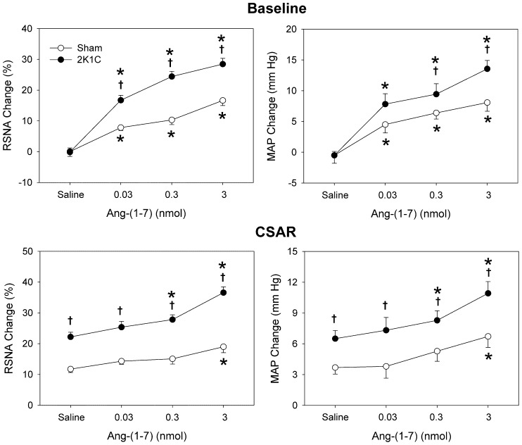
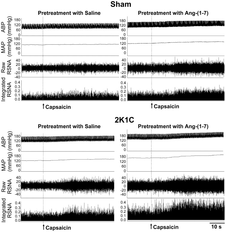
Microinjection of three doses of Ang-(1–7) (0.03, 0.3 and 3 nmol) into the PVN dose-related increased the baseline RSNA and MAP in both 2K1C rats and Sham rats, peaking at about 7 min and lasting at least 12 min. The effects of Ang-(1–7) on RSNA and MAP in 2K1C rats were greater than that in Sham rats (Fig. 1). Representative recordings showed that the CSAR was enhanced in 2K1C rat compared with Sham rat, and PVN microinjection of high dose of Ang-(1–7) enhanced the CSAR compared with saline in both Sham rat and 2K1C rat (Fig. 2). Compared with saline, high dose of Ang-(1–7) significantly enhanced the CSAR in Sham rats, while both moderate and high doses of Ang-(1–7) enhanced the CSAR in 2K1C rats (Fig. 1). In addition, microinjection of high dose of Ang-(1–7) into the anterior hypothalamic area had no significant effects on the RSNA, MAP and CSAR.
Figure 1. Effects of PVN microinjection of saline and three doses of Ang-(1–7) (0.03, 0.3 and 3 nmol) on the baseline RSNA and MAP and CSAR.
The CSAR was evaluated by the RSNA and MAP responses to epicardial application of capsaicin (1 nmol). Values are mean ± SE. * P<0.05 compared with saline. † P<0.05 compared with Sham. n = 6 for each group.
Figure 2. Representative recordings showing the effects of PVN microinjection of saline and Ang-(1–7) (3 nmol) on the CSAR in Sham and 2K1C rats.
The CSAR was evaluated by the RSNA and MAP responses to epicardial application of capsaicin (1 nmol).
Effects of A-779
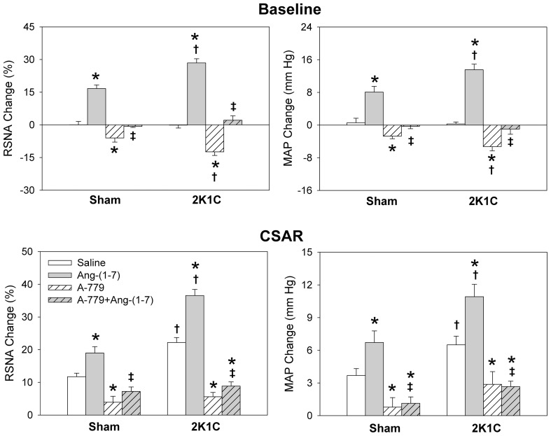
Microinjection of A-779 into the PVN decreased the RSNA and MAP, peaking at about 8 min and lasting at least 30 min, and attenuated the CSAR in both Sham and 2K1C rats. The effects of A-779 were greater in 2K1C rats than that in Sham rats. Pretreatment with A-779 in the PVN abolished the effects of Ang-(1–7) on the RSNA, MAP and CSAR in Sham and 2K1C rats (Fig. 3).
Figure 3. Effects of PVN microinjection of saline, Ang-(1–7) (3 nmol), A-779 (3 nmol), A-779+Ang-(1–7) on the baseline RSNA and MAP and CSAR.
The CSAR was evaluated by the RSNA and MAP responses to epicardial application of capsaicin (1 nmol). Values are mean ± SE. * P<0.05 compared with saline. † P<0.05 compared with Sham. ‡ P<0.05 compared with Ang-(1–7) alone. n = 6 for each group.
Mas receptor protein expression, Ang-(1–7) level and ACE2 activity in PVN
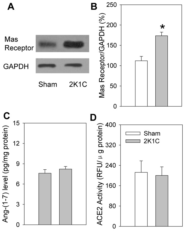
The Mas receptor protein expression in the PVN was significantly increased in 2K1C rats compared with Sham rats. However, there were no significant difference in Ang-(1–7) level and ACE2 activity in the PVN between 2K1C rats and Sham rats (Fig. 4).
Figure 4. Mas receptor protein expression (A & B), Ang-(1–7) level (C) and ACE2 activity (D) in the PVN in Sham and 2K1C rats.
RFU, relative fluorescence units. Values are mean ± SE. * P<0.05 compared with Sham rats. n = 5 for each group.
Effects of SQ22536 and db-cAMP
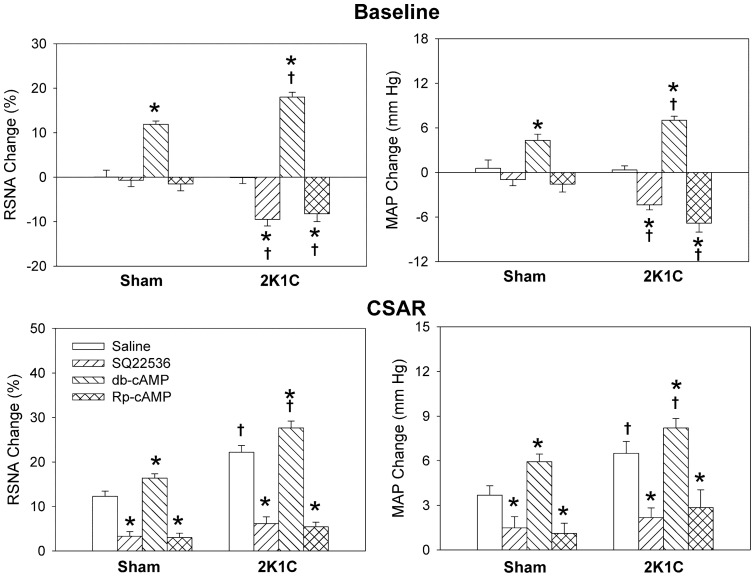
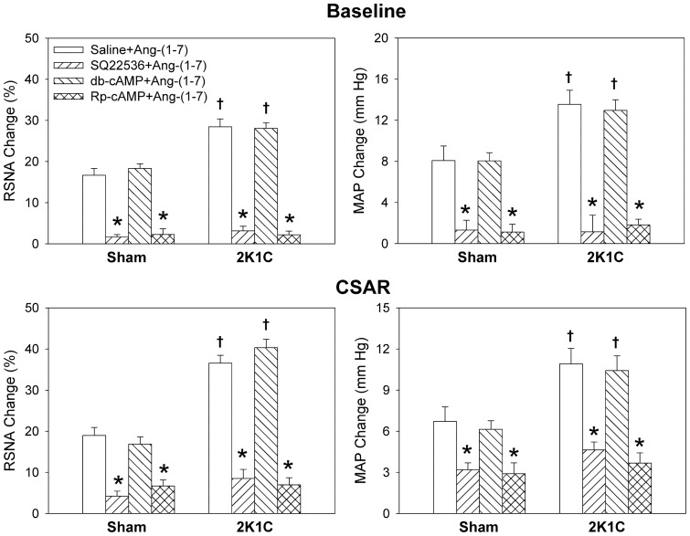
Microinjection of AC inhibitor SQ22536 into the PVN decreased RSNA and MAP in 2K1C rats, and attenuated the CSAR in both Sham and 2K1C rats. A cAMP analogue db-cAMP caused greater increases in RSNA and MAP, and enhancement in CSAR in 2K1C rats than in Sham rats (Fig. 5). On the other hand, pretreatment with SQ22536 in the PVN abolished the effects of Ang-(1–7) on the RSNA, MAP and CSAR in both Sham and 2K1C rats, but db-cAMP pretreatment failed to augment the effects of Ang-(1–7) (Fig. 6).
Figure 5. Effects of the PVN microinjection of saline, SQ22536 (2 nmol), db-cAMP (1 nmol) and Rp-cAMP (1 nmol) on the baseline RSNA and MAP and CSAR.
The CSAR was evaluated by the RSNA and MAP responses to epicardial application of capsaicin (1 nmol). Values are mean ± SE. * P<0.05 compared with saline. † P<0.05 compared with Sham. n = 6 for each group.
Figure 6. Effects of the PVN treatment with saline, SQ22536 (2 nmol), db-cAMP (1 nmol) and Rp-cAMP (1 nmol) on the RSNA, MAP and CSAR responses to Ang-(1–7) (3 nmol).
The CSAR was evaluated by the RSNA and MAP responses to epicardial application of capsaicin (1 nmol). Values are mean ± SE. * P<0.05 compared with saline. † P<0.05 compared with Sham. n = 6 for each group.
Effects of Rp-cAMP
Microinjection of PKA inhibitor Rp-cAMP into the PVN decreased RSNA and MAP in 2K1C rats, and attenuated the CSAR in both Sham and 2K1C rats (Fig. 5). Pretreatment with Rp-cAMP in the PVN abolished the effects of Ang-(1–7) on the RSNA, MAP and CSAR in both Sham and 2K1C rats (Fig. 6).
Discussion
The primary new findings in the present study are that activation of Mas receptors with Ang-(1–7) in the PVN increases sympathetic outflow and blood pressure, and augments the CSAR. Endogenous Ang-(1–7) and Mas receptors contribute to the excessive sympathetic activation and the enhanced CSAR in renovascular hypertensive rats. A cAMP-PKA pathway is involved in the effects of Ang-(1–7) in the PVN.
Abundant Ang-(1–7) immunoreactive staining has been found in the PVN [26]. Microiontophoretic application of Ang-(1–7) into the PVN augments the excitability of the neurons in the PVN [42], which is selectively blocked by Mas receptor antagonist A-779 [43]. Blockade of endogenous Ang-(1–7) by microinjection of A-779 into the PVN reduces renal sympathetic tone in normal rats [30]. In the present study, microinjection of Ang-(1–7) into the PVN caused greater enhancement in the CSAR, and more increases in the RSNA and MAP in 2K1C rats than in Sham rats, which was abolished by A-779. A-779 in the PVN caused greater decreases in RSNA and MAP in 2K1C rats than in Sham rats, and attenuated the CSAR in both Sham and 2K1C rats. These results indicate that Ang-(1–7) in the PVN contributes to the enhanced sympathetic activity and CSAR via Mas receptors in renovascular hypertension. It is known that the CSAR is enhanced in 2K1C-induced hypertensive rats [6], [19] and SHR [10], which is involved in sympathetic activation and hypertension in these animal models [20], [21]. The inhibitory effects of A-779 on the CSAR may be partially responsible for the decreases in RSNA and MAP in renovascular hypertension.
Many studies have shown that Ang-(1–7)/Mas receptor axis is as a counter regulator of the effects of the classic Ang II/AT1 receptor axis-mediated effects [44]–[46]. However, a previous study in our lab has indicated that Ang-(1–7) in the RVLM is as effective as Ang II in sensitizing the CSAR and increasing sympathetic outflow in normal rats [41]. The results is confirmed by a recent finding that endogenous Ang-(1–7) in the RVLM contributes to maintain arterial blood pressure and renal sympathetic nerve activity in both SHR and normal control rats [47]. We have found that Ang II in the PVN augmented the enhanced CSAR and RSNA in 2K1C rats, which were abolished by the pretreatment with AT1 receptor antagonist losartan, and losartan in the PVN normalized the enhanced CSAR and decreased the RSNA and MAP in 2K1C rats [21]. The results in the present study indicate that the roles of Ang-(1–7) are similar to Ang II in modulating the RSNA MAP and CSAR. However, there are some differences between Ang II/AT1 receptors and Ang-(1–7)/Mas receptors in the PVN in regulating RSNA, MAP and CSAR. In normal rats, AT1 receptor antagonist losartan in the PVN had no significant effects on RSNA and MAP [21], but Mas receptors antagonist A-779 decreased RSNA and MAP. The results suggest that Mas receptors rather than AT1 receptors in the PVN are involved in the tonic control of sympathetic activity and blood pressure. Furthermore, losartan had no significant effects on the CSAR in normal rats and normalized the enhanced CSAR in 2K1C rats to the baseline level [21], but A-779 reduced the CSAR in both Sham and 2K1C rats to the lower level than baseline. These results suggest that Ang-(1–7)/Mas receptors rather than Ang II/AT1 receptors in the PVN contribute to the tonic control of the CSAR. Blockade of Mas receptors in the PVN caused greater CSAR-inhibitory effect in 2K1C rats.
We found that the Mas receptor protein expression in the PVN was significantly increased in 2K1C rats compared with Sham rats. However, there were no significant difference in Ang-(1–7) level and ACE2 activity in the PVN between 2K1C rats and Sham rats. These results suggest that the enhanced effects of Ang-(1–7) in the PVN on the RSNA, MAP and CSAR in 2K1C rats arise from the upregulation of Mas receptors in the PVN rather than the production or release of Ang-(1–7) in the PVN. The increased Mas receptor expression in the PVN contributes to the tonic control of RSNA, MAP and CSAR in 2K1C rats.
It is known that nitric oxide (NO) is a key signaling molecule in Ang-(1–7) stimulated neurons that may counteract the signaling events of Ang II [48]. NO in the PVN inhibits the Ang II-mediated increase in sympathetic nerve activity [49]–[52]. The findings in the present study showed that the roles of Ang-(1–7) in the PVN in increasing the RSNA and MAP, and enhancing the CSAR were similar to the roles of Ang II as previously reported [21]. Therefore, it would be impossible that nitric oxide mediate the excitatory effects of Ang-(1–7) in the PVN on the RSNA, MAP and CSAR. The Mas receptor is a kind of G protein-coupled receptor [53]. Ang-(1–7) inhibits vascular growth through the prostacyclin-mediated production of cAMP and activation of cAMP-dependent PKA [33]. Ang-(1–7)-induced activation of ERK1/2 in glomerular mesangial cells is cAMP/PKA-dependent [32]. It is an interesting question whether cAMP/PKA in the PVN is involved in mediating the effects of Ang-(1–7) in the PVN.
In the present study, microinjection of AC inhibitor SQ22536 or PKA inhibitor Rp-cAMP into the PVN decreased RSNA and MAP in 2K1C rats, and attenuated the CSAR in both Sham and 2K1C rats. Pretreatment with SQ22536 or Rp-cAMP in the PVN abolished the effects of Ang-(1–7) on the RSNA, MAP and CSAR in both Sham and 2K1C rats. These results indicate that the cAMP-PKA pathway in the PVN mediates the effects of Ang-(1–7) on the RSNA, MAP and CSAR, and is involved in the sympathetic activation and enhanced CSAR in renovascular hypertension. SQ22536 or Rp-cAMP reduced the CSAR in both Sham and 2K1C rats to the lower level than baseline, suggesting that cAMP in the PVN contributes to the tonic control of the CSAR. The results is supported by the findings that PVN microinjection of a cAMP analogue db-cAMP caused greater increases in RSNA and MAP, and enhancement in CSAR in 2K1C rats than in Sham rats.
In conclusion, activation of Mas receptors with Ang-(1–7) in the PVN increases sympathetic outflow and blood pressure, and augments the CSAR. Endogenous Ang-(1–7) and Mas receptors contribute to the excessive sympathetic activation and the enhanced CSAR in renovascular hypertensive rats. A cAMP-PKA pathway is involved in the excitatory effects of Ang-(1–7) on the sympathetic activity, blood pressure and CSAR in the PVN. Blockade of Mas receptors or the cAMP-PKA pathway in the PVN may be beneficial to attenuate the excessive sympathetic activation in hypertension.
Funding Statement
This work was supported by Chinese National Natural Science Fund (81100182 & 31171095), Natural Science Foundation from Department of Education of Jiangsu Province (11KJB310002 & 10KJB310004), Science and Technology Foundation from Nanjing Medical University (2010NJMUZ23), and a project funded by the Priority Academic Program Development of Jiangsu Higher Education Institutions (PAPD). The funders had no role in study design, data collection and analysis, decision to publish, or preparation of the manuscript.
References
- 1. Hogarth AJ, Mackintosh AF, Mary DA (2007) The effect of gender on the sympathetic nerve hyperactivity of essential hypertension. J Hum Hypertens 21: 239–245. [DOI] [PubMed] [Google Scholar]
- 2. Biaggioni I (2008) Should we target the sympathetic nervous system in the treatment of obesity-associated hypertension? Hypertension 51: 168–171. [DOI] [PubMed] [Google Scholar]
- 3. Rubinger D, Backenroth R, Sapoznikov D (2012) Sympathetic Activation and Baroreflex Function during Intradialytic Hypertensive Episodes. PLoS One 7: e36943. [DOI] [PMC free article] [PubMed] [Google Scholar]
- 4. Ramchandra R, Watson AM, Hood SG, May CN (2010) The response of cardiac sympathetic nerve activity to intravenous irbesartan in heart failure. Am J Physiol Regul Integr Comp Physiol 298: R1056–R1060. [DOI] [PMC free article] [PubMed] [Google Scholar]
- 5. Neumann J, Ligtenberg G, Klein IH, Boer P, Oey PL, et al. (2007) Sympathetic hyperactivity in hypertensive chronic kidney disease patients is reduced during standard treatment. Hypertension 49: 506–510. [DOI] [PubMed] [Google Scholar]
- 6. Han Y, Fan ZD, Yuan N, Xie GQ, Gao J, et al. (2011) Superoxide anions in paraventricular nucleus mediate the enhanced cardiac sympathetic afferent reflex and sympathetic activity in renovascular hypertensive rats. J Appl Physiol 110: 646–652. [DOI] [PubMed] [Google Scholar]
- 7. Fujita M, Ando K, Nagae A, Fujita T (2007) Sympathoexcitation by oxidative stress in the brain mediates arterial pressure elevation in salt-sensitive hypertension. Hypertension 50: 360–367. [DOI] [PubMed] [Google Scholar]
- 8. Stocker SD, Meador R, Adams JM (2007) Neurons of the rostral ventrolateral medulla contribute to obesity-induced hypertension in rats. Hypertension 49: 640–646. [DOI] [PubMed] [Google Scholar]
- 9. Marques FZ, Campain AE, Davern PJ, Yang YH, Head GA, et al. (2011) Genes influencing circadian differences in blood pressure in hypertensive mice. PLoS One 6: e19203. [DOI] [PMC free article] [PubMed] [Google Scholar]
- 10. Fan ZD, Zhang L, Shi Z, Gan XB, Gao XY, et al. (2012) Artificial microRNA interference targeting AT1a receptors in paraventricular nucleus attenuates hypertension in rats. Gene Ther 19: 810–817. [DOI] [PubMed] [Google Scholar]
- 11. Mancia G, Grassi G, Giannattasio C, Seravalle G (1999) Sympathetic activation in the pathogenesis of hypertension and progression of organ damage. Hypertension 34: 724–728. [DOI] [PubMed] [Google Scholar]
- 12. Morise T, Horita M, Kitagawa I, Shinzato R, Hoshiba Y, et al. (2000) The potent role of increased sympathetic tone in pathogenesis of essential hypertension with neurovascular compression. J Hum Hypertens 14: 807–811. [DOI] [PubMed] [Google Scholar]
- 13.Rahn KH, Barenbrock M, Hausberg M (1999) The sympathetic nervous system in the pathogenesis of hypertension. J Hypertens Suppl 17: S11–S14. [PubMed]
- 14. Del CS, Morello F, Rabbia F, Milan A, Naso D, et al. (2007) Antihypertensive drugs and the sympathetic nervous system. J Cardiovasc Pharmacol 50: 487–496. [DOI] [PubMed] [Google Scholar]
- 15. Fisher JP, Fadel PJ (2010) Therapeutic strategies for targeting excessive central sympathetic activation in human hypertension. Exp Physiol 95: 572–580. [DOI] [PMC free article] [PubMed] [Google Scholar]
- 16. Grassi G, Seravalle G, Quarti-Trevano F (2010) The ‘neuroadrenergic hypothesis’ in hypertension: current evidence. Exp Physiol 95: 581–586. [DOI] [PubMed] [Google Scholar]
- 17. Du YH, Chen AF (2007) A “love triangle” elicited by electrochemistry: complex interactions among cardiac sympathetic afferent, chemo-, and baroreflexes. J Appl Physiol 102: 9–10. [DOI] [PubMed] [Google Scholar]
- 18. Malliani A, Montano N (2002) Emerging excitatory role of cardiovascular sympathetic afferents in pathophysiological conditions. Hypertension 39: 63–68. [DOI] [PubMed] [Google Scholar]
- 19. Han Y, Yuan N, Zhang SJ, Gao J, Shi Z, et al. (2011) c-Src in paraventricular nucleus modulates sympathetic activity and cardiac sympathetic afferent reflex in renovascular hypertensive rats. Pflugers Arch 461: 437–446. [DOI] [PubMed] [Google Scholar]
- 20. Zhu GQ, Xu Y, Zhou LM, Li YH, Fan LM, et al. (2009) Enhanced cardiac sympathetic afferent reflex involved in sympathetic overactivity in renovascular hypertensive rats. Exp Physiol 94: 785–794. [DOI] [PubMed] [Google Scholar]
- 21. Chen AD, Zhang SJ, Yuan N, Xu Y, De W, et al. (2011) AT1 receptors in paraventricular nucleus contribute to sympathetic activation and enhanced cardiac sympathetic afferent reflex in renovascular hypertensive rats. Exp Physiol 96: 94–103. [DOI] [PubMed] [Google Scholar]
- 22. Zhong MK, Duan YC, Chen AD, Xu B, Gao XY, et al. (2008) Paraventricular nucleus is involved in the central pathway of cardiac sympathetic afferent reflex in rats. Exp Physiol 93: 746–753. [DOI] [PubMed] [Google Scholar]
- 23. Badoer E (2001) Hypothalamic paraventricular nucleus and cardiovascular regulation. Clin Exp Pharmacol Physiol 28: 95–99. [DOI] [PubMed] [Google Scholar]
- 24. Xu P, Sriramula S, Lazartigues E (2011) ACE2/ANG-(1–7)/Mas pathway in the brain: the axis of good. Am J Physiol Regul Integr Comp Physiol 300: R804–R817. [DOI] [PMC free article] [PubMed] [Google Scholar]
- 25. Santos RA, Haibara AS, Campagnole-Santos MJ, Simoes e Silva AC, Paula RD, et al. (2003) Characterization of a new selective antagonist for angiotensin-(1–7), D-pro7-angiotensin-(1–7). Hypertension 41: 737–743. [DOI] [PubMed] [Google Scholar]
- 26. Block CH, Santos RA, Brosnihan KB, Ferrario CM (1988) Immunocytochemical localization of angiotensin-(1–7) in the rat forebrain. Peptides 9: 1395–1401. [DOI] [PubMed] [Google Scholar]
- 27. Krob HA, Vinsant SL, Ferrario CM, Friedman DP (1998) Angiotensin-(1–7) immunoreactivity in the hypothalamus of the (mRen-2d)27 transgenic rat. Brain Res 798: 36–45. [DOI] [PubMed] [Google Scholar]
- 28. Chappell MC, Brosnihan KB, Diz DI, Ferrario CM (1989) Identification of angiotensin-(1–7) in rat brain. Evidence for differential processing of angiotensin peptides. J Biol Chem 264: 16518–16523. [PubMed] [Google Scholar]
- 29. Metzger R, Bader M, Ludwig T, Berberich C, Bunnemann B, et al. (1995) Expression of the mouse and rat mas proto-oncogene in the brain and peripheral tissues. FEBS Lett 357: 27–32. [DOI] [PubMed] [Google Scholar]
- 30. Silva AQ, Santos RA, Fontes MA (2005) Blockade of endogenous angiotensin-(1–7) in the hypothalamic paraventricular nucleus reduces renal sympathetic tone. Hypertension 46: 341–348. [DOI] [PubMed] [Google Scholar]
- 31. Gironacci MM, Brosnihan KB, Ferrario CM, Gorzalczany S, Verrilli MA, et al. (2007) Increased hypothalamic angiotensin-(1–7) levels in rats with aortic coarctation-induced hypertension. Peptides 28: 1580–1585. [DOI] [PMC free article] [PubMed] [Google Scholar]
- 32. Liu GC, Oudit GY, Fang F, Zhou J, Scholey JW (2012) Angiotensin-(1–7)-induced activation of ERK1/2 is cAMP/protein kinase A-dependent in glomerular mesangial cells. Am J Physiol Renal Physiol 302: F784–F790. [DOI] [PubMed] [Google Scholar]
- 33. Tallant EA, Clark MA (2003) Molecular mechanisms of inhibition of vascular growth by angiotensin-(1–7). Hypertension 42: 574–579. [DOI] [PubMed] [Google Scholar]
- 34. Gan XB, Duan YC, Xiong XQ, Li P, Cui BP, et al. (2011) Inhibition of cardiac sympathetic afferent reflex and sympathetic activity by baroreceptor and vagal afferent inputs in chronic heart failure. PLoS ONE 6: e25784. [DOI] [PMC free article] [PubMed] [Google Scholar]
- 35. Zhang L, Xiong XQ, Fan ZD, Gan XB, Gao XY, et al. (2012) Involvement of enhanced cardiac sympathetic afferent reflex in sympathetic activation in early stage of diabetes. J Appl Physiol 113: 47–55. [DOI] [PubMed] [Google Scholar]
- 36. Becker LK, Etelvino GM, Walther T, Santos RA, Campagnole-Santos MJ (2007) Immunofluorescence localization of the receptor Mas in cardiovascular-related areas of the rat brain. Am J Physiol Heart Circ Physiol 293: H1416–H1424. [DOI] [PubMed] [Google Scholar]
- 37. Dilauro M, Zimpelmann J, Robertson SJ, Genest D, Burns KD (2010) Effect of ACE2 and angiotensin-(1–7) in a mouse model of early chronic kidney disease. Am J Physiol Renal Physiol 298: F1523–F1532. [DOI] [PubMed] [Google Scholar]
- 38. Huentelman MJ, Zubcevic J, Katovich MJ, Raizada MK (2004) Cloning and characterization of a secreted form of angiotensin-converting enzyme 2. Regul Pept 122: 61–67. [DOI] [PubMed] [Google Scholar]
- 39. Xia H, Suda S, Bindom S, Feng Y, Gurley SB, et al. (2011) ACE2-mediated reduction of oxidative stress in the central nervous system is associated with improvement of autonomic function. PLoS One 6: e22682. [DOI] [PMC free article] [PubMed] [Google Scholar]
- 40. Yamazato M, Ferreira AJ, Yamazato Y, ez-Freire C, Yuan L, et al. (2011) Gene transfer of angiotensin-converting enzyme 2 in the nucleus tractus solitarius improves baroreceptor heart rate reflex in spontaneously hypertensive rats. J Renin Angiotensin Aldosterone Syst 12: 456–461. [DOI] [PMC free article] [PubMed] [Google Scholar]
- 41. Zhou LM, Shi Z, Gao J, Han Y, Yuan N, et al. (2010) Angiotensin-(1–7) and angiotensin II in the rostral ventrolateral medulla modulate the cardiac sympathetic afferent reflex and sympathetic activity in rats. Pflugers Arch 459: 681–688. [DOI] [PubMed] [Google Scholar]
- 42. Felix D, Khosla MC, Barnes KL, Imboden H, Montani B, et al. (1991) Neurophysiological responses to angiotensin-(1–7). Hypertension 17: 1111–1114. [DOI] [PubMed] [Google Scholar]
- 43. Ambuhl P, Felix D, Khosla MC (1994) [7-D-ALA]-angiotensin-(1–7): selective antagonism of angiotensin-(1–7) in the rat paraventricular nucleus. Brain Res Bull 35: 289–291. [DOI] [PubMed] [Google Scholar]
- 44. Capettini LS, Montecucco F, Mach F, Stergiopulos N, Santos RA, et al. (2012) Role of Renin-Angiotensin system in inflammation, immunity and aging. Curr Pharm Des 18: 963–970. [DOI] [PubMed] [Google Scholar]
- 45. Gwathmey TM, Alzayadneh EM, Pendergrass KD, Chappell MC (2012) Novel roles of nuclear angiotensin receptors and signaling mechanisms. Am J Physiol Regul Integr Comp Physiol (302) R518–R530. [DOI] [PMC free article] [PubMed] [Google Scholar]
- 46. Zhang F, Hu Y, Xu Q, Ye S (2010) Different effects of angiotensin II and angiotensin-(1–7) on vascular smooth muscle cell proliferation and migration. PLoS One 5: e12323. [DOI] [PMC free article] [PubMed] [Google Scholar]
- 47. Nakagaki T, Hirooka Y, Ito K, Kishi T, Hoka S, et al. (2011) Role of angiotensin-(1–7) in rostral ventrolateral medulla in blood pressure regulation via sympathetic nerve activity in Wistar-Kyoto and spontaneous hypertensive rats. Clin Exp Hypertens 33: 223–230. [DOI] [PubMed] [Google Scholar]
- 48. Zimmerman MC (2011) Angiotensin II and angiotensin-1-7 redox signaling in the central nervous system. Curr Opin Pharmacol 11: 138–143. [DOI] [PMC free article] [PubMed] [Google Scholar]
- 49. Rossi NF, Maliszewska-Scislo M, Chen H, Black SM, Sharma S, et al. (2010) Neuronal nitric oxide synthase within paraventricular nucleus: blood pressure and baroreflex in two-kidney, one-clip hypertensive rats. Exp Physiol 95: 845–857. [DOI] [PMC free article] [PubMed] [Google Scholar]
- 50. Li YF, Wang W, Mayhan WG, Patel KP (2006) Angiotensin-mediated increase in renal sympathetic nerve discharge within the PVN: role of nitric oxide. Am J Physiol Regul Integr Comp Physiol 290: R1035–R1043. [DOI] [PubMed] [Google Scholar]
- 51. Li YF, Patel KP (2003) Paraventricular nucleus of the hypothalamus and elevated sympathetic activity in heart failure: the altered inhibitory mechanisms. Acta Physiol Scand 177: 17–26. [DOI] [PubMed] [Google Scholar]
- 52. Campese VM, Ye S, Zhong H (2002) Downregulation of neuronal nitric oxide synthase and interleukin-1beta mediates angiotensin II-dependent stimulation of sympathetic nerve activity. Hypertension 39: 519–524. [DOI] [PubMed] [Google Scholar]
- 53. Santos RA, Simoes e Silva AC, Maric C, Silva DM, Machado RP, et al. (2003) Angiotensin-(1–7) is an endogenous ligand for the G protein-coupled receptor Mas. Proc Natl Acad Sci U S A 100: 8258–8263. [DOI] [PMC free article] [PubMed] [Google Scholar]


