Summary
Detached GDC was observed by high resolutional industrial microCT, whose resolution was higher than 0.1 mm. Unexpected destruction of the coils (kinking) was detected and unraveling also clearly visualized. Much higher resolution can improve safe and effectivity of GDC procedure.
Key words: micro CT, embolic coil, fine structure
Purpose
Fine structure of coils in aneurysm sac is difficult to observe. With this information, computational fluid dynamics (CFD) will have possibility to analyze flow pattern within emboilized aneurysm. Analysis of regional volume embolization ratio could give us possibility of recurrence. Retrievable coils are not always retrievable, probably because of kink and/or knot
Observing fine structure, if possible three dimensional, should be beneficial for our procedures.
Material and Methods
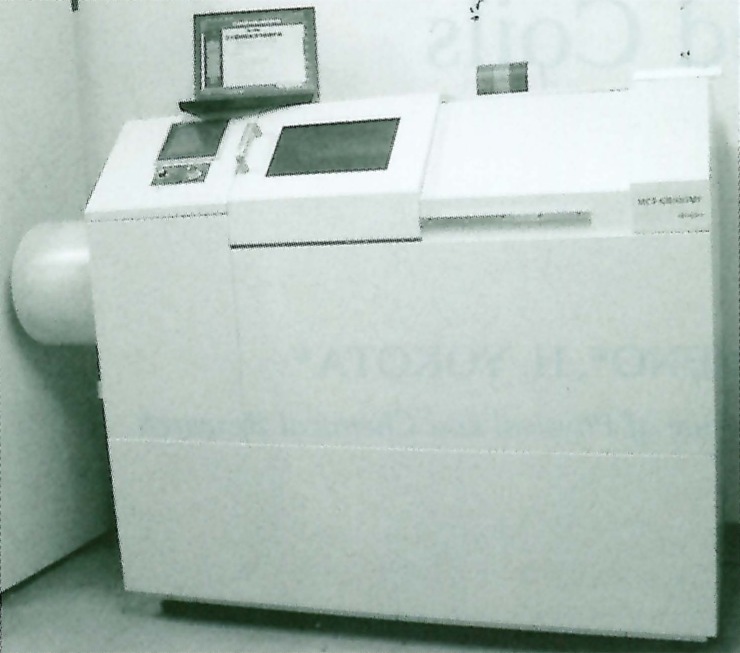
Specimens were observed by a micro-CT (MCT-CB100MF, Hitachi Medico, Tokyo, Japan) (figure 1).
Figure 1.
Micro CT Scanner. Macro CT and A4 size Laptop PC. Samples are placed in the scanning bay from the sliding door with handle. X ray tube us mounted at the left side and the image intensifier in the left.
This micro-CT was designed for industrial materials and had high spatial resolution. In calculation, 3 micrometers (figure Chart or Graph). With this high resolutional CT, we observed autopsy specimens of embolized aneurysm by fluoroscopy and slice images. An embolized aneurysm specimen was observed. Embolization procedure was uneventful. The patient died from other reason from embolization and autopsy specimen was obtained.
Figure 2.
Resolution of the Micro CT in calibration. Image intensifier has two field of view. One is four inch and the other is two. Optical magnification ranges from 1.2 to 10 times.
Then, models of aneurysm placing coils in them were observed. (We have already developed a silicone model system of patients’ aneurysms which have open lumen.) The structure of three dimensional coils and our retrieving wire (Soutenir®, Asahi Intec Seto Japan) was also observed.
The images from micro CT were compared with images from clinical equipments (DSA: Digitex 2400 Shimadzu Kyoto Japan, CT: HiSpeed, General Electric, Milwaukee,US).
Results
Using fluoroscopy of micro-CT, the primary filament of embolic coil could be observed, so kinking of the coil cold be visualized at the peripheral area of deposited coils. In clinical specimen, some kink of the coils were noted by fluoroscopy. The procedure had been uneventful. The aneurysm was packed tightly, so slice data had serious artifacts.
In model study, anchoring of 3D coils could be observed. In good condition of X ray, fine filament of our retrieving device could be observed. Data from clinical equipments were not enough to get detailed structure of coils. (figure 3).
Figure 3.
Micro CT images. Slice image (A) and fluoroscopic image (B, magnified view). Micro CT can visualize even primary filament.
Discussion
Micro-CT has much high spatial resolution to show primary filament of detachable coils. This level of resolution should be effective to find kinking before retrieving coils. Some of clinical fluoroscopy systems have 4.5 inch view. If two inch FOV or three or five times magnification in 4.5 inch II will be available, we will have possibility to observe this kind of fine information. At the same time, this should be a good warning for the risk on retrieving coils. VER (volume embolizarion ratio) is an effective factor to prevent coil compaction. More detailed VER could be calculated by high resolutional study. GDC structure in the aneurysm could be visualized by these high resolutional fluoroscopy and CT.
Conclusions
Coils in aneurysms were observed by micro-CT. With high resolutional fluoroscopy, kink on the detached coil could be observed. Retrieving coils are not always possible to retrieve. Some kink resistant system should be considered and high resolution should be beneficial to detect kinking.


