Abstract
It is well established that the expression of Bacillus thuringiensis (B.t.) toxin genes in higher plants is severely limited at the mRNA level, but the cause remains controversial. Elucidating whether mRNA accumulation is limited transcriptionally or posttranscriptionally could contribute to effective gene design as well as provide insights about endogenous plant gene-expression mechanisms. To resolve this controversy, we compared the expression of an A/U-rich wild-type cryIA(c) gene and a G/C-rich synthetic cryIA(c) B.t.-toxin gene under the control of identical 5′ and 3′ flanking sequences. Transcriptional activities of the genes were equal as determined by nuclear run-on transcription assays. In contrast, mRNA half-life measurements demonstrated directly that the wild-type transcript was markedly less stable than that encoded by the synthetic gene. Sequences that limit mRNA accumulation were located at more than one site within the coding region, and some appeared to be recognized in Arabidopsis but not in tobacco (Nicotiana tabacum). These results support previous observations that some A/U-rich sequences can contribute to mRNA instability in plants. Our studies further indicate that some of these sequences may be differentially recognized in tobacco cells and Arabidopsis.
Gene transfer into plants has become a relatively simple and routine process in many species. However, successful integration of a transgene into the plant genome does not automatically result in expression. For example, introduction of an additional copy of an endogenous gene can trigger cosuppression mechanisms, resulting in silencing of both the introduced and the endogenous genes (for review, see Meyer and Saedler, 1996). In the case of foreign genes transferred into plants, it is not uncommon to find that the introduced gene is incompatible with plant gene-expression mechanisms. Bacillus thuringiensis (B.t.)-toxin genes are perhaps the most widely known example of foreign genes for which it has been difficult to obtain useful levels of expression in transgenic plants (Diehn et al., 1996). Although a variety of mechanisms controlling gene expression have been blamed for poor B.t.-toxin expression, the exact cause remains controversial because of the limited amount of data gathered to address the problem. This is in part due to the inherent difficulties associated with studying a gene expressed at very low levels. It is also due to the fact that, for practical applications in agriculture, this problem can be circumvented by the brute-force approach of constructing highly modified synthetic versions of B.t.-toxin genes that yield high levels of expression. Nevertheless, elucidating the mechanisms that limit B.t.-toxin expression could make the design of new genes more efficient and provide insight about steps in gene expression that are relevant to endogenous plant genes.
A widely observed characteristic of B.t.-toxin gene expression in plants has been that little or no mRNA accumulates, even when transcription is under the control of strong promoters. Because mRNA instability provides a simple explanation, it has been widely assumed that B.t.-toxin transcripts are rapidly degraded in plants. However, this has not been clearly demonstrated, and the limited data that are available have led to contradictory conclusions about the potential role of mRNA stability. Instability of B.t.-toxin transcripts in plants was proposed initially to account for the lack of a correlation between promoter activity and mRNA accumulation in early efforts to express B.t.-toxin genes in plants (Fischhoff et al., 1987; Vaeck et al., 1987). In stably transformed tobacco (Nicotiana tabacum) plants, the majority of B.t.-toxin mRNA was present as transcripts that were shorter than full length (Barton et al., 1987). These short transcripts were assumed to be degradation products, and their presence was used to argue that B.t.-toxin mRNA was unstable. However, data were not presented that would distinguish between mRNA degradation and other mechanisms that could produce the short transcripts (such as premature transcription termination, splicing, or polyadenylation). The proposed instability of B.t.-toxin mRNAs was not tested experimentally in these studies.
It has been demonstrated that the construction of synthetic B.t.-toxin genes with highly modified nucleotide sequences can result in high expression in plants (for review, see Diehn et al., 1996). In those cases in which the effect of extensive sequence modifications on mRNA accumulation was examined, it was found that mRNA levels were increased substantially (Perlak et al., 1991; Adang et al., 1993). The design criteria for the synthetic genes has often included sequence changes targeted at potential mRNA instability elements (Perlak et al., 1990, 1991, 1993; Sutton et al., 1992; Adang et al., 1993; van der Salm et al., 1994). Although in all cases the modifications gave higher expression to varying degrees, the effects of the sequence changes on B.t.-toxin mRNA stability were not tested directly by comparing the stability of the transcripts encoded by the synthetic genes and that of corresponding unmodified genes. Because potential mRNA instability elements were only one of several types of targets for sequence modification, it was not possible to attribute the increased expression to a change in B.t.-toxin transcript stability.
The low accumulation of mRNA from wild-type B.t.-toxin genes in plants has presented a technical challenge for determining the role of mRNA stability in poor B.t.-toxin gene expression. Two previous efforts to assess the stability of B.t.-toxin mRNA in plants led to opposite conclusions after the turnover of cryIA(b) B.t.-toxin transcripts in protoplasts was characterized. Murray and coworkers (1991) showed that cryIA(b) transcripts disappeared faster qualitatively than octopine synthase transcripts after electroporation of plasmids carrying the genes into carrot protoplasts. In contrast, van Aarssen and coworkers (1995) concluded that cryIA(b) mRNA was not unstable in plants, based primarily on a comparison of turnover of in vitro-synthesized cryIA(b) and bar transcripts after electroporation into tobacco mesophyll protoplasts. It is unclear why the two groups reached opposite conclusions regarding cryIA(b) mRNA stability, but the differing results may stem from differences between carrot and tobacco and/or the different experimental methods used.
In the present study we used a well-established experimental system to determine the relative half-lives of mRNAs from a wild-type cryIA(c) and a highly modified synthetic cryIA(c) gene. Reliable half-lives are routinely measured for mRNAs transcribed from genes stably integrated into the nuclear genome using tobacco BY-2 cells (Newman et al., 1993; Ohme-Takagi et al., 1993; Taylor and Green, 1995; Sullivan and Green, 1996; van Hoof and Green, 1996). In this report we demonstrate that wild-type cryIA(c) transcripts are degraded more rapidly than synthetic cryIA(c) transcripts in stably transformed BY-2 cells, with a half-life comparable with that of transcripts known to be unstable in plants. Our results provide direct evidence that mRNA stability can be a factor limiting the expression of foreign genes in plants. We also show that the type of modifications typically used to construct highly expressing synthetic B.t.-toxin genes can function at least in part by stabilizing the transcripts in plants. The distribution of the sequence determinants within the wild-type B.t.-toxin transcripts that limit mRNA accumulation in tobacco cells and Arabidopsis plants differs. The differential recognition of these determinants between these two plant systems may have important implications for the engineering of foreign genes for optimal expression in plants and for the understanding of the mechanisms controlling accumulation of mRNA from endogenous genes.
MATERIALS AND METHODS
Plant Materials
Cells of transformed and untransformed tobacco (Nicotiana tabacum L. cv BY-2, also known as NT-1; Nagata et al., 1992) were grown as described previously (Newman et al., 1993). Gene constructs were introduced into BY-2 cells by Agrobacterium tumefaciens LBA4404-mediated transformation (Newman et al., 1993). Transformed cell lines in the form of calli were screened histochemically for GUS expression (Jefferson et al., 1986). Individual GUS-positive calli were transferred to liquid medium to generate suspension cultures to be used for mRNA half-life measurement. Pools of GUS-positive calli were used for gel-blot analysis of mRNA levels, for protein gel-blot analysis, and for isolation of nuclei for use in run-on transcription assays. Tobacco SR-1 plants were grown and transformed as described previously (Newman et al., 1993).
Arabidopsis ecotype Columbia plants were grown in soil under a 12-h light/12-h dark cycle at 20°C. Transgenic Arabidopsis plants were generated using the vacuum-infiltration method (van Hoof and Green, 1996). Seedlings were grown by germination of seeds on solid Arabidopsis growth medium (Taylor et al., 1993).
Maize (Zea mays cv BMS) cells (a gift from Pioneer Hi-Bred International, Inc., Des Moines, IA) were grown in suspension culture in BMS 237 medium (4.3 g/L Murashige-Skoog salts, 30 g/L Suc, 0.1 g/L myo-inositol, 1.8 mL of 10−2 m 2,4-D, and 2 mL/L 500× Murashige-Skoog vitamins [Sigma], brought to pH 5.6 with KOH) at 28°C in a rotary shaker at 150 rpm.
Synthetic Bacillus thuringiensis (B.t.)-Toxin Gene Construction
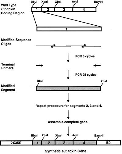
The synthetic cryIA(c) gene was constructed in four segments, each defined by restriction sites as shown in Figure 1, using a two-step PCR approach. Each synthetic segment was generated from a set of modified sequence oligonucleotides (Macromolecular Structure Facility, Michigan State University) spanning each segment. Sets of four oligonucleotides were used to synthesize segments 1 and 2, and sets of six and eight oligonucleotides were used for segments 3 and 4, respectively. The oligonucleotides were designed with 25-bp complementary overlaps so that when annealed, they served as primers and templates for DNA synthesis by PCR. All oligonucleotides were used directly in PCR without purification. PCR was carried out according to the protocol of Dillon and Craig (1990). Eight PCR cycles were performed using the overlapping modified-sequence oligonucleotides in 100-μL reaction volumes. PCR products contained in 1 μL of the first reaction were used as templates for a second set of PCR cycles.
Figure 1.
Schematic representation of the method used to construct the synthetic B.t.-toxin gene. A two-step PCR approach was used to generate synthetic versions of each of four segments of a cryIA(c) gene using sets of overlapping oligonucleotides that incorporated sequence changes according to the criteria described in the text. As shown for one of the four segments, the alternating sense and antisense polarity of the overlapping modified-sequence oligonucleotides indicated by the directions of the arrows allowed the oligonucleotides to anneal to each other and serve as primers for DNA synthesis in PCR. After the first set of 10 PCR cycles, the addition of 30-mer terminal primers followed by a further 25 PCR cycles preferentially amplified those molecules spanning the full segment. PCR products for each segment were cloned and then assembled by standard cloning methods to generate the complete synthetic B.t.-toxin-coding region.
Thirty-base-pair terminal primers complementary to the ends of PCR products spanning the entire gene segment were used to preferentially amplify the full-length molecules over 25 cycles. PCR products of the correct size were cut out of low-melting-point agarose gels and cloned after removal of terminal adenosines into the EcoRV site of a Bluescript II SK(−) vector with a modified multiple cloning site (p948) (Diehn et al., 1998). Clones containing inserts were screened by dideoxy sequencing (Sanger et al., 1977) for PCR products with no or only one to two sequence errors. Sequencing was done with a 7-deaza-dGTP Sequenase sequencing kit (United States Biochemical) to overcome compression artifacts caused by the high G/C content of the modified sequence.
For those synthetic segments for which no clone was found to be entirely free of sequence errors, in vitro mutagenesis (Kunkel et al., 1987) was used to introduce the correct sequence. All segments were then assembled in pUC118 by standard cloning methods using the restriction sites shown in Figure 1 to generate the complete synthetic cryIA(c)-coding region (p1334). The synthetic coding region was sequenced on both strands to verify that the sequence was correct. The nucleotide sequence of the synthetic gene has been deposited in the GenBank Sequence Database. The modified sequence oligonucleotides are described in the annotation section of the GenBank entry. The frame-shift synthetic B.t.-toxin gene described in the text resulted from insertion of a single cytosine 498 bp downstream of the adenosine of the translational start ATG codon (p1329).
Gene Constructions
The plasmid p1204 (Diehn et al., 1998) shown in Figure 2 containing the truncated cryIA(c)-coding region flanked by 2X35S (Diehn et al., 1998) and the 3′ UTR from the pea Rubisco small subunit rbcS-E9 gene was used for transient expression of B.t.-toxin genes in BY-2 cells. The synthetic cryIA(c)-coding region was excised from p1334 (see previous section) with BbuI and BamHI and substituted for the wild-type cryIA(c)-coding region in p1204, which was also cut with BbuI and BamHI to generate p1335 (Fig. 2) for transient expression in BY-2 cells. Expression of the internal reference β-globin gene in BY-2 cells was from p1185 (Fig. 2) (Diehn et al., 1998).
Figure 2.
Schematic diagram of B.t.-toxin and control gene constructions. Expression of wild-type B.t.-toxin, synthetic B.t.-toxin, and globin genes in transient expression assays and stably transformed cell lines and plants was under the control of 2X35S. For transient expression in maize, constructs included an ADH1 intron. Polyadenylation signals were provided by the 3′ UTR from the pea Rubisco small subunit rbcS-E9 gene in all constructs. Plasmid numbers for each construct are indicated at the left.
Similar plasmid constructions were used for transient expression in maize BMS cells but included an ADH1 intron inserted between the promoter and the coding region. For transient expression of the synthetic cryIA(c) gene in BMS cells, the cryIA(c)-coding region and rbcS-E9 3′ UTR contained in a fragment excised from p1335 with BglII and ClaI were inserted into p1138 (Diehn et al., 1998) containing the 2X35S and ADH1 intron cut with BamHI and ClaI to yield p1345 (Fig. 2). The wild-type cryIA(c)-coding region and rbcS-E9 3′ UTR were excised from p995 (Diehn et al., 1998) with BbuI and BamHI and substituted for the synthetic cryIA(c)-coding region in p1345 cut with BbuI and BamHI to yield p1346 (Fig. 2). To generate the globin gene construction for use in BMS cells (p1160), the human β-globin-coding region was excised with NcoI and XbaI from the previously described plasmid p962 (Newman et al., 1993) and inserted into the EcoRV site of a Bluescript II SK(−) vector with a modified multiple cloning site (p948) to yield p963. The β-globin-coding region was then excised from p963 with XbaI and BamHI and substituted for the GUS-coding region in p840, a pUC11 derivative containing a CaMV 35S-GUS-E9 expression construct (details on request) cut with XbaI and BamHI to yield p977 containing the expression cassette CaMV 35S-globin-E9. The β-globin-coding region and rbcS-E9 3′ UTR were excised with BglII and ClaI from p977 and inserted into p1138 cut with BamHI and ClaI to yield p1160 (Fig. 2) containing the expression cassette 2X35S-ADH1 intron-globin-E9.
For transformation into tobacco and Arabidopsis, the wild-type and synthetic 2X35S-cryIA(c)-E9 expression cassettes were transferred to the unique HindIII site in pBI121 as HindIII fragments excised from p1204 and p1335 to yield p1205, containing the wild-type cryIA(c), and p1410, containing the synthetic cryIA(c). The direction of transcription of the cryIA(c) genes was the same as that of the GUS gene in the pBI121 plasmid.
Chimeric wild-type/synthetic cryIA(c) genes were constructed by substituting wild-type segments for the corresponding segments in the synthetic gene. Wild-type segment 1 was excised from p1310, a pUC118 plasmid containing only the wild-type cryIA(c)-coding region, with BbuI and XbaI and inserted into p1334 cut with BbuI and XbaI, yielding p1405. Synthetic segment 2 was added back as an XbaI fragment from p1334 by insertion into p1405 cut with XbaI to give p1440 (wt1-syn2-syn3-syn4). Wild-type segment 2 was excised from p1310 as an XbaI fragment and inserted into p1334 cut with XbaI to yield p1406 (syn1-wt2-syn3-syn4). Wild-type segment 3 was excised with XbaI and AccI from p1310 and inserted into p1334 cut with XbaI and AccI to yield p1407. Synthetic segment 2 was added back as an XbaI fragment from p1334 by insertion into p1407 cut with XbaI to yield p1442 (syn1-syn2-wt3-syn4). Wild-type segment 4 was excised from p1310 AccI and BamHI and inserted into p1334 cut with AccI and BamHI to yield p1411 (syn1-syn2-syn3-wt4). To assemble expression cassettes, all four chimeric coding regions were excised with BbuI and BamHI and substituted for the synthetic cryIA(c)-coding region in p1335 cut with BbuI and BamHI to yield p1450, p1451, p1452, and p1454 containing wild-type segments 2, 4, 1, and 3, respectively, in the chimeric coding regions. For transformation into BY-2 cells and Arabidopsis plants, transformation constructions were made by excision of the four expression cassettes as HindIII fragments and insertion into the HindIII site in pBI121 in the same transcriptional orientation as the GUS gene in pBI121 to give p1505, p1506, p1507, and p1508 containing wild-type segments 1, 2, 3, and 4, respectively, in the chimeric cryIA(c)-coding regions.
Electroporation of Tobacco and Maize Protoplasts
Protoplasts of tobacco BY-2 cells were prepared and transiently transformed by electroporation as described previously (van Hoof and Green, 1996), with the exception that the plasmids introduced into protoplasts carried wild-type cryIA(c), synthetic cryIA(c), or globin expression constructions described above, and 1 h after electroporation, protoplasts were transferred from the 24-well plate used for electroporation to 100-mm plates containing 8 mL of plating medium (80 mL of Nicotiana tabacum medium [Newman et al., 1993], 20 mL of BY-2 culture supernatant, and 400 mm mannitol, brought to pH 5.7 with KOH).
Maize BMS protoplasts were prepared and electroporated by the same method but with the following substitutions. NT wash solution was replaced with BMS wash solution (250 mm mannitol, 50 mm CaCl2, and 5 mm Mes, brought to pH 5.5 with KOH). NT electroporation buffer was replaced with BMS electroporation buffer (200 mm mannitol, 120 mm KCl, 10 mm NaCl, 4 mm CaCl2, and 10 mm Hepes, brought to pH 7.2 with NaOH). NT plating medium was replaced with BMS plating medium (80 mL of BMS 237 medium, 20 mL of BMS culture supernatant, and 300 mm mannitol, brought to pH 5.6 with KOH). Electroporator settings were 250 V and 960 μFD.
RNA Isolation
Total RNA was isolated from BY-2 cells, tobacco protoplasts, maize protoplasts, tobacco leaves, and Arabidopsis plants using the guanidinium thiocyanate protocol described previously (Newman et al., 1993) with the following modifications: When isolating RNA from pooled BY-2 calli, a total of four LiCl precipitations were performed. For RNA isolations from BY-2 and maize protoplasts, the initial grinding step was omitted. In RNA extractions from pooled BY-2 cell lines expressing the chimeric B.t.-toxin genes, frozen cells were lyophilized overnight and then ground by vortexing the cells with 3-mm glass beads until reduced to a fine powder as the first step in the extraction protocol. In all RNA isolations, a phenol-extraction step was added immediately after resuspension of the final LiCl2 pellet.
RNA Gel Blots
For RNA gel-blot analysis, all RNA samples (20 μg/lane) were separated by electrophoresis on 1% agarose/2% formaldehyde gels using 1× Mops buffer (20 mm Mops, 5 mm sodium acetate, 1 mm EDTA, 1 μg/mL ethidium bromide). After capillary transfer to Biotrace HP (Gelman Sciences, Ann Arbor, MI), blots were prehybridized for at least 4 h at 52°C in 5× SSC, 10× Denhardt's solution (Sambrook et al., 1989), 0.1% SDS, 0.1 m potassium phosphate, pH 6.8, and 100 μg/mL sheared and denatured herring-sperm DNA. Blots were hybridized for 16 to 24 h at 52°C in the same solution with 10% (w/v) dextran sulfate and 30% (v/v) deionized formamide added and SDS omitted. RNA isolated from electroporated protoplasts was treated with 1 unit of RQ1 DNase I (Promega) for 15 min at 37°C to remove remaining plasmid DNA before electrophoresis.
DNA Probes
All DNA probes hybridized to RNA gel blots were labeled by the random-primer method (Feinberg and Vogelstein, 1983). Probes hybridized to time-course RNA gel blots for mRNA half-life measurement and chimeric gene RNA blots were double labeled with [α-32P]dCTP and [α-32P]dATP to improve signal detection. All other probes were single labeled with [α-32P]dCTP. Labeled probes were separated from unincorporated nucleotides using Sephadex G-50 medium (Sigma) spin or push columns (Stratagene). All probes were made from restriction fragments isolated as bands cut from low-melting-point agarose gels and used directly in the labeling reactions. Wild-type B.t.-toxin, synthetic B.t.-toxin, and GUS probes consisted of the entire coding regions of each gene. The E9 probe corresponded to a restriction fragment isolated with BamHI and ClaI from plasmid p1425, with a Bluescript derivative containing the rbcS-E9 gene 3′ UTR as an insert. Chimeric B.t.-toxin gene RNA gel blots were hybridized simultaneously with wild-type and synthetic B.t.-toxin double-labeled probes with similar specific activities.
Half-Life Determination
Half-life measurements were made using stably transformed BY-2 cell lines grown as suspension cultures 3 d after subculturing. Fifty-milliliter cultures were treated with 0.5 μg/mL CHX for 2 h, and then the cells were pelleted at 1000 rpm in a centrifuge (model RT-6000D with model H-1000B rotor, Sorvall) for 1 min and resuspended in 50 mL of medium without CHX. Five-milliliter aliquots of cells were harvested immediately before and after the CHX treatment by pelleting at 1000 rpm for 1 min, aspirating the medium, and freezing the pellets in liquid nitrogen. Immediately before CHX removal, a portion of the culture was removed and maintained in the presence of CHX until the end of the time course, when the cells were harvested. The remaining cells were treated with 100 μg/mL ActD and aliquots were harvested at intervals over a 120-min time course. Transcript decay was visualized by RNA gel-blot analysis of the time-course samples. Blots were hybridized with B.t.-toxin probe, and then stripped and rehybridized with the GUS probe. Hybridization signals were quantitated using a phosphor imager (Molecular Dynamics, Sunnyvale, CA), and the values were subjected to linear regression analysis using SigmaPlot software (Jandel Scientific, Corte Madera, CA) to determine mRNA half-lives.
Protein Analysis
Soluble protein extracts were made from Arabidopsis leaves pooled from nine independent lines expressing the wild-type B.t.-toxin gene and 15 independent lines expressing the synthetic B.t.-toxin gene. Leaves were ground in liquid nitrogen and extraction buffer (250 mm NaPO4, pH 7.4, 5 mm EDTA, and 25 μg/mL leupeptin) with a mortar and pestle. Cellular debris were removed by centrifugation in microcentrifuge tubes. Protein concentration of the supernatant was determined by the Bradford assay (Bio-Rad), and proteins (60 μg/lane) were separated by 10% SDS-PAGE (Laemmli, 1970). Fifty nanograms of purified cryIA(b)/cryIA(c) protein (a gift from Novartis, Research Triangle Park, NC) was also run on the gel to serve as a positive control in immunoblot analysis and for estimating the amount of synthetic B.t.-toxin present in the protein extracts. Prestained high-Mr protein size markers (GIBCO-BRL) were also included. Protein was transferred to a PVDF membrane (Immobilon P, Millipore) using a semidry blotting apparatus (BioTrans model B, Gelman Sciences) according to the manufacturer's instructions. Proteins were visualized after transfer to the membrane with Ponceau S (Sigma) staining to verify transfer and equal loading. B.t.-toxin protein was detected by immunoblot analysis (Birkett et al., 1985). Affinity-purified polyclonal goat antibodies raised against crystals isolated from B.t. subsp. kurstaki HD-1 (a gift from Novartis) were used at a dilution of 1:1000. Secondary antibodies linked to horseradish peroxidase (Kirkegaard and Perry Laboratories, Gaithersburg, MD) were used at a dilution of 1:2000.
Insect Bioassays
Bioassays of insecticidal activity of BY-2 cell lines expressing the wild-type or synthetic B.t.-toxin genes were carried out with crude whole-cell extracts using a protocol based on a previously published method (Tailor et al., 1992). Suspension cultures of transformed and untransformed BY-2 lines were pelleted by centrifugation and frozen in liquid nitrogen. Cells were lyophilized for at least 24 h and then ground to a fine powder by vortexing the cells with glass beads. Ten milligrams of ground cell material from each cell line was suspended in 2.5 mL of sterile water and spread onto solid tobacco hornworm medium (Carolina Biological Supply, Burlington, NC) in 100- × 25-mm plates. Tobacco hornworms were hatched from eggs according to the supplier's instructions (Carolina Biological Supply), and 15 hornworms were placed on each plate within 24 h after hatching. Plates were kept in continuous light at 27°C for the duration of the bioassay.
For bioassays of insecticidal activity in transgenic Arabidopsis, T2 seeds were pooled from 16 lines expressing the wild-type B.t.-toxin gene and 16 lines expressing the synthetic B.t.-toxin gene. Pooled transgenic seeds and untransformed seeds were sterilized in 50% bleach solution for 7 min, washed in sterile water, and then plated on primary-selection Arabidopsis growth medium plates (4.3 g/L Murashige-Skoog salts, 1× B5 vitamins, 1% [w/v] Suc, 0.5 g/L Mes, and 0.8% Phytagar, brought to pH 5.7 with KOH) without drugs for germination. After 2 weeks, approximately 100 seedlings were transferred to 150- × 15-mm plates containing two layers of Whatman paper (Fisher Scientific) dampened with sterile water. Within 24 h after hatching, 15 tobacco hornworms were transferred to each of the plates and kept under continuous light at 27°C for the course of the assay.
Isolation of Nuclei from BY-2 Cells
Three independent pools of BY-2 calli transformed with the wild-type B.t.-toxin gene and three independent pools of BY-2 calli transformed with the synthetic B.t.-toxin gene, each containing 90 independent transformed lines, were produced. Pooled calli (7–10 mL packed cell volume) were suspended in 20 mL of NT wash solution and protoplasted as described previously (van Hoof and Green, 1996). For nuclei isolation, all steps were done on ice with prechilled solutions and centrifugation at 4°C. Protoplasts were transferred gently from the protoplasting incubations in 150- × 15-mm plates to 50-mL conical tubes using a wide-bore pipet. Protoplasts were pelleted (700 rpm in the Sorvall centrifuge and rotor) and very gently resuspended in 10 mL of NT wash solution by slowly rolling the tube. The volume was brought to 40 mL with NT wash solution, and the protoplasts were pelleted again as above. To lyse the protoplasts, they were resuspended in nuclei-isolation buffer (De Rocher and Bohnert, 1993) plus 1.0% (v/v) Triton X-100. Nuclei were pelleted at 1000 rpm for 10 min, and the supernatant was removed by aspiration. Pellets were resuspended gently in 500 μL of nuclei-storage buffer (De Rocher and Bohnert, 1993) using a pipeter (P1000, Rainin, Woburn, MA) with a wide-bore tip. Nuclei were divided into 100-μL aliquots, frozen in liquid nitrogen, and stored at −80°C.
Nuclear Run-On Assays
Incorporation of radiolabeled nucleotides into run-on transcripts, isolation of run-on transcripts, preparation of DNA slot blots, and hybridization of the run-on transcripts to the DNA blots were all performed as described previously (De Rocher and Bohnert, 1993), with the following modifications: DNA probes slot-blotted to nitrocellulose (Schleicher & Schuell) to serve as targets for hybridization of run-on transcripts were single stranded to reduce random background hybridization to vector sequences. Single-stranded DNA containing the antisense strand of the probe was prepared by infecting Escherichia coli cultures carrying the appropriate plasmids with VCSM13 helper phage (Stratagene) and isolating the single-stranded DNA from the culture medium. Two-and-one-half micrograms of DNA was blotted for each slot. The following single-stranded phagemid probes were used in the run-on assays. The pea rbcS-E9 3′ UTR common to both the wild-type and synthetic B.t.-toxin transcripts was contained in the p1425 plasmid described above. The GUS-coding region was contained in the plasmid p1414. To control for random background hybridization to vector sequences, Bluescript SK(+) plasmid without any insert was also included on all blots. Hybridization signals were detected and quantitated using a phosphor imager. Background hybridization to vector was subtracted from the B.t.-toxin and GUS hybridization signals.
RESULTS
Design and Construction of a Synthetic B.t.-Toxin Gene Incorporating Modifications That Enhance Expression in Plants
A cryIA(c) gene from B.t. subsp. kurstaki HD-73 (Adang et al., 1985) (kindly provided by Dr. A.I. Aronson, Purdue University, West Lafayette, IN) served as the basis for all B.t.-toxin gene constructs used in the experiments presented here. Specifically, a truncated form of the cryIA(c) gene encoding amino acids 9 through 613, which constitute the insecticidal domain of the 1179-amino acid B.t.-toxin protein (Schnepf and Whiteley, 1985), was used. Insecticidal activity is not dependent on the C-terminal domain, as was demonstrated by deletion analysis in E. coli (Adang et al., 1985), and deletion of this domain is usually necessary to achieve even minimal levels of expression in plants (Barton et al., 1987; Vaeck et al., 1987). The truncated coding region was fused to the sequence encoding the N-terminal 9 amino acids of LacZ, and the translation initiation site was altered to match the plant consensus sequence (Lütcke et al., 1987). Two Pro codons were added to the 3′ end of the coding region for protection of the protein C terminus from protease activity (Bigelow and Channon, 1982; Barton et al., 1987). Signals for polyadenylation present in the 3′ UTR from the pea Rubisco small subunit rbcS-E9 gene (Mogen et al., 1992) provided proper 3′ end processing. This cryIA(c) gene was used as the starting point in the design and construction of a highly modified synthetic gene. The truncated cryIA(c) gene, which contains unmodified wild-type nucleotide sequence, will be referred to in this paper as the wild-type B.t.-toxin gene to distinguish it from the synthetic B.t.-toxin gene.
It has been demonstrated for several B.t.-toxin genes that extensive modification of the coding-region nucleotide sequence can result in increased expression (for review, see Diehn et al., 1996). The A/T richness typical of B.t.-toxin genes tends to occur at the third position of codons, and codons with A or T in the third position tend to be rarely used in plants. Therefore, we chose a strategy that improved the codon usage and simultaneously eliminated the A/T richness. Specifically, we introduced a codon bias typical of maize genes (Wada et al., 1992) without altering the original amino acid sequence. This codon bias was chosen in part because of the extensive data that were available on codon usage in maize genes and the importance of maize and other cereals for agriculture. Moreover, because the codon bias in monocot genes tends to be more stringent than in dicot genes and the set of preferred codons in maize is a subset of those typically used in dicots (Campbell and Gowri, 1990), the codon bias introduced into the B.t.-toxin gene would be compatible with expression in dicot as well as in monocot species.
The strong preference for G or C in the third position in maize genes had the benefit of eliminating by default A/T-rich sequences that might be recognized in plants as unwanted signals for RNA processing and turnover. Other design criteria were to preserve several restriction sites that would be useful in the construction of the synthetic gene, to avoid introducing restriction sites that would interfere with construction or cloning steps, and to avoid introducing A/T-rich sequences and theoretically stable secondary structures. Table I summarizes the results of the sequence modifications and shows that sequence changes were distributed uniformly throughout the coding region. Of 610 codons, 496 were altered, resulting in an increase in G/C content from 38% to 64%.
Table I.
Summary of differences between the wild-type and synthetic B.t.-toxin genes
| Segment | Length | Nucleotides Changed | Codons Changed | Initial G/C Content | Final G/C Content |
|---|---|---|---|---|---|
| bp | no. | % | |||
| 1 | 274 | 82 | 75 /91 | 38 | 66 |
| 2 | 375 | 106 | 96 /125 | 38 | 65 |
| 3 | 531 | 167 | 146 /177 | 37 | 64 |
| 4 | 653 | 190 | 179 /217 | 38 | 63 |
| Total | 1833 | 548 | 496 /610 | 38 | 64 |
After a modified sequence meeting all of the above criteria was designed, the synthetic B.t.-toxin gene was constructed using the PCR-based approach shown in Figure 1. The modified-sequence oligonucleotides ranged in size from 81 to 132 bases in length. Synthesis of segments 1 and 2 was done using four overlapping oligonucleotides (Fig. 1). Segments 3 and 4 were generated using sets of six and eight overlapping oligonucleotides, respectively, because of the greater lengths of these segments. PCR products were cloned and sequenced to identify synthetic B.t.-toxin gene segments with the fewest or no sequence errors. Where necessary, sequence errors were corrected by in vitro mutagenesis. After assembly of all segments, the complete synthetic coding region was resequenced on both strands to verify that the sequence was correct.
Differential mRNA Accumulation Resulting from Sequence Modifications Incorporated into the Synthetic B.t.-Toxin Gene
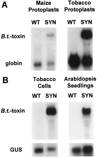
To verify that the design of the synthetic B.t.-toxin gene had the anticipated effect on mRNA accumulation when expressed in plants, plasmids containing either the wild-type or synthetic genes driven by 2X35S were electroporated into protoplasts generated from maize BMS and tobacco BY-2 cell lines. Plasmids containing a human β-globin gene driven by a CaMV 35S promoter were co-electroporated to provide an internal reference for comparison of the relative levels of wild-type and synthetic B.t.-toxin mRNAs (Gil and Green, 1996; Sullivan and Green, 1996). Figure 3A shows the results of representative RNA gel-blot analyses of the electroporation experiments. In all experiments, wild-type B.t.-toxin transcripts were not detected but synthetic B.t.-toxin transcripts accumulated to levels comparable with those of the globin transcripts. The high level of synthetic B.t.-toxin mRNA accumulation in the tobacco BY-2 protoplasts showed, as expected, that the sequence changes incorporated into the synthetic gene were effective in dicot and in monocot species.
Figure 3.
RNA gel-blot analysis of relative expression levels of the synthetic and wild-type B.t.-toxin genes in plant cells. Accumulation of synthetic (SYN) and wild-type (WT) B.t.-toxin mRNAs was compared in transiently transformed maize and tobacco cells and in stably transformed tobacco cells and Arabidopsis plants. A, Plasmids containing wild-type or synthetic B.t.-toxin genes were electroporated into maize BMS and tobacco BY-2 protoplasts. Plasmids containing a human β-globin gene were coelectroporated in both types of experiments to serve as an internal standard. The positions of the B.t.-toxin and globin transcripts are indicated. B, Tobacco BY-2 cells and Arabidopsis plants were stably transformed with constructs containing wild-type or synthetic B.t.-toxin genes and a GUS gene serving as an internal standard. The positions of the B.t.-toxin and GUS transcripts are indicated.
Wild-type and synthetic B.t.-toxin gene expression was also assessed in stably transformed BY-2 cells because this cell line is well established as a system for the measurement of mRNA half-lives (Newman et al., 1993) and could also be used for measuring relative transcription rates of the two genes. Wild-type and synthetic B.t.-toxin genes driven by 2X35S were inserted into pBI121 vector and introduced into BY-2 cells by A. tumefaciens-mediated transformation. The CaMV 35S-driven GUS gene present in the pBI121 plasmid produces a relatively stable transcript in plants (Newman et al., 1993) and served as a co-transformed internal standard for comparison of the expression of the two B.t.-toxin genes in independently transformed cell lines. Kanamycin-resistant calli were screened histochemically for GUS expression, and the calli from more than 90 independent GUS-positive lines were pooled for RNA extraction. Independent lines were pooled to average out possible variations in expression levels among individual lines arising from differences in transgene copy number and from position effects on transgene transcriptional activity as a result of different sites of genomic integration. The left panel in Figure 3B shows a representative RNA gel blot of pools of BY-2 cell lines stably transformed with the wild-type or synthetic B.t.-toxin genes. As in electroporated protoplasts, wild-type B.t.-toxin transcripts were undetectable, whereas accumulation of the transcripts encoded by the synthetic B.t.-toxin gene was comparable with that of transcripts from the internal reference gene.
To verify that the expression pattern seen in transiently transformed protoplasts and stably transformed cell lines was representative of intact plants, we attempted to introduce both B.t.-toxin genes into tobacco plants by A. tumefaciens-mediated transformation (Newman et al., 1993). Shoots regenerated on kanamycin-containing medium were screened histochemically for GUS activity. Although nearly all plants regenerated from the transformation with the wild-type B.t.-toxin gene were GUS positive, almost no plants regenerated from the synthetic B.t.-toxin transformation had detectable GUS activity. The few plants that were GUS positive exhibited no detectable B.t.-toxin transcript accumulation in RNA gel-blot analysis (data not shown). Because initial shoot formation was noticeably delayed for plants regenerated from the synthetic B.t.-toxin transformation, most of these plants were likely to be escapes or to have low transgene expression due to position effects. Repeated transformation of both constructs yielded similar results.
To rule out the possibility of a defect in the synthetic B.t.-toxin transformation construct, the plasmid was rebuilt and the transformation procedure repeated for the wild-type and synthetic genes, with similar results. Parallel transformations with a third construct containing a synthetic B.t.-toxin gene with a frame-shift mutation yielded GUS-positive plants as readily as transformations done with the wild-type B.t.-toxin gene. The frame-shifted synthetic B.t.-toxin gene was recovered during the construction of the synthetic gene, and was the result of a single C insertion, resulting in a premature stop codon about one-third of the way into the coding region. This premature stop codon would prevent expression of an insecticidally active B.t.-toxin protein, since the portion of the protein that would remain untranslated is known to be necessary for activity (Fischhoff et al., 1987).
Because the frame-shifted and synthetic B.t.-toxin transformation constructs were identical except for a single C insertion and the frame-shifted construct could not produce active B.t. toxin, the difference in transformation results may be indicative of a toxic effect of highly expressed active B.t.-toxin protein in regenerating tobacco cells. In contrast, expression of the synthetic B.t.-toxin gene in tobacco calli and suspension cultures had no discernible deleterious effect.
To determine whether this problem was specific for tobacco plants, the same wild-type and synthetic B.t.-toxin transformation constructs were introduced into Arabidopsis by A. tumefaciens-mediated transformation using vacuum infiltration (van Hoof and Green, 1996). The right panel of Figure 3B shows a gel blot of RNA extracted from leaves pooled from 15 independent kanamycin-resistant lines for each construct. As seen in protoplasts and stably transformed tobacco cells, wild-type B.t.-toxin mRNA was undetectable, but synthetic B.t.-toxin mRNA accumulated to an easily detected level. The recovery of Arabidopsis plants expressing the synthetic B.t.-toxin gene indicates that the problem encountered with generation of transgenic tobacco plants is not a general one.
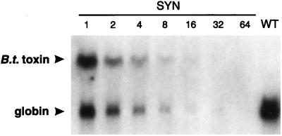
The sequence changes incorporated into the synthetic B.t.-toxin gene substantially increased mRNA accumulation over the wild-type B.t.-toxin gene in protoplasts and in stably transformed tobacco cell lines and Arabidopsis plants. It was not possible to quantitate the magnitude of the increase, because wild-type B.t.-toxin transcripts did not accumulate to detectable levels. However, RNA gel-blot analysis performed on a dilution series of RNA from cells expressing the synthetic B.t.-toxin gene was able to give a minimum estimate of the magnitude of increase by determining how many times the hybridization signal could be diluted before decreasing to background levels. Figure 4 shows the results of such an experiment using total RNA from a transient-expression experiment in tobacco protoplasts that produced the highest hybridization signals above background. The synthetic B.t.-toxin hybridization signal was still detectable above background at a 16-fold dilution, indicating that B.t.-toxin mRNA accumulation was improved at least 16-fold by the sequence modifications. Other experiments with overloaded gels indicated that the difference was at least 64-fold (data not shown).
Figure 4.
Estimation of the minimum difference in mRNA accumulation between wild-type (WT) and synthetic (SYN) B.t.-toxin genes. Tobacco BY-2 protoplasts were electroporated with plasmids containing either wild-type or synthetic B.t.-toxin genes and plasmids containing a human β-globin gene as an internal standard. RNA from protoplasts expressing the synthetic B.t.-toxin gene was diluted in 2-fold steps up to 64-fold with total RNA from untransformed tobacco cells. Each lane was loaded with 20 μg of RNA. The positions of the B.t.-toxin and globin transcripts are indicated by arrowheads. The blot was hybridized with a probe specific for the rbcS-E9 3′ UTR common to all three transcripts. Dilution factors are indicated by numbers above the lanes.
High-Level Accumulation of Functional Protein Produced by the Synthetic B.t.-Toxin Gene
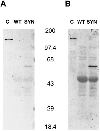
The greater mRNA accumulation from the synthetic B.t.-toxin gene would be expected to be accompanied by a corresponding increase in B.t.-toxin protein accumulation. This was tested by protein gel-blot analysis. Figure 5 shows the results of probing a protein gel blot of extracts from pooled Arabidopsis lines independently transformed with either the wild-type or the synthetic B.t.-toxin genes with anti-cryIA(c) antibodies. B.t.-toxin protein was not detectable in plants expressing the wild-type gene, but a band of approximately the expected size (69 kD) was detected in protein extracts from plants expressing the synthetic B.t.-toxin gene. Based on comparison with signals produced by known quantities of purified cryIA(c) protein (data not shown), we estimate that B.t.-toxin protein accumulated to at least 0.1% of total protein in Arabidopsis plants, comparable to levels previously observed in other species (Diehn et al., 1996). From this result, it is apparent that the maize codon bias used in the synthetic gene is compatible with translation in dicots and results in significant protein accumulation.
Figure 5.
Accumulation of B.t.-toxin protein in plants expressing the wild-type and synthetic B.t.-toxin genes as determined by SDS-PAGE analysis of protein extracts from pools of independent Arabidopsis lines stably transformed with the wild-type (WT) or synthetic (SYN) B.t.-toxin genes. Purified cryIA(c) protein (predicted molecular mass approximately 133 kD) expressed in E. coli was included as a positive control (C) for anti-cryIA(c) antibody binding. Positions of molecular mass standards (in kD) are indicated between the panels. A, Immunoblot incubated with anti-cryIA(c) polyclonal antibodies. B, Immunoblot of duplicate lanes incubated with anti-cryIA(c) polyclonal antibodies and overdeveloped to demonstrate the lack of detectable B.t. toxin in plants transgenic for the wild-type B.t.-toxin gene.
The functionality of the protein expressed in stably transformed Arabidopsis seedlings and BY-2 cells was assessed in bioassays of insecticidal activity against tobacco hornworm. For bioassays of Arabidopsis, hornworm larvae were given as a sole food source seedlings pooled from 16 independent lines transgenic for either the wild-type or synthetic B.t.-toxin genes. Untransformed wild-type seedlings were used as a control for the toxic effects of Arabidopsis seedlings. No hornworms survived on Arabidopsis seedlings expressing the synthetic B.t.-toxin gene 3 d after initiation of feeding, as shown in Table II. Survival was 100% for hornworms feeding on Arabidopsis seedlings expressing the wild-type B.t.-toxin gene or on untransformed seedlings.
Table II.
Results of bioassays of B.t.-toxin activity against tobacco hornworm in tobacco BY-2 cells and Arabidopsis seedlings
| Species | B.t.-Toxin Gene | Cell Line | Larval Survivala |
|---|---|---|---|
| Arabidopsis | Synthetic | 0 /15 | |
| Wild type | 15 /15 | ||
| Controlb | 15 /15 | ||
| Tobacco | Synthetic | SYN1 | 1 /15 |
| SYN2 | 0 /15 | ||
| SYN3 | 0 /15 | ||
| SYN4 | 0 /15 | ||
| Wild type | WT1 | 15 /15 | |
| WT2 | 1 /15 | ||
| Controlc | C1 | 15 /15 |
Larval survival rate after 3 d of feeding on Arabidopsis seedlings and after 7 d of feeding on tobacco BY-2 cells.
Untransformed Arabidopsis seedlings.
Untransformed tobacco BY-2 cells.
To examine B.t.-toxin insecticidal activity in BY-2 cells, crude extracts of stably transformed cell lines expressing either the wild-type or the synthetic B.t.-toxin genes were applied to the surfaces of plates containing solid tobacco hornworm medium. Crude extracts of untransformed BY-2 cells were used as a control for any toxicity of the BY-2 cells. Extracts from all cell lines expressing the synthetic B.t.-toxin gene resulted in almost no surviving hornworms. The effect of extract from the wild-type gene WT1 BY-2 line was indistinguishable from that of extract from an untransformed BY-2 cell line, with 100% survival observed. A second BY-2 line expressing the wild-type gene WT2 exhibited a higher but still very low level of B.t.-toxin mRNA accumulation than the WT1 cell line in RNA gel-blot analysis (data not shown), which was likely the result of positional effects. Extract from the WT2 line resulted in almost no hornworm survival, demonstrating that even low-level expression of a wild-type B.t.-toxin gene can give insecticidal activity against a sensitive species such as tobacco hornworm, as observed previously (Fischhoff et al., 1987; Delanney et al., 1989). The results of the bioassays demonstrated, as expected, that the product of the synthetic B.t.-toxin gene has marked insecticidal activity.
Relative Transcriptional Activities of Wild-Type and Synthetic B.t.-Toxin Genes
The observed disparity in mRNA accumulation between the wild-type and synthetic B.t.-toxin genes could be explained by the presence of sequences in the wild-type B.t.-toxin-coding region that impair transcriptional activity when expressed in plants (Adang et al., 1987). To test this possibility, the relative transcriptional activities of the wild-type and synthetic B.t.-toxin genes were measured by nuclear run-on transcription assays. Three independent pools of more than 90 GUS-positive BY-2 calli stably transformed with either the wild-type or the synthetic B.t.-toxin genes were made. Nuclei were isolated and allowed to incorporate radiolabeled UTP into nascent transcripts. The radiolabeled transcripts were hybridized to B.t.-toxin-specific and GUS-specific DNA probes slot-blotted onto nitrocellulose. The probes used were single-stranded plasmid DNA containing the antisense strand of the rbcS-E9 3′ UTR common to both B.t.-toxin genes or the antisense strand of the GUS-coding region. Single-stranded vector DNA without an insert was included on the blots as a control for background hybridization to vector sequences. Relative transcriptional activities of the wild-type and synthetic B.t.-toxin genes were determined multiple times using the independent pools of calli. Hybridization signals were quantitated using a phosphor imager, and signals representing GUS-transcriptional activity were used as an internal reference for normalization of the transcriptional activities of the B.t.-toxin genes in the different pools.
As shown in Table III, the average ratios of wild-type and synthetic B.t.-toxin transcriptional activities to GUS-transcriptional activity were nearly identical (0.56 ± 0.16 and 0.59 ± 0.13, respectively). The use of the rbcS-E9 3′ UTR ensured that only transcription products extending through the full coding region and into the 3′ UTR were detected, and that products of premature termination would not contribute to the B.t.-toxin hybridization signals. The similarity in the transcriptional activities of the wild-type and synthetic genes indicates that the wild-type B.t.-toxin-coding region is not likely to contain sequences that block transcription initiation, stall transcription, or cause premature termination. Although we cannot formally exclude the possibility that these processes occur but the effects are somehow canceled out, it is clear that the net transcriptional activities of the wild-type and synthetic B.t.-toxin genes are the same. These data indicate that the large difference in mRNA accumulation between the two genes is attributable to a posttranscriptional step in the expression of the wild-type B.t.-toxin gene in plants.
Table III.
Relative transcriptional activities of wild-type and synthetic B.t.-toxin genes
| B.t.-Toxin Gene | Nuclei Poola | Transcriptional Activity | Mean Transcriptional Activity |
|---|---|---|---|
| BTb/GUS | |||
| Wild type | 1 | 0.30 (n = 2) | |
| 2 | 0.53 (n = 2) | 0.56 ± 0.16 | |
| 3 | 0.85 (n = 3) | ||
| Synthetic | 1 | 0.56 (n = 1) | |
| 2 | 0.39 (n = 1) | 0.59 ± 0.13 | |
| 3 | 0.82 (n = 3) | ||
Numbers indicate independent pools of more than 90 independent transformed callus lines made on three different dates.
BT, B.t.-toxin transcripts.
Half-Lives of Wild-Type and Synthetic B.t.-Toxin mRNAs in Plants
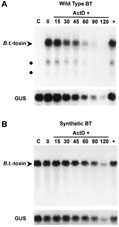
A simple explanation for the failure of wild-type B.t.-toxin mRNA to accumulate, even though there was no significant difference in transcriptional activities of wild-type and synthetic B.t.-toxin genes driven by identical promoters, would be that the wild-type transcripts were unstable. Measurement of the turnover rates of wild-type and synthetic B.t.-toxin mRNAs would provide the most direct evidence for transcript instability. To overcome the lack of detectable wild-type B.t.-toxin mRNA (see WT lanes, Fig. 3) and to allow measurement of mRNA half-life, we took advantage of the effect of the translational inhibitor CHX on wild-type B.t.-toxin mRNA accumulation. Wild-type B.t.-toxin transcripts that are normally undetectable accumulated to detectable levels in the presence of CHX (data not shown). This property is consistent with transcript instability, since this response to CHX has also been observed with other mRNAs known to be unstable (Herrick et al., 1990; Peltz et al., 1991).
BY-2 cells expressing the wild-type B.t.-toxin gene were treated transiently with CHX to generate levels of wild-type B.t.-toxin mRNA accumulation sufficient for detection over a standard half-life time-course experiment after removal of the CHX. Successful application of this approach was recently reported for mRNA half-life measurement in yeast (Zhang et al., 1997). Inhibition of translation with CHX could stabilize transcripts by causing the rapid loss of a labile protein factor that normally destabilizes the mRNA in trans. Alternatively, stabilization could result from blocking a cis-acting mRNA turnover mechanism that requires ongoing translation of the transcript. We determined an optimal minimum concentration of CHX for which the effect on mRNA accumulation was rapidly reversible but still sufficient to make measurement of mRNA half-lives practical (data not shown). CHX had no effect on the accumulation of synthetic B.t.-toxin mRNA, consistent with the possibility that the sequence modifications incorporated in the synthetic gene eliminated instability determinants.
Independent BY-2 cell lines transformed with the wild-type B.t.-toxin gene were screened by RNA gel-blot analysis to identify individual lines with the highest wild-type B.t.-toxin mRNA accumulation in response to CHX treatment (data not shown). Most cell lines accumulated wild-type B.t.-toxin mRNA to barely detectable levels after CHX treatment. Differences in mRNA accumulation between lines were probably caused by position effects. The three lines with the greatest accumulation of wild-type B.t.-toxin mRNA after CHX treatment and three independent cell lines expressing the synthetic B.t.-toxin gene were chosen for time-course experiments. Suspension cultures of these cell lines were treated for 2 h with CHX followed by removal of CHX. Transcription was blocked with ActD (Newman et al., 1993), and cells were harvested at intervals over a 2-h time course. For the sake of maintaining consistent experimental conditions, half-life measurements for synthetic B.t.-toxin mRNA were made with the same transient CHX treatment used for measurement of wild-type mRNA half-life. It should be noted, however, that synthetic B.t.-toxin mRNA half-life was unaffected by CHX, as determined by half-life measurements made with and without CHX treatment (data not shown). The accumulation and half-life of GUS mRNA, which served as the internal reference, was similarly unaffected by CHX.
The results of RNA gel-blot analysis of time-course experiments are shown in Figure 6. The effect of the transient CHX treatment on wild-type B.t.-toxin mRNA accumulation can be seen by comparing the C and 0 lanes in Figure 6A. In contrast, CHX treatment had no effect on mRNA accumulation of either the synthetic B.t.-toxin or GUS mRNAs (C and 0 lanes in Fig. 6B and lower part of Fig. 6A). The disappearance of wild-type B.t.-toxin mRNA during the time course was visibly faster than that of the GUS mRNA (Fig. 6A). Short transcripts (Fig. 6A) resulting from polyadenylation within the coding region (Diehn et al., 1998) also diminished in amount faster than the GUS transcripts. In contrast, synthetic B.t.-toxin mRNA (Fig. 6B) disappeared at a rate similar to that of the GUS mRNA. Hybridization signals were quantitated and subjected to linear-regression analysis to calculate mRNA half-lives. Of the three lines expressing the wild-type B.t.-toxin gene initially selected for half-life analysis, the hybridization signals obtained in time-course experiments for one of these lines proved to be too close to background for acceptable quantitation. Two independent half-life determinations were made for each of the remaining two cell lines expressing the wild-type B.t.-toxin gene. Synthetic B.t.-toxin mRNA half-life determinations were made for three independent cell lines. All time-course RNA gel blots were reprobed to determine the GUS mRNA half-life in each cell line.
Figure 6.
Measurement of relative half-lives of wild-type and synthetic B.t.-toxin transcripts (BT). Wild-type and synthetic B.t.-toxin mRNA decay was measured over a time course in stably transformed tobacco BY-2 suspension cell cultures. Cells were harvested before transient CHX treatment (C), after the 2-h treatment followed by removal of CHX (0), at intervals after the addition of ActD (15, 30, 45, 60, 90, and 120 min), and after continuous treatment with CHX and no ActD (+). A, RNA gel-blot analysis of a time-course experiment using a cell line expressing the wild-type B.t.-toxin gene. Full-length wild-type B.t.-toxin transcripts are indicated by the arrowhead, and short polyadenylated B.t.-toxin transcripts are indicated by dark circles. The blot was stripped and rehybridized with a probe specific for GUS mRNA. B, RNA gel-blot analysis of a time-course experiment using a cell line expressing the synthetic B.t.-toxin gene. Full-length synthetic B.t.-toxin transcripts are indicated by the arrowhead. The blot was stripped and rehybridized with a probe specific for GUS mRNA.
The absolute half-lives of wild-type and synthetic B.t.-toxin mRNAs were distinctly different, as can be seen in Table IV. As described above, the genes encoding these transcripts have identical flanking sequences and differ only in the sequences of their coding regions. Wild-type B.t.-toxin transcripts were turned over more rapidly than synthetic B.t.-toxin transcripts, with an average absolute half-life of 19.5 min compared with 66 min. We have found in previous studies that half-lives of different mRNAs can be compared most reliably when expressed as relative half-lives using the half-lives of a reference mRNA common to all cell lines as an internal standard (Newman et al., 1993; Ohme-Takagi et al., 1993). The relative half-life of 0.33 min for the wild-type B.t.-toxin transcripts using GUS as the reference transcript places them among the most rapidly degraded mRNAs for which measurements have been made in BY-2 cells (Newman et al., 1993; Taylor and Green, 1995; Gil and Green, 1996; Sullivan and Green, 1996). After normalization to GUS mRNA half-life, the relative turnover rate of wild-type B.t.-toxin transcripts was approximately 3.6 times faster than that of synthetic B.t.-toxin transcripts, demonstrating that the sequence modifications included in the synthetic gene resulted in transcript stabilization.
Table IV.
Relative mRNA half-lives of wild-type and synthetic B.t.-toxin transcripts (BT) in stably transformed tobacco BY-2 cells
| B.t.-Toxin Gene | Cell Line | B.t.-Toxin mRNA Half-Life | GUS mRNA Half-Life | Relative Half-Life | Average Relative Half-Life |
|---|---|---|---|---|---|
| min | BT/GUS | ||||
| Wild type | WT1 | 17 | 64 | 0.27 | |
| 22 | 63 | 0.35 | |||
| WT2 | 15 | 47 | 0.32 | 0.33 | |
| 24 | 62 | 0.38 | |||
| Synthetic | SYN1 | 68 | 65 | 1.05 | |
| SYN2 | 45 | 40 | 1.13 | 1.19a | |
| SYN3 | 85 | 61 | 1.39 | ||
se ± 0.10.
Distribution of Sequences in the Wild-Type B.t.-Toxin Gene That Limit mRNA Accumulation in Plants
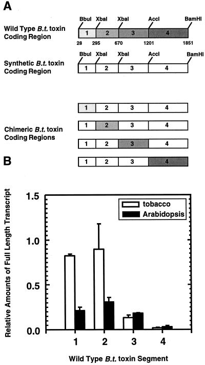
The lack of wild-type B.t.-toxin mRNA accumulation in plants must be caused by sequence elements present in the coding region, because the high level of mRNA accumulation produced by the synthetic B.t.-toxin gene was the result of sequence changes that were limited to the coding region. The extensive nature of the sequence changes precluded identification of specific regions of the B.t.-toxin gene that contained these elements. Therefore, we sought to determine if the deleterious sequences were localized to one or multiple sites in the wild-type B.t.-toxin-coding region. High accumulation of stable mRNA made it possible to use the synthetic B.t.-toxin gene as a reporter gene for identifying regions of the wild-type gene containing sequences limiting mRNA accumulation. A series of chimeric B.t.-toxin-coding regions was constructed, as shown in Figure 7A. Restriction sites conserved in both B.t.-toxin genes were used to substitute segments of the wild-type-coding region individually for the corresponding regions of the synthetic coding region. The chimeric B.t.-toxin genes were introduced by A. tumefaciens-mediated transformation into tobacco BY-2 cells and Arabidopsis plants.
Figure 7.
Effects of wild-type B.t.-toxin sequences on synthetic B.t.-toxin mRNA accumulation. A, Schematic diagram of chimeric gene constructs used to test the effects of wild-type B.t.-toxin sequences on mRNA accumulation. The synthetic B.t.-toxin-coding region was divided into four segments using restriction sites conserved between the wild-type and synthetic coding regions. Each segment of the wild-type gene was substituted individually for the corresponding segment of the synthetic gene. B, Histogram summarizing the results of RNA gel-blot analysis of chimeric B.t.-toxin gene expression relative to the synthetic B.t.-toxin gene in pools of stably transformed tobacco BY-2 cell lines (white bars) and Arabidopsis plants (black bars). Hybridization signals for full-length B.t.-toxin mRNAs were quantitated and normalized using GUS-hybridization signals as an internal standard. Hybridization signals of the synthetic B.t.-toxin transcripts in tobacco and Arabidopsis were set equal to one to allow comparison of relative expression levels between tobacco and Arabidopsis. Data are presented as means and ses for three sets of tobacco and four sets of Arabidopsis pools.
The effect of wild-type B.t.-toxin sequences on synthetic B.t.-toxin mRNA accumulation was used as an indicator of the presence of detrimental sequence elements in the wild-type segments. For tobacco, three independent pools containing at least 50 independent transformed BY-2 cell lines were made for each of the chimeric constructs. For Arabidopsis, four independent pools containing seedlings from 10 to 15 independent lines were made for each construct. Relative mRNA accumulation levels for each of the chimeric B.t.-toxin genes were determined by gel-blot analysis of RNA isolated from the pools of BY-2 and Arabidopsis lines. Hybridization signals for full-length chimeric B.t.-toxin transcripts were quantitated and normalized to synthetic B.t.-toxin mRNA accumulation using GUS-hybridization signals as an internal standard.
To allow comparison of relative expression levels of the chimeric genes between tobacco and Arabidopsis, hybridization signals of the synthetic B.t.-toxin transcripts in tobacco and Arabidopsis were each set equal to one. As shown in Figure 7, when substituted for the corresponding segment of the synthetic B.t.-toxin gene, each of the four segments of the wild-type B.t.-toxin gene caused lowered mRNA accumulation relative to the full synthetic gene in Arabidopsis. In contrast, only the 3rd and 4th wild-type gene segments caused reduced chimeric mRNA accumulation in tobacco cells. Segment 4 had the greatest effect, decreasing mRNA accumulation approximately 10-fold relative to the synthetic gene in both Arabidopsis and tobacco. None of the segments reduced the mRNA levels to below the limit of detection, indicating that the very low mRNA levels from the full wild-type B.t.-toxin gene resulted from combined effects of sequence elements present in different segments of the gene. Sequences in wild-type B.t.-toxin gene segments 1 and 2 that reduced chimeric gene mRNA accumulation by 70% to 80% in Arabidopsis were relatively ineffective in tobacco cells, indicating possible species-specific differences in recognition of sequence elements affecting mRNA accumulation. Alternatively, the observed differences may reflect differences in sequence-element recognition in cultured cells and whole plants.
Based on the identification of three polyadenylation sites within the wild-type B.t.-toxin-coding region described in the accompanying report (Diehn et al., 1998), it could be expected that products of aberrant polyadenylation might also be observed in the chimeric genes if segment junctions do not interrupt the regulatory sequences. Short transcripts consistent in size with polyadenylation at the three identified sites were observed for the chimeric genes containing wild-type segments 2, 3, and 4 in both tobacco and Arabidopsis (data not shown). The chimeric gene containing wild-type segment 3 produced small amounts of additional short transcripts that were the same in both tobacco and Arabidopsis and were not analyzed further. Although the accumulation of the chimeric transcript containing segment 4 was quite low, as mentioned above, most of the mRNA produced in both tobacco and Arabidopsis was consistent with polyadenylation at the segment 4 poly(A+) site mapped in the accompanying paper (Diehn et al., 1998). The similarity in transcript processing in both species suggests that the difference in the effects of segments 1 and 2 in tobacco and Arabidopsis may be primarily attributable to differential recognition of instability determinants. In contrast to polyadenylation signals, the differences in the effect of some wild-type B.t.-toxin sequences on mRNA accumulation in tobacco and Arabidopsis indicate that at least some sequence elements detrimental to mRNA accumulation may be recognized differently among dicots. This differential recognition, together with the inability to express the synthetic B.t.-toxin gene in transgenic tobacco plants in contrast to transgenic Arabidopsis, argues that there may be inherent differences between species that are relevant to optimizing the expression of B.t.-toxin genes.
DISCUSSION
Even though the incompatibility of wild-type B.t.-toxin genes and plant gene-expression mechanisms has been known for more than 10 years, and speculation on the potential causes has been extensive, the mechanistic basis for the incompatibility has remained unknown. Extensive sequence modifications have been used in the construction of synthetic B.t.-toxin genes to alleviate the incompatibility. These changes have resulted in high protein and mRNA accumulation without providing any direct information about why the wild-type genes were so poorly expressed to begin with. In this study we show that wild-type B.t.-toxin sequences do not inhibit transcriptional activity. Moreover, we provide the first direct evidence to our knowledge for rapid degradation of wild-type B.t.-toxin mRNA in stably transformed plant cells and show that the sequence modifications typically used to obtain high expression with synthetic B.t.-toxin genes increase transcript stability. In the accompanying paper (Diehn et al., 1998), we also demonstrate for the first time to our knowledge that a wild-type B.t.-toxin-coding region contains sequences that are recognized as polyadenylation signals in plants, thereby interfering with expression of full-length B.t.-toxin mRNAs.
A Synthetic B.t.-Toxin Gene Designed for Use as a Comparative Control and as a Reporter Gene
Our approach for assessing the possible transcriptional or posttranscriptional causes of low B.t.-toxin mRNA accumulation in plants took advantage of the fact that wild-type and synthetic B.t.-toxin genes produce very different levels of mRNA when driven by identical promoters. This allowed us to compare wild-type B.t.-toxin transcriptional activity and mRNA stability with a closely related, highly expressed gene with different primary sequence but encoding an identical protein and flanked by identical regulatory sequences. This eliminated any possible contribution to differences in transcriptional activity or mRNA turnover that might arise from comparison of unrelated genes and made it possible to ascribe observed differences specifically to the sequences contained in the wild-type B.t.-toxin-coding region.
One approach used in the design of synthetic B.t.-toxin genes has been to modify A/T-rich sequences resembling known sequences involved in RNA processing or stability by converting codons in the target sequences to synonymous codons typically used in plant genes (Perlak et al., 1990, 1991, 1993; Sutton et al., 1992; van der Salm et al., 1994). It has been suggested that the abundance of rare codons in B.t.-toxin genes could cause inefficient translation, resulting in poor expression in plants (Vaeck et al., 1987; Murray et al., 1991; Perlak et al., 1991; Wong et al., 1992), although this remains unproven. Based on this latter hypothesis, however, other synthetic B.t.-toxin genes giving high expression in plants have been constructed that primarily incorporate sequence modifications intended to convert rare codons to plant-preferred codons (Adang et al., 1993; Fujimoto et al., 1993; Koziel et al., 1993). Our primary design criterion was conversion to a maize codon bias that effectively combined both of these approaches by virtue of the resulting high G/C content that eliminated A/T-rich sequence elements. This maize codon bias proved to be fully compatible with expression in dicots, resulting in high mRNA levels (Fig. 3) and significant levels of functional protein (Fig. 5; Table II).
Lack of Evidence That Wild-Type B.t.-Toxin Sequences Block Transcription
In a previous study, the transcriptional activities of cryIA(b) and neomycin phosphotransferase (nptII) genes under the control of two different promoters were compared in nuclear run-on transcription assays using nuclei isolated from tobacco mesophyll protoplasts (Van Aarssen et al., 1995). No significant difference in transcriptional activities was detected that could account for the at least 50-fold difference in mRNA accumulation between the cryIA(b) and nptII genes, assuming that the inherent rates of transcriptional initiation from the two different promoters were similar. This previous study used a probe for the nuclear run-on assays that hybridized to the 5′ 714 nucleotides of the cryIA(b)-coding region. Premature transcription termination or inhibition of elongation downstream of the probed region could not be detected by this probe. Therefore, the authors could conclude only that transcriptional initiation was not affected by the wild-type B.t.-toxin sequences. Our run-on transcription studies extend previous work by using the rbcS-E9 3′ UTR common to both the wild-type and synthetic B.t.-toxin genes as a probe. This allowed us to compare the relative transcriptional activities through the full coding regions of the two B.t.-toxin genes. The lack of any significant difference in transcriptional activity between the wild-type and synthetic genes provides strong evidence that the differential mRNA accumulation from the two genes was not caused by transcriptional initiation or elongation but, rather, to posttranscriptional events.
Instability of the Wild-Type B.t.-Toxin mRNA
Two previous efforts to assess the stability of B.t.-toxin mRNA in plants led to opposite conclusions after the turnover of cryIA(b) transcripts in protoplasts was characterized (Murray et al., 1991; van Aarssen et al., 1995). Our results are in agreement with those of Murray and coworkers (1991), who monitored the disappearance of cryIA(b) and octopine synthase mRNAs after electroporation of plasmids carrying the genes into carrot protoplasts. It was shown that cryIA(b) transcripts disappeared faster qualitatively than octopine synthase transcripts, leading to the proposal that the cryIA(b) mRNA was unstable. In the same study, similar results were obtained using a cryIIIA gene with only 50.2% nucleotide sequence similarity to the cryIA(b) gene, suggesting that instability might be a general feature of B.t.-toxin transcripts. Because the cryIA(b) and cryIA(c) genes are 90% identical at the nucleotide level, the transcripts from these two genes could also be expected to have similar stability characteristics.
The results of our studies extend those of Murray et al. (1991) by carrying out the analysis in stably transformed tobacco cells and by measuring mRNA half-lives. We also compared the half-life of the wild-type cryIA(c) mRNA with that of mRNA from a modified synthetic cryIA(c) gene that produces high levels of mRNA. The range in absolute half-lives from 15 to 24 min observed for wild-type cryIA(c) mRNA places it among the most rapidly degraded mRNAs for which half-lives have been determined in BY-2 cells. In contrast to Murray et al. (1991), we did not observe a smear of low-Mr transcripts that accumulated over time while the full-length transcript disappeared. The lack of apparent decay intermediates is typical of most eukaryotic transcripts, indicating that after decay is initiated it proceeds rapidly to completion.
We were able to show quantitatively that the wild-type cryIA(c) transcripts were unstable relative to synthetic cryIA(c) transcripts. The relative half-lives of wild-type and synthetic cryIA(c) mRNAs differed by a factor of approximately 3.6 after using the half-life of GUS transcripts as an internal standard to normalize for differences in growth and metabolic rates of independent transformed cell lines. Although this represents a substantial difference in turnover rates, we consider this to be a minimum estimate of the difference in stabilities, because it may not be possible to remove 100% of the CHX before the start of the time course. Accordingly, some residual stabilization of wild-type cryIA(c) mRNA may occur, and the half-lives we measure may be an underestimate of the decay rate in the complete absence of CHX. Nevertheless, the 3.6-fold difference in half-lives that we were able to measure provides direct evidence that the sequence modifications used in the synthetic gene increased mRNA stability.
Our results are in contrast to those obtained by van Aarssen and coworkers (1995), who concluded that cryIA(b) mRNA was not unstable in plants based on a comparison of turnover of in vitro-synthesized capped and polyadenylated cryIA(b) and bar transcripts after electroporation into tobacco mesophyll protoplasts. Because the measured half-lives of the two transcripts in protoplasts were not significantly different, it was concluded that the lack of cryIA(b) mRNA relative to bar mRNA in transformed tobacco plants was due to nuclear events limiting cytoplasmic mRNA accumulation and that cryIA(b) transcripts were not especially unstable. The differing results of these two previous studies may stem from the different experimental methods used. Turnover of in vitro-synthesized transcripts in electroporated plant cells has not been demonstrated to be representative of mRNA turnover processes in whole plants. Unfortunately, data on cryIA(b) and bar mRNA half-lives in protoplasts from transgenic tobacco plants expressing either gene that were cited as evidence for biological significance of the electroporation experiments were not published, making it difficult to assess their relevance (van Aarssen et al., 1995). An intriguing explanation for the lack of a difference in the half-lives of in vitro-synthesized cryIA(b) and bar transcripts in electroporated protoplasts may be that proper assembly of hnRNPs and mRNPs may be necessary to engage normal mRNA-decay mechanisms. Such proper assembly may not occur if in vitro-synthesized RNA is introduced into cells by electroporation.
The sequence modifications incorporated into the synthetic B.t.-toxin gene resulted in mRNA stabilization and dramatically increased mRNA accumulation. In addition, aberrant polyadenylation within the coding region (Diehn et al., 1998) was eliminated, as demonstrated by the absence of short transcripts in gel blots of RNA from lines expressing the synthetic B.t.-toxin gene (Fig. 6B). As with the full-length wild-type B.t.-toxin mRNAs, the short transcripts accumulated to detectable levels only after treatment with CHX. Although accumulation of the short polyadenylated transcripts was too low for accurate quantitation of half-lives, their disappearance during time-course experiments was qualitatively similar to that of the full-length transcripts, indicating that they were unstable as well. The relative contributions of mRNA stability and aberrant polyadenylation to low wild-type B.t.-toxin mRNA accumulation are difficult to estimate precisely. Although the wild-type transcript decayed 3.6 times faster than the synthetic transcript, this does not account for the more than 16-fold difference in mRNA accumulation. It seems most likely that aberrant polyadenylation within the B.t.-toxin-coding region (Diehn et al., 1998), resulting in the production of unstable short transcripts, also plays a major role in limiting the level of B.t.-toxin mRNA. In addition, it is possible that the wild-type B.t.-toxin transcript is even more unstable than our measurements indicated if any residual CHX remained during our experiment and stabilized the mRNA.
Deleterious Effects of B.t.-Toxin Sequences Differ in Arabidopsis and Tobacco
Expression of the chimeric wild-type and synthetic B.t.-toxin genes in Arabidopsis demonstrated that sequence elements capable of limiting wild-type B.t.-toxin mRNA accumulation were dispersed over the coding region, whereas in tobacco deleterious sequences were prominent only in segments 3 and 4 (Fig. 7). These results extend the findings of a previous study testing the effect of wild-type B.t.-toxin sequences on protein accumulation. Sequences limiting protein expression were previously shown to be present in two regions of a cryIA(b) gene (Perlak et al., 1991). Two chimeric genes in which either the 5′ one-third or the 3′ two-thirds of a fully modified cryIA(b) gene was substituted with the corresponding region of the wild-type gene both gave protein levels in tobacco plants greater than that observed from the wild-type gene but less than from the fully modified gene. The relative mRNA levels were not determined, so it was not possible to conclude if the inhibitory sequences present in both regions of the wild-type gene exerted their effects by inhibiting translation, by limiting mRNA accumulation, or by a combination of both. Our results show that wild-type B.t.-toxin mRNA accumulation is limited by sequence determinants located at multiple sites in the coding region.
It has been shown previously that some A/U-rich sequences can cause rapid mRNA decay in tobacco (Ohme-Takagi et al., 1993), and the instability of the wild-type B.t.-toxin mRNA relative to the G/C-rich synthetic version is consistent with that observation. In particular, AUUUA repeats are known to act as mRNA-instability determinants in higher eukaryotes (Ohme-Takagi et al., 1993; Chen and Shyu, 1995), and previous efforts to enhance B.t.-toxin expression have included AUUUA sequences as targets for sequence modification. Segments 1, 2, 3, and 4 of the wild-type gene contained zero, three, two, and four AUUUA elements, respectively. The number of AUUUA elements was not proportional to the effect of each wild-type B.t.-toxin gene segment on chimeric B.t.-toxin gene mRNA accumulation. This is not surprising, because the context of AUUUA sequences is known to influence their function (Lagnado et al., 1994; Walker et al., 1995; Zubiaga et al., 1995) and additional instability sequences may exist within the B.t.-toxin-coding region. Accordingly, modification strategies targeting AUUUA sequences alone may not be effective.
Rapid turnover of wild-type B.t.-toxin mRNA could be due to specific sequence elements or inefficient translation caused by rare codons, resulting in exposure of the transcript to mRNA-degradation machinery. Substitution of wild-type segments 1 or 2 into the synthetic B.t.-toxin gene had little or no effect on the accumulation of the chimeric transcripts in tobacco. Because 45 of 91 and 61 of 125 codons in wild-type segments 1 and 2, respectively, are the least frequently used of synonymous codons in maize genes (Wada et al., 1992), it is clear that the presence of rare codons per se does not interfere with mRNA accumulation. These results are consistent with the results of previous experiments in which a 26-codon cryIA(c) gene sequence rich in rare codons was inserted in-frame in one and two copies into a phytohemagglutinin gene with no effect on transcript stability (van Hoof and Green, 1996). Therefore, increased mRNA accumulation that results from wholesale conversion of codon bias to be plant-like may be caused not by increased translational efficiency but by default elimination of deleterious RNA sequence elements.
In summary, we have demonstrated that undesirable interactions with mRNA stability and polyadenylation mechanisms can severely limit B.t.-toxin gene expression in higher plants. This emphasizes that fundamental research into posttranscriptional mechanisms in plants could provide information that could enhance the rational engineering of B.t.-toxin and other foreign genes for optimal expression.
ACKNOWLEDGMENTS
We thank Dr. Dan Vernon for comments on the manuscript, Linda Danhof for assistance with Arabidopsis transformation, Dr. Pedro Gil for construction of p995, Dr. Christie Howard for construction of p1425, Kurt Stepnitz for photography, and Marlene Cameron for assistance with graphics.
Abbreviations:
- 2X35S
doubly enhanced cauliflower mosaic virus 35S promoter
- ActD
actinomycin D
- ADH1
alcohol dehydrogenase 1
- BMS
Black Mexican Sweet
- BY-2
Bright Yellow 2
- CaMV
cauliflower mosaic virus
- CHX
cycloheximide
- UTR
untranslated region
Footnotes
This work was supported by grants from the Department of Energy, the U.S. Department of Agriculture, Project Green, Michigan State University Research Excellence Funds, Midwest Plant Biotechnology Consortium, Consortium for Plant Biotechnology Research, and matching funds from Ciba-Geigy, Sandoz, Pioneer, Anheuser-Busch, ICI, Agrigenetics, and DowElanco. S.H.D. and E.J.D.R. were supported in part by a National Institutes of Health predoctoral traineeship and a U.S. Department of Agriculture postdoctoral fellowship, respectively.
The accession number for the sequence reported in this article is AF023672.
LITERATURE CITED
- Adang MJ, Brody MS, Cardineau G, Eagan N, Roush RT, Shewmaker CK, Jones A, Oakes JV, McBride KE. The reconstruction and expression of a Bacillus thuringiensis cryIIIA gene in protoplasts and potato plants. Plant Mol Biol. 1993;21:1131–1145. doi: 10.1007/BF00023609. [DOI] [PubMed] [Google Scholar]
- Adang MJ, Firoozabady E, Klein J, DeBoer D, Sekar V, Kemp JD, Murray EE, Rocheleau TA, Rashka K, Staffeld G, and others (1987) Expression of a Bacillus thuringiensis insecticidal crystal protein gene in tobacco plants. In CJ Arntzen, C Ryan, eds, Molecular Strategies for Crop Protection: UCLA Symposia on Molecular and Cellular Biology, New Series, Vol 46. Alan R. Liss, New York, pp 345–353
- Adang MJ, Staver MJ, Rocheleau TA, Leighton J, Barker RF, Thompson DV. Characterization of full-length and truncated plasmid clones and the crystal protein of Bacillus thuringiensis subsp. kurstaki HD-73 and their toxicity to Manduca sexta. Gene. 1985;36:289–300. doi: 10.1016/0378-1119(85)90184-2. [DOI] [PubMed] [Google Scholar]
- Barton KA, Whiteley HR, Yang N-S. Bacillus thuringiensis δ-endotoxin expressed in transgenic Nicotiana tabacum provides resistance to lepidopteran insects. Plant Physiol. 1987;85:1103–1109. doi: 10.1104/pp.85.4.1103. [DOI] [PMC free article] [PubMed] [Google Scholar]
- Bigelow CC, Channon M (1982) In GD Fasman, ed, CRC Handbook of Biochemistry and Molecular Biology, Ed 3: Proteins, Vol 1. CRC Press, Boca Raton, FL, pp 209–243
- Birkett CR, Foster KE, Gull K. Use of monoclonal antibodies to analyse the expression of a multi-tubulin family. FEBS Lett. 1985;187:211–218. doi: 10.1016/0014-5793(85)81244-8. [DOI] [PubMed] [Google Scholar]
- Campbell CH, Gowri G. Codon usage in higher plants, green algae, and cyanobacteria. Plant Physiol. 1990;92:1–11. doi: 10.1104/pp.92.1.1. [DOI] [PMC free article] [PubMed] [Google Scholar]
- Chen C-YA, Shyu A-B. AU-rich elements: characterization and importance in mRNA degradation. Trends Biochem Sci. 1995;20:465–470. doi: 10.1016/s0968-0004(00)89102-1. [DOI] [PubMed] [Google Scholar]
- Delannay X, LaVallee BJ, Proksch RK, Fuchs RL, Sims SR, Greenplate JT, Marrone PG, Dodson RB, Augustine JJ, Layton JG and others. Field performance of transgenic tomato plants expressing the Bacillus thuringiensis var. kurstaki insect control protein. Bio/Technology. 1989;7:1265–1269. [Google Scholar]
- De Rocher EJ, Bohnert HJ. Development and environmental stress employ different mechanisms in the expression of a plant gene family. Plant Cell. 1993;5:1611–1625. doi: 10.1105/tpc.5.11.1611. [DOI] [PMC free article] [PubMed] [Google Scholar]
- Diehn SH, Chiu W-L, De Rocher EJ, Green PJ. Premature polyadenylation at multiple sites within a Bacillus thuringiensis toxin gene-coding region. Plant Physiol. 1998;117:1433–1443. doi: 10.1104/pp.117.4.1433. [DOI] [PMC free article] [PubMed] [Google Scholar]
- Diehn SH, De Rocher EJ, Green PJ (1996) Problems that can limit the expression of foreign genes in plants: lessons to be learned from B.t.-toxin genes. In JK Setlow, ed, Genetic Engineering: Principles and Methods, Vol 18. Plenum Press, New York, pp 83–99 [DOI] [PubMed]
- Dillon PJ, Craig AR. A rapid method for the construction of synthetic genes using the polymerase chain reaction. BioTechniques. 1990;9:298–300. [PubMed] [Google Scholar]
- Feinberg AP, Vogelstein B. A technique for radiolabeling DNA restriction endonuclease fragments to high specific activity (addendum) Anal Biochem. 1983;137:266. doi: 10.1016/0003-2697(84)90381-6. [DOI] [PubMed] [Google Scholar]
- Fischhoff DA, Bowdish KS, Perlak FJ, Marrone PG, McCormick SM, Niedermeyer JG, Dean DA, Kusano-Kretzmer K, Mayer EJ, Rochester DE and others. Insect tolerant transgenic tomato plants. Bio/Technology. 1987;5:807–813. [Google Scholar]
- Fujimoto H, Itoh K, Yamamoto M, Kyozuka J, Shimamoto K. Insect resistant rice generated by introduction of a modified δ-endotoxin gene of Bacillus thuringiensis. Bio/Technology. 1993;11:1151–1155. doi: 10.1038/nbt1093-1151. [DOI] [PubMed] [Google Scholar]
- Gil P, Green PJ. Multiple regions of the Arabidopsis SAUR-AC1 gene control transcript abundance: the 3′ untranslated region functions as an mRNA instability determinant. EMBO J. 1996;15:1678–1686. [PMC free article] [PubMed] [Google Scholar]
- Herrick D, Parker R, Jacobson A. Identification and comparison of stable and unstable mRNAs in Saccharomyces cerevisiae. Mol Cell Biol. 1990;10:2269–2284. doi: 10.1128/mcb.10.5.2269. [DOI] [PMC free article] [PubMed] [Google Scholar]
- Jefferson RA, Burgess SM, Hirsh D. β-Glucuronidase from Escherichia coli as a gene-fusion marker. Proc Natl Acad Sci USA. 1986;83:8447–8451. doi: 10.1073/pnas.83.22.8447. [DOI] [PMC free article] [PubMed] [Google Scholar]
- Koziel MG, Beland GL, Bowman C, Carozzi NB, Crenshaw R, Crossland L, Dawson J, Desai N, Hill M, Kadwell S and others. Field performance of elite transgenic maize plants expressing an insecticidal protein derived from Bacillus thuringiensis. Bio/Technology. 1993;11:194–200. [Google Scholar]
- Kunkel TA, Roberts JD, Zakour RA. Rapid and efficient site-specific mutagenesis without phenotypic selection. Methods Enzymol. 1987;154:367–382. doi: 10.1016/0076-6879(87)54085-x. [DOI] [PubMed] [Google Scholar]
- Laemmli UK. Cleavage of structural proteins during the assembly of the head of bacteriophage T4. Nature. 1970;227:680–685. doi: 10.1038/227680a0. [DOI] [PubMed] [Google Scholar]
- Lagnado CA, Brown CY, Goodall GJ. UUUA is not sufficient to promote poly(A) shortening and degradation of an mRNA: the functional sequence within AU-rich elements may be UUAUUUA(U/A)(U/A) Mol Cell Biol. 1994;14:7984–7995. doi: 10.1128/mcb.14.12.7984. [DOI] [PMC free article] [PubMed] [Google Scholar]
- Lütcke HA, Chow KC, Mickel FS, Moss KA, Kern HF, Scheele GA. Selection of AUG initiation codons differs in plants and animals (abstract) EMBO J. 1987;6:43–48. doi: 10.1002/j.1460-2075.1987.tb04716.x. [DOI] [PMC free article] [PubMed] [Google Scholar]
- Meyer P, Saedler H. Homology-dependent gene silencing in plants. Annu Rev Plant Physiol Plant Mol Biol. 1996;47:23–48. doi: 10.1146/annurev.arplant.47.1.23. [DOI] [PubMed] [Google Scholar]
- Mogen BD, MacDonald MH, Leggewie G, Hunt AG. Several distinct types of sequence elements are required for efficient mRNA 3′ end formation in a pea rbcS gene. Mol Cell Biol. 1992;12:5406–5414. doi: 10.1128/mcb.12.12.5406. [DOI] [PMC free article] [PubMed] [Google Scholar]
- Murray EE, Rocheleau T, Eberle M, Stock C, Sekar V, Adang M. Analysis of unstable RNA transcripts of insecticidal crystal protein genes of Bacillus thuringiensis in transgenic plants and electroporated protoplasts. Plant Mol Biol. 1991;16:1035–1050. doi: 10.1007/BF00016075. [DOI] [PubMed] [Google Scholar]
- Nagata T, Nemoto Y, Hasezawa S. Tobacco BY-2 cell line as the “HeLa” cell in the cell biology of higher plants. Int Rev Cytol. 1992;132:1–30. [Google Scholar]
- Newman TC, Ohme-Takagi M, Taylor CB, Green PJ. DST sequences, highly conserved among plant SAUR genes, target reporter transcripts for rapid decay in tobacco. Plant Cell. 1993;5:701–714. doi: 10.1105/tpc.5.6.701. [DOI] [PMC free article] [PubMed] [Google Scholar]
- Ohme-Takagi M, Taylor CB, Newman TC, Green PJ. The effect of sequences with high AU content on mRNA stability in tobacco. Proc Natl Acad Sci USA. 1993;90:11811–11815. doi: 10.1073/pnas.90.24.11811. [DOI] [PMC free article] [PubMed] [Google Scholar]
- Peltz SW, Brewer G, Bernstein P, Hart P, Ross J. Regulation of mRNA turnover in eukaryotic cells. Crit Rev Eukaryotic Gene Expression. 1991;1:99–126. [PubMed] [Google Scholar]
- Perlak FJ, Deaton RW, Armstrong TA, Fuchs RL, Sims SR, Greenplate JT, Fischhoff DA. Insect resistant cotton plants. Bio/Technology. 1990;8:939–943. doi: 10.1038/nbt1090-939. [DOI] [PubMed] [Google Scholar]
- Perlak FJ, Fuchs RL, Dean DA, McPherson SL, Fischhoff DA. Modification of the coding sequence enhances plant expression of insect control protein genes. Proc Natl Acad Sci USA. 1991;88:3324–3328. doi: 10.1073/pnas.88.8.3324. [DOI] [PMC free article] [PubMed] [Google Scholar]
- Perlak FJ, Stone TB, Muskopf YM, Petersen LJ, Parker GB, McPherson SA, Wyman J, Love S, Reed G, Biever D and others. Genetically improved potatoes: protection from damage by Colorado potato beetles. Plant Mol Biol. 1993;22:313–321. doi: 10.1007/BF00014938. [DOI] [PubMed] [Google Scholar]
- Sambrook J, Fritsch EF, Maniatis T. Molecular Cloning: A Laboratory Manual. Cold Spring Harbor, NY: Cold Spring Harbor Laboratory Press; 1989. [Google Scholar]
- Sanger F, Nicklen S, Coulson AR. DNA sequencing with chain-terminating inhibitors. Proc Natl Acad Sci USA. 1977;74:5463–5467. doi: 10.1073/pnas.74.12.5463. [DOI] [PMC free article] [PubMed] [Google Scholar]
- Schnepf HE, Whiteley HR. Delineation of a toxin-encoding segment of a Bacillus thuringiensis crystal protein gene. J Biol Chem. 1985;260:6273–6280. [PubMed] [Google Scholar]
- Sullivan ML, Green PJ. Mutational analysis of the DST element in tobacco cells and transgenic plants: identification of residues critical for mRNA instability. RNA. 1996;2:308–315. [PMC free article] [PubMed] [Google Scholar]
- Sutton DW, Havstad PK, Kemp JD. Synthetic cryIIIA gene from Bacillus thuringiensis improved for high expression in plants. Transgenic Research. 1992;1:228–236. doi: 10.1007/BF02524753. [DOI] [PubMed] [Google Scholar]
- Tailor R, Tippett J, Gibb G, Pells S, Pike D, Jordan L, Ely S. Identification and characterization of a novel Bacillus thuringiensis δ-endotoxin entomocidal to coleopteran and lepidopteran larvae. Mol Microbiol. 1992;6:1211–1217. doi: 10.1111/j.1365-2958.1992.tb01560.x. [DOI] [PubMed] [Google Scholar]
- Taylor CB, Bariola PA, DelCardayré SB, Raines RT, Green PJ. RNS2: a senescence-associated RNase of Arabidopsis that diverged from the S-RNases before speciation. Proc Natl Acad Sci USA. 1993;90:5118–5122. doi: 10.1073/pnas.90.11.5118. [DOI] [PMC free article] [PubMed] [Google Scholar]
- Taylor CB, Green PJ. Identification and characterization of genes with unstable transcripts (GUTs) in tobacco. Plant Mol Biol. 1995;28:27–38. doi: 10.1007/BF00042035. [DOI] [PubMed] [Google Scholar]
- Vaeck M, Reynaerts A, Hofte H, Jansens S, De Beuckeleer M, Dean C, Zabeau M, Van Montagu M, Leemans J. Transgenic plants protected from insect attack. Nature. 1987;328:33–37. [Google Scholar]
- van Aarssen R, Soetaert P, Stam M, Dockx J, Gosselé V, Seurinck J, Reynaerts A, Cornelissen M. cry IA(b) transcript formation in tobacco is inefficient. Plant Mol Biol. 1995;28:513–524. doi: 10.1007/BF00020398. [DOI] [PubMed] [Google Scholar]
- van der Salm T, Bosch D, Honee G, Feng L, Munsterman E, Bakker P, Stiekema WJ, Visser B. Insect resistance of transgenic plants that express modified Bacillus thuringiensis cryIA(b) and cryIC genes: a resistance management strategy. Plant Mol Biol. 1994;26:51–59. doi: 10.1007/BF00039519. [DOI] [PubMed] [Google Scholar]
- van Hoof A, Green PJ. Premature nonsense codons decrease the stability of phytohemagglutinin mRNA in a position-dependent manner. Plant J. 1996;10:415–424. doi: 10.1046/j.1365-313x.1996.10030415.x. [DOI] [PubMed] [Google Scholar]
- Wada K, Wada Y, Ishibashi F, Gojobori T, Ikemur T. Codon usage tabulated from the GenBank genetic sequence data. Nucleic Acids Res. 1992;20:2111–2118. doi: 10.1093/nar/20.suppl.2111. [DOI] [PMC free article] [PubMed] [Google Scholar]
- Walker EL, Weeden NF, Taylor CB, Green PJ, Coruzzi GM. Molecular evolution of duplicate copies of genes encoding cytosolic glutamine synthetase in Pisum sativum. Plant Mol Biol. 1995;6:1111–1125. doi: 10.1007/BF00020456. [DOI] [PubMed] [Google Scholar]
- Wong EY, Hironaka CM, Fischhoff DA. Arabidopsis thaliana small subunit leader and transit peptide enhance the expression of Bacillus thuringiensis proteins in transgenic plants. Plant Mol Biol. 1992;20:81–93. doi: 10.1007/BF00029151. [DOI] [PubMed] [Google Scholar]
- Zhang S, Welch EM, Hogan K, Brown AH, Peltz SW, Jacobson A. Polysome-associated mRNAs are substrates for the nonsense-mediated mRNA decay pathway in Saccharomyces cerevisiae. RNA. 1997;3:234–244. [PMC free article] [PubMed] [Google Scholar]
- Zubiaga AM, Belasco JG, Greenberg ME. The nonamer UUAUUUAUU is the key AU-rich sequence motif that mediates mRNA degradation. Mol Cell Biol. 1995;15:2219–2230. doi: 10.1128/mcb.15.4.2219. [DOI] [PMC free article] [PubMed] [Google Scholar]


