

Abstract
Calcineurin plays a key role in morphogenesis, pathogenesis and drug resistance in most fungi. However, the function of calcineurin genes in Puccinia striiformis f. sp. tritici (Pst) is unclear. We identified and characterized the calcineurin genes PsCNA1 and PsCNB1 in Pst. Phylogenetic analyses indicate that PsCNA1 and PsCNB1 form a calcium/calmodulin regulated protein phosphatase belonging to the calcineurin heterodimers composed of subunits A and B. Quantitative RT-PCR analyses revealed that both PsCNA1 and PsCNB1 expression reached their maximum in the stage of haustorium formation, which is one day after inoculation. Using barely stripe mosaic virus (BSMV) as a transient expression vector in wheat, the expression of PsCNA1 and PsCNB1 in Pst was suppressed, leading to slower extension of fungal hyphae and reduced production of urediospores. The immune-suppressive drugs cyclosporin A and FK506 markedly reduced the germination rates of urediospores, and when germination did occur, more than two germtubes were produced. These results suggest that the calcineurin signaling pathway participates in stripe rust morphogenetic differentiation, especially the formation of haustoria during the early stage of infection and during the production of urediospores. Therefore PsCNA1 and PsCNB1 can be considered important pathogenicity genes involved in the wheat-Pst interaction.
Introduction
Calcineurin, a serine-threonine-specific calcium/calmodulin-dependent protein phosphatase with two subunits (CNA and CNB), regulates a variety of physiological processes, such as growth, morphogenesis, pathogenicity, and membrane stress responses through the calcium signaling pathway in eukaryotes [1], [2]. The first fungal calcineurin genes were reported in 1991 from the budding yeast Saccharomyces cerevisiae [3], [4] and the filamentous fungus Neurospora crassa [5]. Many homologs of CNA or/and CNB have been found in medicinal fungi [6] and plant pathogens such as Botrytis cinerea [7] and Magnaporthe oryzae [8], [9]. Recent studies have confirmed that calcineurin controls virulence, hyphal elongation and multiple stress responses in the human pathogens Candida dubliniensis [10], Cryptococcus neoformans [11], [12], Candida albicans [13] and Aspergillus fumigatus [14], [15]. Similar findings have also been reported for the phytopathogens Ustilago maydis [16], Cochliobolus miyabeanus [17], and Sclerotinia sclerotiorum [18]. The calcineurin pathway also plays a role in drug resistance to azoles in C. albicans [19], [20], and in C. dubliniensis [10]. Inhibition of calcineurin can decrease fungal growth and arrest tissue invasion [21]. This opens possibilities to develop new antifungal agents targeting the calcineurin pathway in fungi [6].
RNA induced gene silencing or RNA interference (RNAi) is a complex natural phenomenon and a powerful reverse genetics tool for the analysis of gene function in eukaryotes [22]–[26]. In plants, virus-induced gene silencing (VIGS) was developed for rapid functional analysis of plant genes using viruses to deliver silencing constructs [27]–[29]. It has widely been applied in dicots such as Arabidopsis [30], tobacco [31] and tomato [32]–[33], and monocots such as barley [34] and wheat [35]–[38]. In fungi, RNAi technology has been deployed in more than 40 species including plant and human pathogens [39]. Nguyen et al. [40] developed a high-throughput RNA-silencing vector for M. oryzae to identify an involvement of calcineurin genes in colony pigmentation, sporulation, appressorium formation, and pathogenicity. However, for there is still no applicable transformation system available there are currently no techniques on hand for silencing genes in obligate biotrophic fungi directly. Host-induced gene silencing (HIGS) is a newly developed RNAi technology to indirectly silence parasite genes by expressing an RNAi construct in vivo in the host [41]. Host induced RNAi of three target genes suppressed their expression in the planthopper Nilaparvata lugens after feeding on rice plants [42]. Recent studies confirm the hypothesis that fungal genes can be suppressed in planta during interaction of the fungus with the host. Tinoco et al. [43] silenced the reporter gene GUS in Fusarium verticillioides by expressing GUS dsRNA in tobacco. HIGS was also successfully used in obligate biotrophic fungi. Using a BSMV-VIGS system expressing the target dsRNA in wheat, Nowara et al. [44] showed that the fungal genes GTF1 and GTF2 in Blumeria graminis play a role in haustorium formation and elongation of secondary hyphae. Yin et al. [45] also developed a BSMV-based HIGS approach to identify gene function in the biotrophic rust fungus Puccinia striiformis f. sp. tritici (Pst).
Wheat stripe rust, caused by the Basidiomycete Pst, is an important disease in wheat worldwide. As an obligate biotrophic pathogen infecting wheat leaves, Pst undergoes a high degree of morphological and physiological differentiation from urediospore to germ tube, invasive hypha and haustorium, a special structure for nutrient uptake from the host [46]–[48]. A few studies reported that the calcium signaling pathway is involved in the initial infection and biotrophic growth of rust fungi [49], [50]. Some homologs involved in calcium signaling such as CDPK were identified in Pst [49]. Recently, the PST-130 genome has been sequenced [51]. The sequences provided information to clone Pst genes involved in calcium signaling. In this study, we describe cloning, sequencing and transcription analysis of two calcineurin subunits from Pst, designated PsCNA1 and PsCNB1. HIGS analysis using the BSMV-VIGS system and drug tests indicate a vital function in rust growth, development and sporulation.
Results
PsCNA1 and PsCNB1 encode calcineurin homologs
One of the expressed sequence tags (ESTs) from a full-length cDNA library of Pst [52] was found to be highly similar to PtCNA from Puccinia triticina (PTTG_07903) and PgCNA from Puccinia graminis f. sp. tritici (PGTG_14891). Another two ESTs from cDNA libraries contructed by Zhang et al. [49] and Ma et al. [53] are almost identical to the CNB genes from the other two wheat rusts (PTTG_02210 and PGTG_04308). Further sequencing of these clones from the Chinese Pst race CYR31, provided full-length cDNA sequences of PsCNA1 and PsCNB1 (Genbank accession numbers JX424819 and JX424820, respectively). The full length cDNA sequence for PsCNA1 is 2,680 bp with an open reading frame (ORF) of 2,097 bp encoding a 698 amino acid (AA) protein, which consists of two calcineurin A domains and six Serine/threonine-protein phosphatase domains with a calculated molecular mass of 76.74 kDa (Fig. S1). The PsCNB1 cDNA is 770 bp in length with an ORF of 528 bp encoding a 175 AA protein, with a molecular mass of 19.77 kDa, which has four calcium-binding EF-hand motifs and a N-myristoylation site (Fig. S2).
The levels of conservation of PsCNA1 and PsCNB1 are indicated in comparison with homologs from other fungi and some model organisms in Figure S1 and Figure S2, respectively. PsCNA1 is 90% identical to the calcineurin A subunit of P. triticina and 84% identical to P. graminis f. sp. tritici CNA. PsCNA1 is conserved among other fungi analyzed with 54% to 75%. PsCNB1 exhibits strong similarity to calcineurin B proteins from other organisms. It is 100% identical to PtCNB, 99% identical to PgCNB, and 60% to 83% identical to CNB genes from other fungi analyzed.
Phylogenetic analysis revealed that PsCNA1 and PsCNB1 cluster with other Basidiomycete fungi. Especially the three Puccinia sp. were most close to each other (Fig. 1). However, PsCNA1 was closer to P. graminis f. sp. tritici than to P. triticina while PsCNB1 was closer to P. triticina than to P. graminis f. sp. tritici.
Figure 1. Phylogenetic analyses of CNA and CNB genes.

A: AfCNA (Aspergillus fumigatus, XP_753703), BfCNA (Botryotinia fuckeliana, XP_001558972), CcCNA (Coprinopsis cinerea, XP_001838986), CnCNA (Cryptococcus neoformans var. grubii, AAB97372), DmCNA (Drosophila melanogaster, NP_727985), HsCNA (Homo sapiens, NP_000936), LbCNA (Laccaria bicolor, XP_001884713), NcCNA (Neurospora crassa, XP_961193), NfCNA (Neosartorya fischeri, XP_001259754), PgCNA (Puccinia graminis f. sp. tritici, EFP89050), PpCNA (Postia placenta, XP_002470453), PsCNA (Puccinia striiformis f. sp. tritici, JX424819), PtCNA (Puccinia triticina, PTTG_07903), RnCNA (Rattus norvegicus, BAA14083), ScCNA1 (Saccharomyces cerevisiae, SCRG_04371), ScCNA2 (Saccharomyces cerevisiae, SCRG_01842), SsCNA (Sclerotinia sclerotiorum, XP_001597594), UmCNA (Ustilago maydis, AAP48999). B: AfCNB (Aspergillus flavus, XP_002378292), BfCNB (Botryotinia fuckeliana, XP_001555369), CiCNB (Coccidioides immitis, XP_001248933), CnCNB (Cryptococcus neoformans var. neoformans, XP_57033), GzCNB (Gibberella zeae, XP_387580), HsCNB (Homo sapiens, NP_000936), LbCNB (Laccaria bicolor, XP_001884421), MoCNB (Magnaporthe oryzae, ADD84607), NcCNB (Neurospora crassa, CAA73345), PbCNB (Paracoccidioides brasiliensis, XP_002795006), PgCNB, (Puccinia graminis tritici, EFP78352), PsCNB (Puccinia striiformis f. sp. tritici, JX424820), PtCNB (Puccinia triticina, PTTG_02210), RsCNB (Rattus sp., BAA03318), ScCNB (Saccharomyces cerevisiae, SCRG_03838). The unrooted phylograms were constructed based on NJ analysis. Confidence of groupings was estimated by using 1,000 bootstrap replicates. Numbers next to the branching point indicate the percentage of replicates supporting each branch.
Suppressors block Pst germination
The immuno-suppressants cyclosporin A (CsA) and FK506 inhibit calcineurin activity and affect its function just like mutants of CNA or/and CNB in several fungi [6]. In order to test whether these two drugs affect the function of PsCNA1 and PsCNB1, stripe rust urediospores were incubated with these drugs and germination was monitored. After 10 hours, microscopic analyses indicated that germination rate was reduced to 40.5% for FK506 (3 µM) and 66.5% for CsA (3 µM) treatment, compared to water (Table 1). Germ tubes of Pst were limited in their elongation by treatment with of FK506 (1 µM) or CsA (0.1 µM) compared to the control (Fig. 2C, 2B, 2A). Urediospores also frequently produced two or three more irregular germ tubes than the control (Fig. 2D, 2E, 2F).
Table 1. Germination rates of Pst (mean±SE).
| Treatments | Percent Germination (10 hours) |
| Water | 98.7±0.2U |
| FK506(3 µM) | 40.5±4.7U |
| CsA (3 µM) | 66.5±3.1U |
Values are significantly different at P = 0.05 according to the Tukey's test.
Figure 2. Immuno-suppressive drugs assay of Pst urediospores.

Pst race CYR31 was treated with water (A, D), CsA (0.1 µM, B, E), or FK506 (1 µM, C, F). Treatment with either drug can limit the elongation of Pst germ tubes and block further differentiation. Analyses were performed using the light microscope; scale bar, 50 µm.
Expression profiles of PsCNA1 and PsCNB1
To gain insight into the possible function of PsCNA1 and PsCNB1 in Pst, we investigated the expression of PsCNA1 and PsCNB1 (mRNA abundance) in different stages of Pst using quantitative PCR (qRT-PCR). PsCNA1 and PsCNB1 had similar expression profiles (Fig. 3). Transcript levels of both genes were drastically increased at 1.0 dpi, but quickly decreased to 3-fold, and 0.5-fold, respectively, at 11 dpi (Fig. 3). The maximum accumulation of transcript was 58 fold for PsCNA1 and 38 fold for PsCNB1. However, kinetics of transcript accumulation differed between the two genes. The transcripts for PsCNB1 drastically increased over time up to 1 dpi, whereas transcript level of PsCNA1 only showed a dramatic increase at 1 dpi.
Figure 3. Transcript levels of PsCNA1 and PsCNB1 during Pst differentiation stages.

RNA samples were isolated from leaves of wheat cultivar Suwon 11 inoculated with Pst race CYR31 at the indicated time points. Expression levels of PsCNA1/PsCNB1 were estimated by the ΔΔCt method with the elongation factor gene of Pst as endogenous reference for normalization. S: urediospore, GS: germinated urediospore, dpi: days post inoculation.
HIGS for PsCNA1 and PsCNB1
In order to determine the best inoculation day for Pst inoculation, virus symptoms were scored by visual assessment at four time point (8, 10, 12, 14 dpi) for the BSMV:γ:0-as vector (data not shown). Only three out of eighteen seedlings showed slight virus symptoms at 8 dpi. Symptoms increased at 10 dpi, and almost all seedlings showed 100% virus infection at 12 dpi. At 14 dpi leaves showed heavy symptoms with large yellow areas.Therefore, 12 dpi with BSMV was chosen for rust inoculation.
In order to identify HIGS efficiency (knockdown rates), transcript levels of PsCNA1 and PsCNB1 were scored in inoculated silenced plants at 8 dpi by qRT-PCR. Results showed that silencing was detected for both BSMV vectors. PsCNA1 transcript level exhibited an average of 24% expression in BSMV:γ:PsCNA1-as infected plants. However, PsCNB1 transcript level showed only an average of 18% reduction. HIGS for both genes lost most effectiveness at 16 dpi (PsCNA1: 49% expression, PsCNB1: 110% expression) (Fig. 4).
Figure 4. Transcript levels of PsCNA1 and PsCNB1 after HIGS during Pst differentiation stages.

RNA samples were isolated from leaves infected with BSMV of wheat cultivar Suwon 11 inoculated with Pst race CYR31. Transcript levels of PsCNA1 and PsCNB1 were estimated by the comparative ΔΔCt method with elongation factor gene of Pst as the endogenous reference for normalization. dpi: days post inoculation.
Silencing calcineurin blocks Pst growth and development in wheat leaves
To determine cytological changes associated with fungal growth on plants, silenced for PsCNA1 or PsCNB1 wheat leaves inoculated with race CYR31 were examined microscopically. Two time points (2 and 5 dpi) were compared. No significant differences in Pst development and hyphal growth were observed between control plants and plants carrying PsCNA1 or PsCNB1 knock-down constructs at 2 dpi (Table 2; Fig. 5A, 5B, 5C). However, at 5 dpi hyphal length on average in BSMV:γ:PsCNA1-as and BSMV:γ:PsCNB1-as infected wheat leaves were much shorter than those observed in controls (Table 2).
Table 2. Histological observation during HIGS (mean±SE).
| TreatmentsV | Number of hyphal branches at 2 dpiW | Number of haustoria at 2 dpiX | Hyphal length at 5 dpi (µm)Y | Number of uredia at 16 dpiZ |
| BSMV:γ:0-as | 2.00±0.28 | 3.21±0.32 | 283.94±10.41 | 99±7 |
| BSMV:γ:PsCNA1-as | 1.86±0.30 | 2.62±0.35 | 183.11±12.78 | 60±8 |
| BSMV:γ:PsCNb1-as | 1.82±0.17 | 2.59±0.17 | 197.41±13.09 | 68±5 |
Abbreviations: dpi, day post inoculation; SE, Standard Error.
Leaves infected inoculated with BSMV:γ:0-as (empty vector), BSMV:PsCNA1-as and BSMV:PsCNB1-as followed by inoculation with CYR31.
Distance from the base of the substomatal vesicles to the hyphal tips.
Values are not significantly different at P = 0.05 according to the Tukey's test.
Values are significantly different at P = 0.05 according to the Tukey's test.
Figure 5. Histological observation of Pst growth using calcofluor staining.

All wheat leaves were inoculated with Pst strain CYR31; fluorescence-microscopic analyses were done 2 days post inoculation. A: BSMV:γ:0-as (empty viral vector control); B: BSMV:γ:PsCNA1-as (silencing for PsCNA1); C: BSMV:γ:PsCNB1-as (silencing for PsCNB1); IH: infection hyphae; HM: haustorial mother cell; SV: substomatal vesicle. Scale bars: 50 µm.
Reduction in rust sporulation after silencing Pst calicineurin
We also scored sporulation and found that the number of uredia was reduced on silencing plants (Fig. 6). Sporulation of Pst on silencing plants occurred two days later (12 dpi) than on the control plants (10 dpi). Statistical analyses 16 dpi determined an average number of 99 uredia for control plants, 60 uredia for BSMV:γ:PsCNA1-as, and 68 uredia for BSMV:γ:PsCNB1-as (Table 2). Uredia in silencing plants were smaller in size with open cavities that were shorter, and contained fewer spores (Fig. 7).
Figure 6. Uredia on silenced leaves 16 days after Pst inoculation.
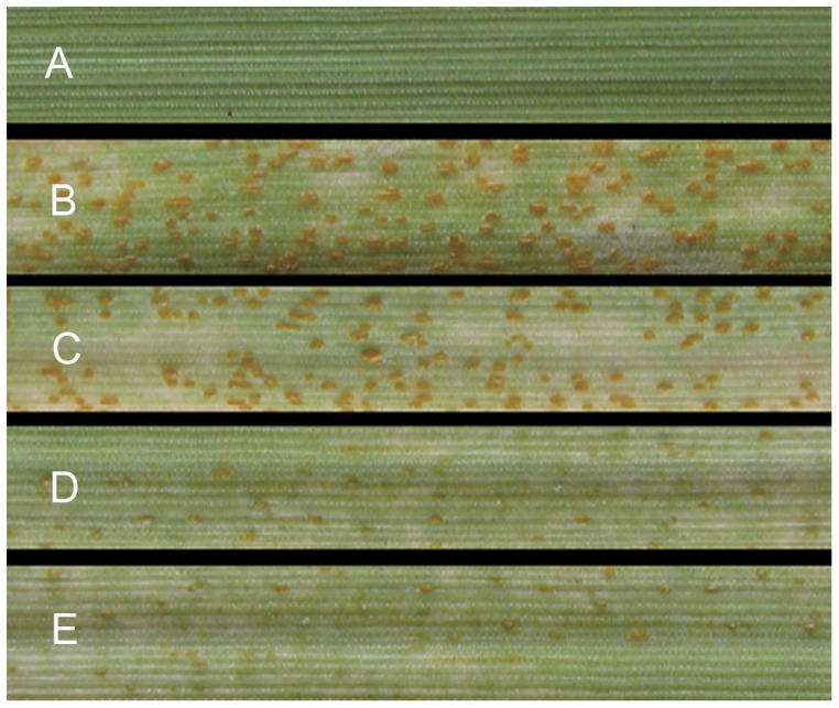
Pst development on wheat leaves after HIGS. A: No virus and Pst (healthy leaf control); B: No virus (normal infection with Pst); C: BSMV:γ:0-as (empty viral vector control); D: BSMV:γ:PsCNA1-as (Pst infection after silencing PsCNA1); E: BSMV:γ:PsCNB1-as (Pst infection after silencing PsCNB1).
Figure 7. SEM photograph of uredia after HIGS at 16 dpi.

Sorus of silenced PsCNA1/PsCNB1 of Pst infected leaves inoculated with CYR31. Scanning electron micrographs by 600×, scale bar: 20 µm. A: BSMV:γ:0-as (empty viral vector control); B: BSMV:γ:PsCNA1-as (silencing PsCNA1); C: BSMV:γ:PsCNB1-as (silencing PsCNB1).
Discussion
In this study we describe the isolation and characterization of two calcineurin genes from the wheat stripe rust fungus Pst. Phylogenetic analyses of eukaryotic CNA and CNB genes clearly show that PsCNA1 and PsCNB1 are closely related to calcineurin genes from other basidiomycetes. The calcineurin A/B protein family appears to be conserved in size and structure. Most CNA genes encode a protein of more than 500 amino acids and most CNB proteins contain about 175 amino acids [54]. All CNA proteins share high homology within the N-terminal 50 to 420 amino acid residues, but with significant variations in the C-terminal part. With a size of almost 700 aa PsCNA1 is considerably larger than other CNA proteins (Fig. S1). In contrast, PsCNB1 shares length, four conserved EF-hands, and a N-myristoylation site with other CNB proteins (Fig. S2). Especially the three Puccinia sp. have conserved amino acid residues at the N-myristoylation site and the first three EF motifs. Juvvadi et al. [15] found that CnaB is required for localization of CnaA to the septum and that the two calcineurin subunits are required to control hyphal growth and septation in A. fumigatus. PsCNB1 is up-regulated earlier than PsCNA1, which might indicate that expression of PsCNB1 is necessary for expression or stability of PsCNA1. Whether PsCNB1 is indeed necessary for stabilization of PsCNA1 and whether the two gene products co-operate still needs more research.
Harel et al. [18] reported that the transcript level of CNA in S. sclerotiorum is 2.5-fold higher in sclerotia than in infection hyphae. In contrast, our results from qRT-PCR show that transcript levels of PsCNA1 and PsCNB1 were much higher at 1 dpi, which corresponds to an early infection stage such as invasive hypha and initial haustorium formation. These differences in expression may be due to the fact that RNAs from S. sclerotiorum were prepared from mycelium grown on artificial medium, while RNAs of Pst were prepared from infected host plants (except germinated urediospores). Another reason may be different functions of calcineurin in the basidiomycete Pst and the ascomycete S. sclerotiorum.
Complexes of CsA with Cyclophilin A or/and FK506 with FK506-binding protein 12 (FKBP12) interfere with calcineurin binding to other phosphoprotein substrates in eukaryotes [1], [6]. The two inhibitors have been applied in numerous fungi to illustrate functions of calcineurin. CsA (10 µg/ml) induced cell-division patterns indistinguishable from mutants in calcineurin in U. maydis [16]. Both CsA and FK506 suppress growth of C. neoformans in vitro at 37°C [6], [11], and a similar phenomenon is seen in C. dubliniensis [10]. With FK506 and CsA treatment, A. fumigatus reveals stunted hyphae, more branches and defects of the conidiophore [55]. The immuno-suppressants CsA and FK506 led to germination defects in Pst. More than two or branched germ tubes appeared, which is a similar phenotypes as inhibitor treatment in U. maydis [16], or mutants of calcineurin in Ustilago hordei [56] or A. fumigatus [15].
Host induced gene silencing (HIGS) has succeeded to identify functions of parasite genes as an efficient reverse genetic tool in several fungi, insects and nematodes [41]. HIGS has emerged as parasite-derived resistance (PDR) to develop durable resistance in agricultural industry [41]. Virus induced gene silencing (VIGS) mediated by the barley stripe mosaic virus (BSMV) has been successfully developed in wheat [35], [36], and recently applied to HIGS with B. graminis [44] and Pst [45]. The BSMV-VIGS system is developed to express double stranded RNA (dsRNA) of targets from Blumeria and Puccinia genes in plants to trigger RNA silencing. We use BSMV-VIGS as viral vector to deliver Pst silencing constructs for calcineurin genes in order to silence them in Pst through the host. The suppression was almost 76% for PsCNA1, but only 18% for PsCNB1 at 8 dpi (Fig. 4). Effective silencing was evident by the reduced number of uredia (Table 2. and Fig. 6), and smaller uredia with less open areas (Fig. 7). Our results illustrate different knock-down efficiencies for Pst HIGS by BSMV vectors with the two calcineurin genes. Silencing efficiency is variable for different genes or even the same gene. This has been shown for thirty-seven genes in M. oryzae [40], eleven genes of Pst [45], inf1 in Phytophthora infestans [57] and GUS in F. verticillioides [43]. However, the phenotype of PsCNB1 was similar to that of PsCNA1 knock downs. An explanation might be that PsCNA1 or/and PsCNB1 act together to regulate rust sporulation. Less silencing of PsCNB1 still led to an indistinguishable phenotype compared to the strong silencing of PsCNA1. This result might suggest that PsCNA1 and PsCNB1 are indispensable for each other because they have to compose a protein-complex for their function. PsCNA1 and PsCNB1 in Pst might join in the elongation or expansion of hyphae and sporulation.
Baulcombe [26] reported that plants have feedback mechanisms in RNA silencing. Fungi seem to exhibit a similar regulatory phenomenon. GUS expression and activity resumed to normal levels after seven generations in F. verticillioides [39]. Expressions of gfp and inf1 are partially revcoverd in the Oomycete P. infestans [57]. Our results for Pst also showed recovery of expression of target genes after RNA silencing. The expression of silenced PsCNA1/PsCNB1 genes increased up to almost normal expression levels during Pst development in planta (Fig. 4). This might be due to self-repair or self-protection mechanisms for an RNAi dynamic balance. In developing hyphae and haustoria increasing normal RNA might break the balance of RNA silencing. These phenomena must be considered as drawback of RNAi [39], [58].
The silencing signal can be propagated to the offspring in plants and fungi. VIGS by BSMV vectors can be transferred to the next generation in wheat and barley [38]. Other VIGS vectors still also transmit silencing signals to next generation seedlings. Gene silencing by VIGS-ALSV (Apple Latent Spherical Virus) is from 33% of first progeny seedlings to 55% of subsequent progeny in soybean [27]. Silencing gfp was maintained in subsequent generations of Moniliophthora perniciosa [59]. We did not directly show BSMV heredity to next generation Pst urediospores. But after HIGS to PsCNA1 and PsCNB1, we observed that the new urediospores also had similar defects with strange branched tubes (Fig. 8B, 8C) to that by the immuno-suppressors FK506 and CsA (Fig. 2E, 2F). These results illustrated that PsCNA1 and PsCNB1 take important function in the morphodifferentiation, hyphal development and sporulation in wheat stripe rust.
Figure 8. SEM photograph of germinated Pst urediospores after HIGS at 16 dpi.

New urediospores of silenced PsCNA1/PsCNB1 of Pst CYR31 in wheat leaves after 6 hour mist moisture. SEM micrographs for A: BSMV:γ:0-as (empty viral vector control), 1500×; B: BSMV:γ:PsCNA1-as (silencing PsCNA1), 2000×; C: BSMV:γ:PsCNB1-as (silencing PsCNB1), 2000×. Scale bars: 10 µm.
Although HIGS can be a valuable tool in identifying functions of fungal genes, the question is how the transfer of the silencing signals takes place. Fungal haustoria and the extrahaustorial matrix between host and fungus are specialized places to exchange nutrients and information [47], [48]. Nowara et al. [44] regarded the exosomal pathway as the means of secreted dsRNA or siRNA tranfer from the host plant wheat into B. graminis. By this way multivesicular bodies and exosomes transport RNAs to the extrhaustorial matrix. Yin et al [45] postulated that RNA silencing signals also extend from the expressing host cell into haustoria of Pst. Another possibility would be that the VIGS-BSMV vectors can cross the extrahaustorial matrix into haustoria in Pst. In this case the rust fungus needs a complete silencing machinery to accomplish degradation of RNAs. Although Argonaute-like proteins (AGO) have been identified in Pst (GenBank accession: AEM61140.1) [51], future work could answer these questions.
Materials and Methods
Strains and RNA isolation
Chinese Pst race CYR31 was inoculated and propagated on wheat cultivar Suwon 11 as described previously [53]. For isolating RNA from infected plants, infected wheat leaves were harvested at 0, 0.5, 1, 3, 7, 9 and 11 days post inoculation (dpi). All material was frozen in liquid nitrogen and stored at −80°C.
Total RNA was extracted using the Qiagen Plant RNeasy kit (Qiagen, Hilden, Germany) from sampled urediospores, germinated urediospores, and infected wheat leaves. First-strand cDNA was synthesized from 1 µg total RNA of each sample using the SMART™ reverse transcription Kit (Clontech Laboratories, Inc., Mountain View, CA) according to the manufacturer's instructions.
Isolation and sequence analysis of PsCNA1 and PsCNB1
Screening the Pst cDNA library constructed by Ling et al. [52], one EST clone (480 bp) was found to be homologous to CNA genes. Another two EST clones (zyh1090 and mjb959; GenBank accession: ES322265 and GR305110, respectively) with homology to CNB genes were obtained from a Pst cDNA library constructed by Zhang et al. [49] and a cDNA library from wheat leaves inoculated with Pst constructed by Ma et al. [53]. Two primers PsCNA-S/AS and PsCNB-S/AS (Table 3) were designed to get the full length cDNA sequence of PsCNA1 and PsCNB1 from Pst. The clones of PsCNA1 and PsCNB1 were sequenced on an ABI PRISM 3130XL Genetic Analyzer (Applied Biosystems, Carlsbad CA). Sequences were analyzed using NCBI BLAST (http://blast.ncbi.nlm.nih.gov/Blast.cgi), BLAST searches against the database of P. graminis f. sp. tritici and P. triticina (http://www.broadinstitute.org/annotation/genome/puccinia_group/GenomesIndex.html), and ORF Finder (http://www.ncbi.nlm.nih.gov/gorf/gorf.html). The alignments of the deduced protein sequences and phylogenetic trees were computed using MEGA4 and ClustalX version 1.83 as described by Guo et al. [60]. PsCNA1 and PsCNB1 sequences have been deposited in GenBank (GenBank accession number JX424819 and JX424820, respectively).
Table 3. Primers used in this study.
| Primers | Sequence (5′ to 3′) |
| PsCNA-S | ATGTCGACCGGCCTACCCGAAT |
| PsCNA-AS | TTAAGATGCAGAACCAGAACCAGATG |
| PsCNB-S | ATGGGTCAAACCGGATCACAAC |
| PsCNB-AS | CTAAAATAGGGCCTCGAGTGTC |
| PsCNA-1VS | ATATTAATTAACAGCAGACGCAGGAGAA |
| PsCNA-1VAS | TATGCGGCCGCCTGGACTAGTAGGTACGG |
| PsCNB-1VS | ATATTAATTAAATGAAGTTGGATAGGGAC |
| PsCNB-1VAS | TATGCGGCCGCCCTCGACTGCTGAATGC |
| PsCNA-1RTS | CCTCAAAGGCGTCAACCGT |
| PsCNA-1RTAS | CGAGCAACCTCTGACGTGG |
| PsCNB-1RTS | TTGATTACCAGTCACGAGCAG |
| PsCNB-1RTAS | CGCTATCGCTGGAATCTGTA |
| PsCNB-2RTS | TGATGAAGATGGAGGAGGAAC |
| PsCNB-2RTAS | GTCTTATCAACGATCTGTTGGAGT |
| PsActin-1RTS | TTGGATTCTGGAGATGGTGTC |
| PsActin-1RTAS | CTCTTCGGCGGTGGTAGTGA |
| PsEF-1RTS | TTCGCCGTCCGTGATATGAGACAA |
| PsEF-1RTAS | ATGCGTATCATGGTGGTGGAGTGA |
Quantitative RT-PCR
To analyze the transcript levels of the two subunits of Pst calcineurin, relative quantification of gene expression was performed using quantitative RT-PCR (qRT-PCR) on an ABI prism 7500 sequence detection system (Applied Biosystems, Carlsbad, CA). Transcript abundance was assessed with three independent biological replicates. Amplification was performed as follows: 95°C for 1 min, followed by 40 cycles of 10 s at 95°C, 20 s at 60°C and 40 s at 72°C. This was followed by melting curve analysis. The transcript levels of PsCNA1 and PsCNB1 were calculated by the 2−ΔΔCT method with the EF1 gene of Pst as endogenous reference for normalization as described by Guo et al. [60]. The following primers were used for qRT-PCR PsCNA1 (PsCNA-1RT S/AS, Table 3), PsCNB1 (PsCNB-1RT S/AS, Table 3). Relative quantification of PsCNA1 and PsCNB1 was computed for the different stages in comparison to that at zero hour incolulation with Pst urediospores.
Construction of BSMV-based VIGS vectors and VIGS assay
BSMV-VIGS vectors are based on the constructs by Holzberg et al. [35]. To avoid non-specific silencing of wheat genes, the target regions of VIGS vectors were blasted for homologs to all wheat sequences in the NCBI database and designed to be rust-specific. Selected PsCNA1 and PsCNB1 gene fragments were amplified by PCR from Pst cDNA using primers with restriction enzymes NotI and PacI sites (Primers: PsCNA-1VS/AS and PsCNB-1VS/AS, respectively; Table 3). Amplicons were ligated into the BSMV γ vector generating BSMV:γ:PsCNA1-as and BSMV:γ:PsCNB1-as. The native BSMV:γ:0-as was used as negative control.
Two-leaf wheat seedlings were used for virus inoculation by rubbing the first leaf as described by Yin et al [45]. Seedlings were incubated in the greenhouse after spraying with water (25°C for 16 hours light and 20°C for 8 hours dark). After inoculation with Pst urediospores, plants were incubated at 20°C for 16 hours light and 16°C for 8 hours dark. Primers (PsCNA-1RT S/AS and PsCNB-2RT S/AS, respectively; Table 3) were used for assaying the transcript levels of PsCNA1 and PsCNB1. Control seedlings were infected with the BSMV:γ:0-as vector and also inoculated with Pst. Total RNA was extracted from leaves of 18 wheat seedlings at two time points (8 and 16 days) after rust inoculation.
Histological obervtion of Pst growth in wheat leaves
Wheat leaves were sampled at 2 and 5 days and stained with Calcofluor White. The infected leaves were examined with the microscope (Olympus BX-51) to observe Pst haustoria and infection hyphae under UV light. Wheat leaves infected Pst (16 dpi) were observed by scanning electron microscopy (JEM1230).
Inhibitor assays using CsA or FK506
CsA (Sigma, USA) was diluted to be 1 mM mother solution in 95% ethyl alcohol. FK506 (Sigma, USA) was diluted to be 1 mM mother solution in DMSO. CsA and FK506 were added to 5 ml of sterile water with Pst urediospores to reach final concentrations of 3 µM and 0.1 µM for CsA, or 3 µM and 1 µM for FK506. Spore suspensions were incubated at 4°C for germination with sterile water-incubated urediospores as control. Germination was examined under the microscope after 10 hours.
Supporting Information
Comparison of Ps CNA1 to other homologous CNA proteins. CnCNA (Cryptococcus neoformans var. grubii, AAB97372), PgCNA, (Puccinia graminis tritici, EFP89050), PsCNB (Puccinia striiformis f. sp. tritici, JX424819), PtCNA (Puccinia triticina, PTTG_07903), SsCNA (Sclerotinia sclerotiorum, XP_001597594), UmCNA (Ustilago maydis, AAP48999). The solid arrow lines show the STPHPHTASE (Serine/threonine-protein phosphatase domains) domains and the dashed line shows the Cacineurin A Domain. Shaded regions show the same AA.
(TIF)
Comparison of Ps CNB1 to other homologous CNB proteins. BfCNB (Botryotinia fuckeliana, XP_001555369), CnCNB (Cryptococcus neoformans var. neoformans, XP_57033), MoCNB (Magnaporthe oryzae, ADD84607), NcCNB (Neurospora crassa, CAA73345), PgCNB, (Puccinia graminis tritici, EFP78352), PsCNB (Puccinia striiformis f. sp. tritici, JX424820), PtCNB (Puccinia triticina, PTTG_02210), ScCNB (Saccharomyces cerevisiae, SCRG_03838). The first solid arrow line show Myristrylation site, the other solid arrow lines show the EF-hands motifs. Shaded regions indicate the same AA.
(TIF)
Acknowledgments
We thank Professor Xianming Chen (Washington State University) and Tobias Link (Univerität Hohenheim) for help with the manuscript.
Funding Statement
This study was financially supported by the National Basic Research Program of China (no. 2013CB127700), the National Natural Science Foundation of China (no. 30930064 and 31171795), the 111 Project from the Ministry of Education of China (No. B07049) and the Fundamental Research Funds for the Central Universities of China (QN2009035). The funders had no role in study design, data collection and analysis, decision to publish, or preparation of the manuscript.
References
- 1. Rusnak F, Mertz P (2000) Calcineurin: form and function. Physiol Rev 80: 1483–1521. [DOI] [PubMed] [Google Scholar]
- 2. Stie J, Fox D (2008) Calcineurin regulation in fungi and beyond. Eukaryot Cell 7: 177–186. [DOI] [PMC free article] [PubMed] [Google Scholar]
- 3. Cyert MS, Kunisawa R, Kaim D, Thorner J (1991) Yeast has homologs (CNA1 and CNA2 gene products) of mammalian calcineurin, a calmodulin-regulated phosphoprotein phosphatase. Proc Natl Acad Sci USA 88: 7376–7380. [DOI] [PMC free article] [PubMed] [Google Scholar]
- 4. Kuno T, Tanaka H, Mukai H, Chang CD, Hiraga K, et al. (1991) cDNA cloning of a calcineurin B homologue in Saccharomyces cerevisiae . Biochem Biophys Res Commun 180: 1159–1163. [DOI] [PubMed] [Google Scholar]
- 5. Higuchi S, Tamura J, Giri PR, Polli JW, Kincaid RL (1991) Calmodulin-dependent protein phosphatase from Neurospora crassa Molecular cloning and expression of recombinant catalytic subunit. J Biol Chem 266: 18104–18112. [PubMed] [Google Scholar]
- 6. Steinbach WJ, Reedy JL, Cramer RA Jr, Perfect JR, Heitman J (2007) Harnessing calcineurin as a novel anti-infective agent against invasive fungal infections. Nat Rev Microbiol 5: 418–430. [DOI] [PubMed] [Google Scholar]
- 7. Viaud M, Brunet-Simon A, Brygoo Y, Pradier JM, Levis C (2003) Cyclophilin A and calcineurin functions investigated by gene inactivation, cyclosporin A inhibition and cDNA arrays approaches in the phytopathogenic fungus Botrytis cinerea . Mol Microbiol 50: 1451–1465. [DOI] [PubMed] [Google Scholar]
- 8. Zelter A, Bencina M, Bowman BJ, Yarden O, Read ND (2004) A comparative genomic analysis of the calcium signaling machinery in Neurospora crassa, Magnaporthe grisea, and Saccharomyces cerevisiae . Fungal Genet Biol 41: 827–841. [DOI] [PubMed] [Google Scholar]
- 9. Choi JH, Kim Y, Lee YH (2009) Functional analysis of MCNA, a gene encoding a catalytic subunit of calcineurin, in the rice blast fungus Magnaporthe oryzae . J Microbiol Biotechnol 19: 11–16. [PubMed] [Google Scholar]
- 10. Chen YL, Brand A, Morrison EL, Silao FGS, Bigol UG, et al. (2011) Calcineurin controls drug tolerance, hyphal growth, and virulence in Candida dubliniensis. . Eukaryot Cell 10: 803–819. [DOI] [PMC free article] [PubMed] [Google Scholar]
- 11. Kozubowski L, Aboobakar EF, Cardenas ME, Heitman J (2011) Calcineurin colocalizes with P-Bodies and stress granules during thermal stress in Cryptococcus neoformans . Eukaryot Cell 10: 1396–1402. [DOI] [PMC free article] [PubMed] [Google Scholar]
- 12. Cruz MC, Fox DS, Heitman J (2001) Calcineurin is required for hyphal elongation during mating and haploid fruiting in Cryptococcus neoformans . EMBO J 20: 1020–1032. [DOI] [PMC free article] [PubMed] [Google Scholar]
- 13. Bader T, Bodendorfer B, Schröppel K, Morschhäuser J (2003) Calcineurin is essential for virulence in Candida albicans . Infect Immun 71: 5344–5354. [DOI] [PMC free article] [PubMed] [Google Scholar]
- 14. Steinbach WJ, Cramer RA Jr, Perfect BZ, Asfaw YG, Sauer TC, et al. (2006) Calcineurin controls growth, morphology, and pathogenicity in Aspergillus fumigatus . Eukaryot Cell 5: 1091–1103. [DOI] [PMC free article] [PubMed] [Google Scholar]
- 15. Juvvadi PR, Fortwendel JR, Rogg LE, Burns KA, Randell SH, et al. (2011) Localization and activity of the calcineurin catalytic and regulatory subunit complex at the septum is essential for hyphal elongation and proper septation in Aspergillus fumigatus . Mol Microbiol 82: 1235–1259. [DOI] [PMC free article] [PubMed] [Google Scholar]
- 16. Egan JD, Garcia-Pedrajas MD, Andrews DL, Gold SE (2009) Calcineurin is an antagonist to PKA protein phosphorylation required for postmating filamentation and virulence, while PP2A is required for viability in Ustilago maydis . Mol Plant Microbe Interact 22: 1293–1301. [DOI] [PubMed] [Google Scholar]
- 17. Ahn IP, Suh SC (2007) Calcium/calmodulin-dependent signaling for prepenetration development in Cochliobolus miyabeanus infecting rice. J Gen Plant Pathol 73: 113–120. [Google Scholar]
- 18. Harel A, Bercovich S, Yarden O (2006) Calcineurin is required for sclerotial development and pathogenicity of Sclerotinia sclerotiorum in an oxalic acid-independent manner. Mol Plant Microbe Interact 19: 682–693. [DOI] [PubMed] [Google Scholar]
- 19. Uppuluri P, Nett J, Heitman J, Andes D (2008) Synergistic effect of calcineurin inhibitors and fluconazole against Candida albicans biofilms. Antimicrob Agents Chemother 52: 1127–1132. [DOI] [PMC free article] [PubMed] [Google Scholar]
- 20. Hameed S, Dhamgaye S, Singh A, Goswami SK, Prasad R (2011) Calcineurin signaling and membrane lipid homeostasis regulates iron mediated multidrug resistance mechanisms in Candida albicans . PLoS One 6: e18684. [DOI] [PMC free article] [PubMed] [Google Scholar]
- 21. Cowen LE, Steinbach WJ (2008) Stress, drugs, and evolution: the role of cellular signaling in fungal drug resistance. Eukaryot Cell 7: 747–764. [DOI] [PMC free article] [PubMed] [Google Scholar]
- 22. Rissland OS, Lai EC (2011) RNA silencing in Monterey. Development 138: 3093–3102. [DOI] [PubMed] [Google Scholar]
- 23. Novina CD, Sharp PA (2004) The RNAi revolution. Nature 430: 161–164. [DOI] [PubMed] [Google Scholar]
- 24. Dykxhoorn DM, Lieberman J (2005) The silent revolution: RNA interference as basic biology, research tool, and therapeutic. Annu Rev Med 56: 401–423. [DOI] [PubMed] [Google Scholar]
- 25. Nakayashiki H (2005) RNA silencing in fungi: mechanisms and applications. FEBS Lett 579: 5950–59507. [DOI] [PubMed] [Google Scholar]
- 26. Baulcombe D (2004) RNA silencing in plants. Nature 431: 356–363. [DOI] [PubMed] [Google Scholar]
- 27. Senthil-Kumar M, Mysore KS (2011) New dimensions for VIGS in plant functional genomics. Trends Plant Sci 16: 656–665. [DOI] [PubMed] [Google Scholar]
- 28. Burch-Smith TM, Anderson JC, Martin GB, Dinesh-Kumar SP (2004) Applications and advantages of virus-induced gene silencing for gene function studies in plants. Plant J 39: 734–746. [DOI] [PubMed] [Google Scholar]
- 29. Yuan C, Li C, Yan L, Jackson AO, Liu Z, et al. (2011) A high throughput barley stripe mosaic virus vector for virus induced gene silencing in monocots and dicots. PLoS One 6: e26468. [DOI] [PMC free article] [PubMed] [Google Scholar]
- 30. Burch-Smith TM, Schiff M, Liu Y, Dinesh-Kumar SP (2006) Efficient virus-induced gene silencing in Arabidopsis. Plant Physiol 142: 21–27. [DOI] [PMC free article] [PubMed] [Google Scholar]
- 31. Hiriart JB, Aro EM, Lehto K (2003) Dynamics of the VIGS-mediated chimeric silencing of the Nicotiana benthamiana ChlH gene and of the tobacco mosaic virus vector. Mol Plant Microbe Interact 16: 99–106. [DOI] [PubMed] [Google Scholar]
- 32. Liu Y, Schiff M, Dinesh-Kumar SP (2002) Virus-induced gene silencing in tomato. Plant J 31: 777–786. [DOI] [PubMed] [Google Scholar]
- 33. Fu DQ, Zhu BZ, Zhu HL, Jiang WB, Luo YB (2005) Virus-induced gene silencing in tomato fruit. Plant J 43: 299–308. [DOI] [PubMed] [Google Scholar]
- 34. Bruun-Rasmussen M, Madsen CT, Jessing S, Albrechtsen M (2007) Stability of Barley stripe mosaic virus-induced gene silencing in barley. Mol Plant Microbe Interact 20: 1323–1331. [DOI] [PubMed] [Google Scholar]
- 35. Holzberg S, Brosio P, Gross C, Pogue GP (2002) Barley stripe mosaic virus-induced gene silencing in a monocot plant. Plant J 30: 315–327. [DOI] [PubMed] [Google Scholar]
- 36. Scofield SR, Huang L, Brandt AS, Gill BS (2005) Development of a virus-induced gene-silencing system for hexaploid wheat and its use in functional analysis of the Lr21-mediated leaf rust resistance pathway. Plant Physiol 138: 2165–2173. [DOI] [PMC free article] [PubMed] [Google Scholar]
- 37. Wang XJ, Tang CL, Zhang HC, Xu JR, Liu B, et al. (2011) TaDAD2, a negative regulator of programmed cell death, is important for the interaction between wheat and the stripe rust fungus. Mol Plant Microbe Interact 24: 79–90. [DOI] [PubMed] [Google Scholar]
- 38. Bennypaul HS, Mutti JS, Rustgi S, Kumar N, Okubara PA, et al. (2012) Virus-induced gene silencing (VIGS) of genes expressed in root, leaf, and meiotic tissues of wheat. Funct Integr Genomics 12: 143–156. [DOI] [PubMed] [Google Scholar]
- 39. Salame TM, Ziv C, Hadar Y, Yarden O (2011) RNAi as a potential tool for biotechnological applications in fungi. Appl Microbiol Biotechnol 89: 501–512. [DOI] [PubMed] [Google Scholar]
- 40. Nguyen QB, Kadotani N, Kasahara S, Tosa Y, Mayama S, et al. (2008) Systematic functional analysis of calcium-signalling proteins in the genome of the rice-blast fungus, Magnaporthe oryzae, using a high-throughput RNA-silencing system. Mol Microbiol 68: 1348–1365. [DOI] [PubMed] [Google Scholar]
- 41. Nunes CC, Dean RA (2012) Host-induced gene silencing: a tool for understanding fungal host interaction and for developing novel disease control strategies. Mol Plant Pathol 13: 519–529. [DOI] [PMC free article] [PubMed] [Google Scholar]
- 42. Zha W, Peng X, Chen R, Du B, Zhu L, et al. (2011) Knockdown of midgut genes by dsRNA-transgenic plant-mediated RNA interference in the hemipteran insect Nilaparvata lugens . PLoS One 6: e20504. [DOI] [PMC free article] [PubMed] [Google Scholar]
- 43. Tinoco MLP, Dias BBA, Dall'Astta RC, Pamphile JA, Aragao FJ (2010) In vivo trans-specific gene silencing in fungal cells by in planta expression of a double-stranded RNA. BMC Biol 8: 27. [DOI] [PMC free article] [PubMed] [Google Scholar]
- 44. Nowara D, Gay A, Lacomme C, Shaw J, Ridout C, et al. (2010) HIGS: host-induced gene silencing in the obligate biotrophic fungal pathogen Blumeria graminis . Plant Cell 22: 3130–3141. [DOI] [PMC free article] [PubMed] [Google Scholar]
- 45. Yin C, Jurgenson JE, Hulbert SH (2011) Development of a host-induced RNAi system in the wheat stripe rust Fungus Puccinia striiformis f. sp. tritici . Mol Plant Microbe Interact 24: 554–561. [DOI] [PubMed] [Google Scholar]
- 46. Wang CF, Huang LL, Buchenauer H, Han QM, Zhang HC, et al. (2007) Histochemical studies on the accumulation of reactive oxygen species (O2 − and H2O2) in the incompatible and compatible interaction of wheat-Puccinia striiformis f. sp. tritici . Physiol Mol Plant Pathol 71: 230–239. [Google Scholar]
- 47. Voegele RT, Mendgen KW (2003) Rust haustoria: nutrient uptake and beyond. New Phytol 159: 93–100. [DOI] [PubMed] [Google Scholar]
- 48. Voegele RT, Mendgen KW (2011) Nutrient uptake in rust: how sweet is parasitic life? Euphytica 179: 41–55. [Google Scholar]
- 49. Zhang YH, Qu ZP, Zheng WM, Liu B, Wang XJ, et al. (2008) Stage-specific gene expression during urediniospore germination in Puccinia striiformis f. sp tritici . BMC Genomics 9: 203. [DOI] [PMC free article] [PubMed] [Google Scholar]
- 50. Hu G, Linning R, McCallum B, Banks T, Cloutier S, et al. (2007) Generation of a wheat leaf rust, Puccinia triticina, EST database from stage-specific cDNA libraries. Mol Plant Pathol 8: 451–467. [DOI] [PubMed] [Google Scholar]
- 51. Cantu D, Govindarajulu M, Kozik A, Wang MN, Chen XM, et al. (2011) Next generation sequencing provides rapid access to the genome of Puccinia striiformis f. sp. tritici, the causal agent of wheat stripe rust. PLoS One 6: e24230. [DOI] [PMC free article] [PubMed] [Google Scholar]
- 52. Ling P, Wang MN, Chen XM, Campbell KG (2007) Construction and characterization of a full-length cDNA library for the wheat stripe rust pathogen (Puccinia striiformis f. sp. tritici). BMC Genomics 8: 145. [DOI] [PMC free article] [PubMed] [Google Scholar]
- 53. Ma JB, Huang XL, Wang XJ, Chen XM, Qu ZP, et al. (2009) Identification of expressed genes during compatible interaction between stripe rust (Puccinia striiformis) and wheat using a cDNA library. BMC Genomics 10: 586. [DOI] [PMC free article] [PubMed] [Google Scholar]
- 54. Fox DS, Heitman J (2002) Good fungi gone bad: the corruption of calcineurin. Bioessays 24: 894–903. [DOI] [PubMed] [Google Scholar]
- 55. Juvvadi PR, Fortwendel JR, Pinchai N, Perfect BZ, Heitman J, et al. (2008) Calcineurin localizes to the hyphal septum in Aspergillus fumigatus: implications for septum formation and conidiophore development. Eukaryot Cell 7: 1606–1610. [DOI] [PMC free article] [PubMed] [Google Scholar]
- 56. Cervantes-Chavez JA, Ali S, Bakkeren G (2011) Response to environmental stresses, cell-wall integrity, and virulence are orchestrated through the calcineurin pathway in Ustilago hordei . Mol Plant Microbe Interact 24: 219–232. [DOI] [PubMed] [Google Scholar]
- 57. Whisson SC, Avrova AO, Van West P, Jones JT (2005) A method for double-stranded RNA-mediated transient gene silencing in Phytophthora infestans . Mol Plant Pathol 6: 153–163. [DOI] [PubMed] [Google Scholar]
- 58. Nakayashiki H, Nguyen QB (2008) RNA interference: roles in fungal biology. Curr Opin Microbiol 11: 494–502. [DOI] [PubMed] [Google Scholar]
- 59. Caribé dos Santos AC, Sena JAL, Santos SC, Dias CV, Pirovani CP, et al. (2009) dsRNA-induced gene silencing in Moniliophthora perniciosa, the causal agent of witches' broom disease of cacao. Fungal Genet Biol 46: 825–836. [DOI] [PubMed] [Google Scholar]
- 60. Guo J, Dai XW, Xu JR, Wang YL, Bai P, et al. (2011) Molecular characterization of a Fus3/Kss1 type MAPK from Puccinia striiformis f. sp. tritici, PsMAPK1. PLoS One 6: e21895. [DOI] [PMC free article] [PubMed] [Google Scholar]
Associated Data
This section collects any data citations, data availability statements, or supplementary materials included in this article.
Supplementary Materials
Comparison of Ps CNA1 to other homologous CNA proteins. CnCNA (Cryptococcus neoformans var. grubii, AAB97372), PgCNA, (Puccinia graminis tritici, EFP89050), PsCNB (Puccinia striiformis f. sp. tritici, JX424819), PtCNA (Puccinia triticina, PTTG_07903), SsCNA (Sclerotinia sclerotiorum, XP_001597594), UmCNA (Ustilago maydis, AAP48999). The solid arrow lines show the STPHPHTASE (Serine/threonine-protein phosphatase domains) domains and the dashed line shows the Cacineurin A Domain. Shaded regions show the same AA.
(TIF)
Comparison of Ps CNB1 to other homologous CNB proteins. BfCNB (Botryotinia fuckeliana, XP_001555369), CnCNB (Cryptococcus neoformans var. neoformans, XP_57033), MoCNB (Magnaporthe oryzae, ADD84607), NcCNB (Neurospora crassa, CAA73345), PgCNB, (Puccinia graminis tritici, EFP78352), PsCNB (Puccinia striiformis f. sp. tritici, JX424820), PtCNB (Puccinia triticina, PTTG_02210), ScCNB (Saccharomyces cerevisiae, SCRG_03838). The first solid arrow line show Myristrylation site, the other solid arrow lines show the EF-hands motifs. Shaded regions indicate the same AA.
(TIF)