Abstract
This study evaluated a method to maintain the optimal image quality in clinical practice for image quality management in a picture archiving and communication system (PACS) that uses typical technology for digital medical images. This study conducted a survey of 25 hospitals in Seoul and metropolitan areas that had installed PACS to examine the reality of image quality management. Sixteen diagnostic monitors were used as calibration tools to compare and analyze the external illuminance uniformity and grayscale standard display function (GSDF) values at each frequency. According to the survey results, most of the hospitals did not have any particular rules or standardized methods for image quality control. In a PACS, the calibration frequency was examined within the allowable limits of error for each week and month. The calibration was not affected by the difference in brightness of the environment for reading an image. The GSDF measurement values were quite different from the standard values. In conclusion, to improve the image quality of the digital system, it is important to make good use of the system and maintain the image quality. Therefore, it is critical to capitalize on the method suggested in this study and maintain the optimal image quality to guarantee a high level of observer satisfaction.
Keywords: PACS, Korean hospital, Image quality
Introduction
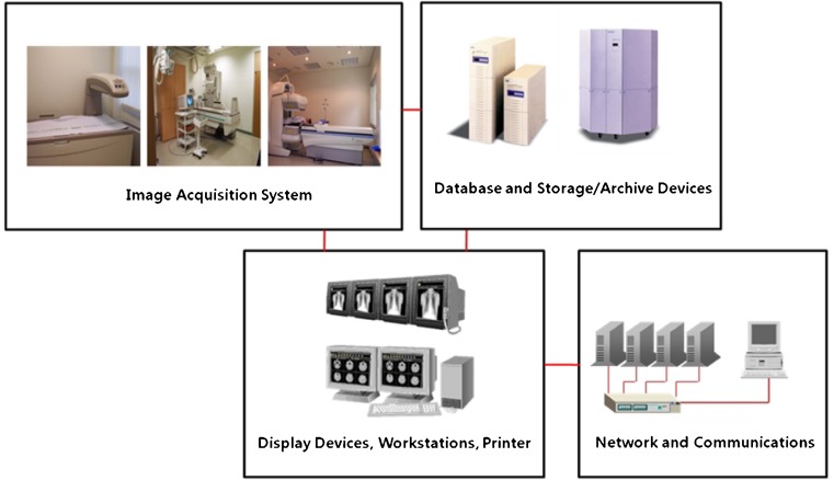
The advances in information and communication technology have allowed people to live in more comfortable circumstances. This trend has caused changes in medical care, education, finance, and other fields. Just as the case where the film camera has been replaced by the digital camera owing to its convenience and compatibility with computers, the medical field has experienced changes [1]. In previous film images, the spatial resolution and gray have analog values based on film exposure and processing. As a result, the dynamic range and optimal exposure range of film are limited by the maximum optical density allowed for the film. On the other hand, a digital image utilizes a discrete numerical matrix to express an image [2]. In a digital image, the contrast is expressed by the difference in numerical pixel value compared to that of other areas of the image. Digital images are used quite frequently because computer technology has been adopted for radiography systems in the field of imaging diagnosis. This has improved not only the quality of diagnosis but also the image quality because the image data are processed by a computer. In addition, the use of digital images enables function and quantitative analyses, which can increase the accuracy of diagnosis. This way, imaging diagnosis is being increasingly digital [3]. The typical example is the use of a picture archiving and communication system (PACS) that replaces the existing film. In PACS, the image is saved in digital form, rather than as film, before the doctors read the transmitted image. PACS has many advantages of convenience in keeping, transmitting, and reading a film along with the eco-friendly factor in that it is not necessary to develop film or dispose old film [4]. As shown in Fig. 1, PACS is generally composed of an image acquisition system, database and image storage/archive device, display device, workstation and printer, and network and communication device [5]. Consequently, PACS has either been adopted or is being considered for adoption by many teaching hospitals in South Korea including the majority of university hospitals [6].
Fig. 1.
Schematic diagram of PACS
Despite the many strong points, PACS has caused some problems, such as the reduced quality of reading and diagnosis with a PACS image, decreasing work efficiency, and an inability to exchange data between medical institutions due to the installation and use of equipment with low levels of technology or equipment that does not meet international standards. In addition, management of the image quality is the most important factor for improving the diagnosis efficiency [7]. When an existing film is used, management of the image quality is dependent on how the X-ray generator, film, screen, and film developer are managed. Therefore, a better image quality is maintained by making visible effects in improving the recognition and attitudes of equipment managers as well as in the management of a range of parameters on a regular basis. On the other hand, for image quality management in PACS, it is possible to adjust and apply the image in a variety of ways through an arithmetical transformation of discrete data. Innovative technological changes in saving, transmitting, and inquiring such an image have led to many corresponding requirements, such as support with the resources of the equipment and technical support. This has raised the importance of image quality management to maintain the reproducibility and consistency of the image. Unfortunately, the reality is that the image quality is not managed properly due to a lack of data and experience of clinical managers, even though image quality management in PACS requires considerable expertise and information technology. Against this background, this study evaluated a method to maintain the optimal image quality in clinical practice for image quality management in PACS that provides a typical medical image in digital form.
Materials and Method
The monitors were classified as those for clinical and reading purposes. In the case of monitor for clinical purposes, 25 hospitals in Seoul and metropolitan areas where PACS had been installed were surveyed from August 2010 to December 2010 to determine the reality of image quality management. In the case of the monitor for reading, 16 monitors were installed in the hospital that we worked for. The questionnaire survey was performed to determine whether radiation workers understood the importance of quality control before suggesting systematic and objective measures for quality control of PACS and whether the image quality was managed appropriately.
For clinical purpose monitor, the survey was conducted on 100 radiologists who had directly related work. The survey was carried out to check the status of monitor installation for each hospital after the monitor had been classified as either reading or clinical purposes. The survey was also conducted to determine the level of satisfaction depending on the brightness of the reading room and type of monitor. Furthermore, the survey was conducted to check the degree of fatigue from the monitor and the status of image quality management for each monitor currently used in each hospital. The survey items related to image quality management of the monitor included recognition of the monitor image quality control, learning path of monitor image quality control, cause of nonperformance of image quality control by size of the hospital, performance frequency of image quality control, and proper frequency of image quality control of the monitor. A calibration experiment was carried out for the reading purpose monitor.
The measurements were taken each week and month to determine if the calibration had changed. Eight sets of monitors with bright external illuminance and eight sets of monitors with dark external illuminance were calibrated on a weekly and monthly basis (for 2 months from May 19 to July 19) to determine the difference in monitor calibration depending on the changes in the environment for reading. In addition, tools and programs provided by the monitor manufacturer were used to conduct the calibration to meet the recommendations for a reading purpose monitor as much as possible. According to the recommendations by the monitor’s manufacturer, it is acceptable if a quadrangle can be recognized from 5 to 95 or 9 to 5 % in the Society of Motion Picture and Television Engineers (SMPTE) pattern. The relationship between the lifetime and uniformity was examined using Medical Pro 2.01 software to measure the lifetime of each monitor. In the monitor sets with the maximum and minimum lifetime, uniformity was measured using the VeriLUM grayscale pod and VeriLUM 4.2.
Lastly, the grayscale standard display function (GSDF) was analyzed by the external illuminance. The external illuminance was classified as 7.24, 0.34, and 0.28 for analysis. The GSDF, which is the standard that regulates how an image is displayed visually on certain equipment, was measured because it relates an image to a given illuminance and provides an objective and quantitative system. First, the VeriLUM grayscale pod was placed at a point 40 cm (16 in.) away from the plane of the monitor in parallel with the monitor to measure the external illuminance (ambient light). To measure the GSDF input value, the GSDF test image level 17 was monitored on the screen of the PACS viewing program before measuring the luminance for each level and entering the digital driving level (DDL). The measurement values were recorded in each step after attaching a shield to shut off the external illuminance to the monitor and tool to the center of each level. To measure the GSDF output, the program was used to obtain the measured DDL in each step. Subsequently, the p value was mapped to the DDL to reconcile the device characteristic curve with the GSDF curve in the luminance range. For statistical analysis, a survey was conducted on the status of monitor installation by the hospital, the level of satisfaction depending on brightness of the reading room and type of monitor, the degree of fatigue by the monitor, and the image quality management for the monitors used in each hospital. Based on the survey results, frequency analysis and crosstab analysis were performed to compare the difference in the number of cases. In the calibration experiment for the reading purpose monitor, the mean of the measurement values was calculated from the frequency of calibration.
SPSS version 16.0 (SPSS Inc., Chicago, IL) was used for statistical analysis. A paired t test was used to determine the difference in monitor calibration depending on the changes in the reading environment, whereas an ANOVA test was conducted for GSDF comparative analysis using the uniformity measurement value and illuminance of the monitor that depended on the difference in lifetime. A Duncan test was conducted as a post hoc analysis. A p value <0.05 was considered significant.
Results
Management of Monitor Image Quality
Before examining the management of the monitor image quality, a survey was conducted on the PACS image quality management in 25 hospitals that had used PACS to examine the installation status of the monitor for reading purposes and the monitor for clinical purposes. The examination of the status of reading purpose monitor installation showed that most of the 25 hospitals surveyed for this study had used a high-resolution monitor regardless of their size. Although 7.7 % used an LCD monitor as reading purpose monitor, it can be assumed that an LCD monitor was used for reading a color image in ultrasonography or endoscopy (Table 1). The level of satisfaction with such a high-resolution monitor was generally high in all hospitals surveyed. A survey was conducted on the satisfaction level depending on the brightness of the reading room. In the existing film system, the darker the room was, the higher the level of satisfaction level. On the other hand, when PACS was used, the satisfaction level was found to be affected only slightly by the brightness of the reading room (Table 2). The status of the installation of the clinical purpose monitor did not show any difference according to the size of the hospital. A flat CRT monitor was used the most by 52.0 % of hospitals followed in order by a general CRT monitor (28 %) and LCD monitor (20 %, Table 3). This study targeted radiologists to determine the level of satisfaction with the monitor they were using. The overall satisfaction level was 44 % regardless of the type of monitor, but there was a large difference in satisfaction depending on the type of monitor. With respect to the general CRT monitor, the installation rate was 28 % with a dissatisfaction level of 42 %, which shows a low installation rate but high dissatisfaction level. On the contrary, with regard to the LCD monitor, the installation rate was low, but the satisfaction level was high (60 %, Table 4). The degree of fatigue was similar regardless of the monitor used. Overall, the respondents in 60 % of the hospitals surveyed for this study felt some degree of fatigue. As the degree of fatigue is believed to be related to the image quality of the monitor, this study focused on the management of image quality. The awareness of management of the monitor image quality in clinical practice was examined. Most of the respondents (76 %) reported an awareness but 24 % answered that they were “unfamiliar” with the management. These results show that an awareness of the need to manage the monitor image quality is still insufficient (Table 5).
Table 1.
Status of installation of a reading purpose monitor according to the size of the hospital
| Size of hospital | LCD monitor | High-resolution monitor |
|---|---|---|
| Hospital | 0 (0.0) | 4 (100) |
| General hospital | 1 (7.7) | 12 (92.3) |
| University hospital | 0 (0.0) | 8 (100) |
| Total (average) | 1 (4.0) | 24 (96.0) |
Interaction effect using frequency analysis model and crosstabs model. Unit is the number of case/percent
Table 2.
Satisfaction level of a reading purpose monitor depending on the brightness of the reading room
| Brightness of reading room | Very satisfactory | Satisfactory | Fair | Unsatisfactory |
|---|---|---|---|---|
| Dark | 3 (42.9) | 6 (35.3) | 0 (0.0) | 0 (0.0) |
| Fair | 4 (57.1) | 7 (41.2) | 0 (0.0) | 0 (0.0) |
| Bright | 0 (0.0) | 4 (28.5) | 1 (100) | 0 (0.0) |
| Total (%) | 7 (100) | 17 (100) | 1 (100) | 0 (0.0) |
Interaction effect using frequency analysis model and crosstabs model. Unit is the number of case/percent
Table 3.
Status of installation of a clinical purpose monitor according to the size of the hospital
| Size of hospital | General CRT monitor | LCD monitor | Flat CRT monitor |
|---|---|---|---|
| Hospital | 1 (25.05) | 1 (25.0) | 2 (50.0) |
| General hospital | 5 (38.5) | 2 (15.4) | 6 (46.2) |
| University hospital | 1 (12.5) | 2 (25.0) | 5 (62.5) |
| Total (average) | 7 (28.0) | 5 (20.0) | 13 (52.0) |
Interaction effect using frequency analysis model and crosstabs model. Unit is the number of case/percent
Table 4.
Radiologist’s satisfaction level of the clinical purpose monitor
| Clinical purpose monitor | Satisfactory | Fair | Unsatisfactory |
|---|---|---|---|
| General CRT monitor | 3 (42.9) | 1 (14.3) | 3 (42.9) |
| LCD monitor | 3 (60.0) | 2 (40.0) | 0 (0) |
| Flat CRT monitor | 5 (38.5) | 8 (61.5) | 0 (0) |
| Total (average) | 11 (44.0) | 11 (44.0) | 3 (12.0) |
Interaction effect using frequency analysis model and crosstabs model. Unit is number and case/percent
Table 5.
Degree of fatigue by the monitor and the awareness of image quality management
| Degree of fatigue | Very tired | Tired | Fair | Cannot feel |
| Total (%) | 1 (4.0) | 14 (56.0) | 8 (32.0) | 2 (8.2) |
| Awareness | Well aware of | Aware of | Fair | Not aware of |
| Total (%) | 1 (4.0) | 10 (40.0) | 8 (32.0) | 6 (24.0) |
Interaction effect using frequency analysis model and crosstabs model. Unit is number and percentage
This study examined how the respondents, who were aware of the management, became aware of information on the management of monitor image quality. According to the survey results, 48 % of respondents said that they became interested in image quality management as they felt the need themselves. The remainder said that they become interested in the management by using a product or reading a book. On the other hand, there is still insufficient information on management of image quality in the symposium or mass media (Table 6). An investigation of whether the image quality of the monitor is managed properly among hospitals showed that only 48 % of 25 hospitals surveyed had managed the image quality while the remaining 52 % had not.
Table 6.
How to learn about management of the monitor image quality
| Through product company | Through book | On one’s own | Through mass media | Through symposium | |
|---|---|---|---|---|---|
| Total (%) | 11 (44.0) | 2 (8.0) | 12 (48.0) | 0 (0.0) | 0 (0.0) |
Interaction effect using Frequency analysis model and Crosstabs model. Unit is number and percentage
First, the reasons for the nonmanagement of the image quality among the hospitals were examined. Although no difference was shown according to the size of the hospital, 23.1 % of respondents from general hospitals and university hospitals said that they did not see the necessity of image quality management, which demonstrated that they had insufficient awareness of image quality management. Notwithstanding the awareness of the need to manage the image quality, the hospitals could not manage the image quality because of the method or conditions (Table 7).
Table 7.
Cause of nonmanagement of the image quality according to the size of the hospital
| Size of hospital | Do not knowthe method | Do not have a tool for management | Conditions are not allowed to manage | Do not feel the need | For other reasons |
|---|---|---|---|---|---|
| Hospital | 1 (50) | 0 (0.0) | 1 (50.0) | 0 (0.0) | 0 (0.0) |
| General hospital | 1 (16.7) | 1 (16.7) | 1 (16.7) | 2 (33.3) | 1 (16.7) |
| University hospital | 0 (0.0) | 1 (20.0) | 0 (0.0) | 1 (20.0) | 3 (60.0) |
| Total (average) | 2 (15.4) | 2 (15.4) | 2 (15.4) | 3 (23.1) | 4 (30.8) |
Interaction effect using frequency analysis model and crosstabs model. Unit is the number of case/percent
Next, a survey was conducted on the hospitals that had managed image quality based on the image quality management frequency. More respondents said that image quality was managed “frequently” than on a regular basis. In addition, 16 % answered that management on a regular basis meant management once a month. The level of awareness of image quality management of a monitor was low, while the image quality management was insufficient (Table 8).
Table 8.
Frequency of image quality management
| No management | Once per week | Once per month | Once per quarter | Frequently | |
|---|---|---|---|---|---|
| Total (%) | 13 (52.0) | 2 (8.0) | 4 (16.0) | 1 (4.0) | 5 (20.0) |
Interaction effect using frequency analysis model and crosstabs model. Unit is number and percentage
The proper frequency of image quality management that had been considered by many in general was investigated based on the results of the survey mentioned above. According to the investigation, many answered that once per month was the appropriate frequency for managing the image quality of reading purpose and clinical purpose monitors. On the other hand, a considerable number of the respondents answered “others,” which demonstrated that the respondents had insufficient awareness of regular image quality management (Table 9).
Table 9.
Proper frequency of image quality management for a reading purpose monitor and clinical purpose monitor
| Clinical purpose monitor | Once per week | Once per month | Once per year | Others | Total (%) |
|---|---|---|---|---|---|
| General CRT Monitor | 4 (57.1) | 1 (14.3) | 1 (14.3) | 1 (14.3) | 7 (100) |
| LCD monitor | 1 (20.0) | 2 (40.0) | 0 (0.0) | 2 (40.0) | 5 (100) |
| Flat CRT monitor | 2 (28.0) | 5 (32.0) | 1 (8.0) | 5 (38.5) | 13 (100) |
| Total (%) | 7 (28.0) | 8 (32.0) | 2 (8.0) | 8 (32.0) |
Interaction effect using frequency analysis model and crosstabs model. Unit is the number of cases and percentage
Calibration Experiment for Reading Purpose Monitor
When the calibration frequency was weekly, the values of the white, black, and quality level, which were displayed on a monitor, were included in the set standards, whereas the SMPTE was also included in the range (Table 10). When the frequency was monthly, the values of white, black, and quality levels, which were displayed on the monitor, were included in the set standards, whereas the SMPTE was also included in the range (Table 11).
Table 10.
Weekly monitor calibration
| Calibration | Average | Target | Warning tolerance | Error tolerance | |
|---|---|---|---|---|---|
| A | White (cd/m2) | 301.23 | 300 | ±3.0 % | ±6.0 % |
| Black (cd/m2) | 0.001 | 0 | ±1.0 % | ±2.0 % | |
| Quality level (%) | 99.23 | 100 | ±5.0 | ±10.0 | |
| SMPTE | OK | OK | |||
| B | White (cd/m2) | 300.4 | 300 | ±3.0 % | ±6.0 % |
| Black (cd/m2) | 0.001 | 0 | ±1.0 % | ±2.0 % | |
| Quality level (%) | 99.012 | 100 | ±5.0 | ±10.0 | |
| SMPTE | OK | OK | |||
Target: set standard for white, black, and quality level that are displayed on the monitor. Warning tolerance: allowable limit. Error tolerance: error limit. Interaction effect using frequency analysis model and crosstabs model
Table 11.
Monthly monitor calibration (average of two sets)
| Calibration | Average | Target | Warning tolerance | Error tolerance | |
|---|---|---|---|---|---|
| C | White (cd/m2) | 301.767 | 300 | ±3.0 % | ±6.0 % |
| Black (cd/m2) | 0.047 | 0 | ±1.0 % | ±2.0 % | |
| Quality level (%) | 99.361 | 100 | ±5.0 | ±10.0 | |
| SMPTE | OK | OK | |||
| D | White (cd/m2) | 303.406 | 300 | ±3.0 % | ±6.0 % |
| Black (cd/m2) | 0.002 | 0 | ±1.0 % | ±2.0 % | |
| Quality level (%) | 99.295 | 100 | ±5.0 | ±10.0 | |
| SMPTE | OK | OK | |||
Target: set standard for white, black, and quality level that are displayed on the monitor. Warning tolerance: allowable limit. Error tolerance: error limit. Interaction effect using frequency analysis model and crosstabs model
Regarding the monitor calibration difference depending on the changes in the environment for reading, the white, black, and quality levels, which were displayed on the monitor, in both the bright and dark environment were included in the set standards, whereas the SMPTE was also included in the range. The difference between the two environments was not significant (p > 0.05, Table 12). According to the measurement values of uniformity of the monitor depending on the difference in lifetime, the uniformity was found to be better when the lifetime was a minimum than a maximum (Table 13, p < 0.05).
Table 12.
Monitor calibration depending on the changes in the environment for reading an image
| Calibration | Average | Target | Warning tolerance | Error tolerance | p | |
|---|---|---|---|---|---|---|
| Bright Environment | White (cd/m2) | 300.998 | 300 | ±3.0 % | ±6.0 % | 0.251 |
| Black (cd/m2) | 0.024 | 0 | ±1.0 % | ±2.0 % | 0.435 | |
| Quality level (%) | 99.296 | 100 | ±5.0 | ±10.0 | 0.550 | |
| SMPTE | OK | OK | ||||
| Dark Environment | White (cd/m2) | 301.90 | 300 | ±3.0 % | ±3.0 % | 0.224 |
| Black (cd/m2) | 0.001 | 0 | ±1.0 % | ±2.0 % | 0.385 | |
| Quality level (%) | 99.153 | 100 | ±5.0 | ±10.0 | 0.657 | |
| SMPTE | OK | OK | ||||
Target: set standard for white, black, and quality level that are displayed on the monitor. Warning tolerance: allowable limit. Error tolerance: error limit. Interaction effect using paired t test model
Table 13.
Measurement values of the monitor’s uniformity depending on the difference in lifetime
| Lifetime | Center luminance | Top left luminance | Top right luminance | Bottom left luminance | Bottom right luminance | p | |
|---|---|---|---|---|---|---|---|
| Minimum value (D) | 3,602 | 306.19 | 300.22 (−1.9 %) | 302.04 (−1.4 %) | 302.08 (−1.3 %) | 304.93 (−0.4 %) | 0.025* |
| Maximum value (C) | 4,965 | 303.63 | 316.09 (4.1 %) | 314.26 (3.5 %) | 315.50 (3.9 %) | 316.16 (4.1 %) | 0.040* |
Interaction effect using ANOVA test model and Duncan test model. Unit is the number and in candela per square meter
*p < 0.05
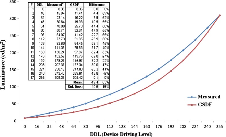
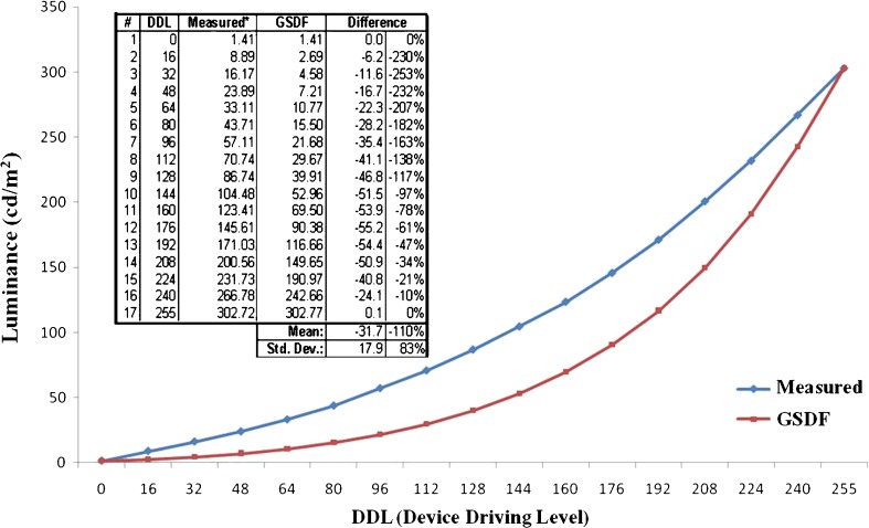
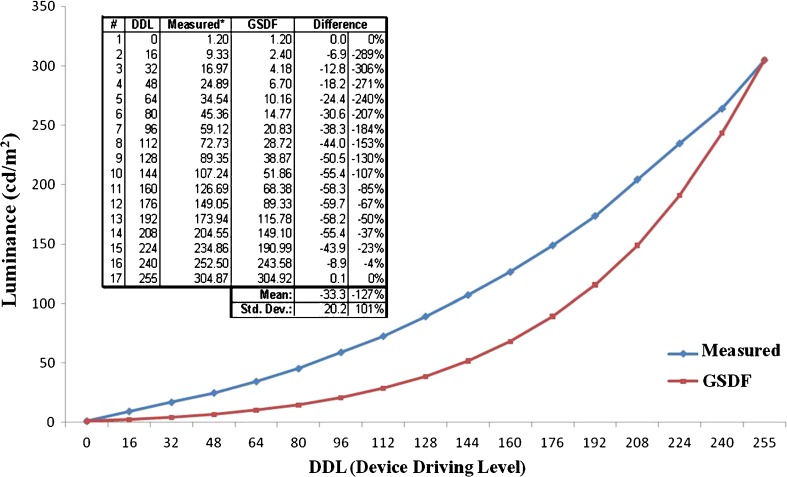
In comparative analysis of the GSDF, measurements in the bright environment with high external illuminance (ambient light) and the dark environment with low external illuminance (ambient light) were conducted before comparing the measurements. The comparison showed no significant difference (p < 0.05). Furthermore, in the GSDF measurement experiment, the ambient light value and DDL value were measured to determine the difference between the measurement value and standard value before entering them into the program. The results revealed a significant difference between the standard and measurement values for all illuminance values (Figs. 2, 3, and 4).
Fig. 2.
GSDF measurement in an external illuminance (ambient light) of 7.24
Fig. 3.
GSDF measurement in an external illuminance (ambient light) of 0.34
Fig. 4.
GSDF measurement in an external illuminance (ambient light) of 0.28
Discussion
The recent trend is that hospitals in South Korea are being increasingly provided with an environment for digital radiography. CT and MR have enabled digital imaging for a long time, and have no problems being applied to digital radiography.
In general, digital signal obtained from digital radiography can be measured, specialized, and transmitted to be reproduced in an objective and accurate manner. Nevertheless, visual analysis of such signal tends to be dependent on the changing characteristics of the system that displays an image of the signal. Recently, there was a case where the images generated from the same signal provided information and characteristics in a completely different visual shape and in different display equipment [8].
In the field of medical imaging, it is important to consider how a given digital image is displayed. For example, it is important to ensure visual consistency between the viewing image on a workstation monitor and viewing it on films in a view box. Without any standards that regulate how such images are displayed visually, a digital image, which has a good diagnostic value when displayed on one piece of equipment, might be viewed differently or could have significantly lower diagnostic value when displayed on other viewing programs [9]. To display an image as objectively as possible, a calibration tool from the manufacturing company was used to measure the display white, display black, quality level, and SMPTE pattern before comparing and entering the external factors, such as calibration frequency, external illuminance, and time of monitor use. Most of the hospitals surveyed did not have quality control practices based on certain rules or standardized methods, which demonstrated a lack of awareness of quality control. These results demonstrated that relevant materials had not been provided through the symposium, publication, or other mass media. Furthermore, in regard to the satisfaction level depending on the brightness for reading an image among various environmental factors, there was no difficulty in reading an image regardless of the brightness in the PACS system, which is unlike the system where the image could be read depending on the brightness of the view box when film was used. Although an awareness of the monitor’s lifetime can be taken lightly, it is expected that effective management can be possible if such a monitor’s lifetime is widely recognized and publicized as a part of quality control. With respect to monitor calibration, the GSDF provides details on the objective and quantitative system, where digital images are mapped to the luminance in a given range [10]. As a result, the most critical factor for quality control is to illustrate the relationship between the digital value and display luminance in higher visual consistency. Quality control is very important and requires careful management in a range of fields.
Conclusions
A survey on quality control among current medical institutions found that such medical institutions lacked quality control. Against this background, this experiment was conducted to suggest a more systematic and objective method.
The monitor calibrated weekly had a lower error rate than that calibrated monthly, but the difference was within the range of allowable error. Therefore, it would be more efficient to calibrate monthly or quarterly. There was a significant difference in the satisfaction level of brightness when the survey was conducted on the satisfaction level depending on the environment for reading an image. Fewer errors were found in the monitor calibrated in the dark environment than in the bright environment, and the error rate was low in the monitor calibrated in a dark environment. On the other hand, the calibration values were similar to the values obtained in the bright environment, which demonstrated that the environment for reading an image does not have a significant effect on calibration.
The mean lifetime of the monitors was 4,232 h, which means that a monitor can be used 7.7 h per day (in the hospital opened in February 2001). In most cases, when the monitor had a shorter lifetime, it showed a fewer uniformity errors. On the other hand, some monitors had rather high errors in uniformity regardless of the lifetime. Therefore uniformity of the monitor could be influenced significantly by other factors (calibration, movement of measuring device, contamination level of monitor, etc.) in addition to the lifetime. Since the results were not out of the margin of error (±15 %), it is believed that a measurement on a regular basis would be beneficial to image quality management for a monitor.
In the GSDF measurement experiment, the ambient light and DDL values were measured to determine the difference between the measurement and standard values. These values were entered into the program, which showed a significant difference between the standard and measurement values. The causes of such a difference included the error in measuring the ambient light value and the error in measuring the DDL value. The ambient light value is basically measured at a point, 40 cm away from the center of the monitor. Despite the measurement with the distance kept constant, the value cannot be expressed in a single value because it is affected by factors, such as the location of external illuminance and the location of the artificial grayscale pod. This means that it is difficult to conduct a precise measurement. Similarly, it is important to select the correct location for measuring level 17 to obtain the DDL value. Hence, the measurement would be influenced significantly by the skill of the technician. Against this backdrop, this survey was conducted to examine the status of image quality management of a monitor. An awareness of the necessity to manage the image quality is important, but there is no standardized method. This paper suggests an effective method for managing the image quality of a monitor.
Contributor Information
Jae-Hwan Cho, Email: 8452404@hanmail.net.
Hae-Kag Lee, Email: lhk7083@sch.ac.kr.
Kyung-Rae Dong, Email: krdong@hanmail.net.
Woon-Kwan Chung, Phone: +82-62-2307166, FAX: +82-62-2329218, Email: wkchung@chosun.ac.kr.
References
- 1.Digital Imaging and Communications in Medicine (DICOM): Base Standard. National Electrical Manufacturers Association. Rosslyn, VA, USA. 2008
- 2.Fujita H, Ueda K, Morishita J. Basic imaging properties of a computed radiographic system with photostimulable phosphors. Med Phys. 1989;16:52–59. doi: 10.1118/1.596402. [DOI] [PubMed] [Google Scholar]
- 3.Axelsson B, Boden K, Fransson S. A comparison of analogue and digital techniques in upper gastrointestinal examinations: absorbed dose and diagnostic quality of the images. Eur Radiol. 2000;10:1351–1354. doi: 10.1007/s003300000327. [DOI] [PubMed] [Google Scholar]
- 4.Park YH. Consideration about medical treatment’s utility through input of image. J Korean Soc Picture Archiving Commun Syst. 2004;10:125–132. [Google Scholar]
- 5.Shin MJ, Song KS, Auh YH, Lim KT, Lee JH, Lee SH. PACS development in Asan medical center. KMIAA. 1997;3:1–3. [Google Scholar]
- 6.Ro DW, Kim BH, Choo SW, Lee WJ, Lee KS, Kim BK. Status of full PACS at Sammung medical center. KMIAA. 1998;4:1–4. [Google Scholar]
- 7.Bramble JM, Huang HK, Murphy MD. Image data compression. Invest Radiol. 1998;23:707–712. doi: 10.1097/00004424-198810000-00001. [DOI] [PubMed] [Google Scholar]
- 8.Crochiere RE, Oppenheim AV, Dudgeon DE, Mersereau RM, Portnoff MR, Tribolet JM, Zue VW, Allen J. Digit Signal Proc RLE Prog Rep. 1974;112:79–82. [Google Scholar]
- 9.Huh YJ, Jeon IS, Heo MS, Lee SS, Choi SC, Park TW, Kim JD. A comparative study on the accuracy of digital subtraction radiography according to the acquisition methods of reconstructed images. Korean J Oral Maxillofac Radiol. 2002;32:107–111. [Google Scholar]
- 10.Jones DM. Utilization of DICOM GSDF to modify lookup tables for images acquired on film digitizers. J Digit Imaging. 2006;19:167–171. doi: 10.1007/s10278-005-9241-z. [DOI] [PMC free article] [PubMed] [Google Scholar]


