Abstract
Purpose:
To describe preoperative factors, long-term (>3 years) postoperative outcome and cost of traumatic cataracts in children in predominantly rural districts of western India.
Subjects:
Eighty-two traumatic cataracts in 81 children in a pediatric ophthalmology department of a tertiary eye-care center.
Materials and Methods:
Traumatic cataracts operated in 2004–2008 were reexamined prospectively in 2010–2011 using standardized technique. Cause and type of trauma, demographic factors, surgical intervention, complications, and visual acuity was recorded.
Statistical Analysis:
Data analysis done by using SPSS (Statistical package for social sciences) version 17.0 We have used Chi-square test, Fisher's exact test, paired t-test to find the association between the final vision and various parameters at 5% level of significance; binary logistic regression was performed for visual outcome ≥6/18 and ≥6/60.
Results:
The children were examined in a 3–7 year follow-up (4.35 ± 1.54). Average age at time of surgery was 10.4 ± 4.43 years (1.03 to 18). Fifty (61.7%) were boys. Forty (48.8%) were blunt and 32 (39%) were sharp trauma. The most common cause was wooden stick 23 (28.0%) and sharp thorn 14 (17.1%). Delay between trauma and presentation to hospital ranged from same day to 12 years after the injury with median of 4 days. The mean preoperative visual acuity by decimal notation was 0.059 ± 0.073 and mean postoperative visual acuity was 0.483 ± 0.417 (P < 0.001). Thirty-eight (46.3%) had best corrected visual acuity (BCVA) ≥6/18 and 51 (62.2%) had BCVA ≥ 6/60. In univariable analysis, visual outcome (≥6/18) depended on type of surgery (P = 0.002), gender (P = 0.028), and type of injury (P = 0.07)–sharp trauma and open globe injury had poorer outcomes; but not on age of child, preoperative vision, and type of surgeon. On multivariable binary logistic regression, only gender was significant variable. Of the 82 eyes, 18 (22%) needed more than one surgery. The parents spent an average of  2250 ($45) for the surgery and 55 (66.4%) were from lower socio-economic class.
Conclusion:
The postoperative visual outcomes varied and less than half achieved ≥ 6/18.
Keywords: Trauma, pediatric cataract, visual outcome
Traumatic cataracts form a separate category of cataracts as they present with other ocular morbidity like corneal tears, iris injury, vitreous hemorrhage, and retinal tears; and they are to some extent, preventable.[1] While studies of pediatric cataract in South Asia report traumatic cataracts as a significant component (25–38%), there are few studies detailing the etiology and types of trauma.[2–5] The causes of trauma may be different in rural regions where a majority of the population of India resides. A recent large study looked at the causes and outcomes of traumatic cataracts in India in all age groups, but was limited to 6 weeks follow-up.[6] A search of published literature in PubMed revealed no study regarding long-term outcome of traumatic cataracts in children in the developing world. In addition, most published series report outcome of traumatic cataract surgery in retrospective study design. The objective of our current study is 2-fold: to report pre- and perioperative factors (demographics, socio-economic status, cause of trauma, and type of surgery) and to report 3- to 7-year outcome of traumatic cataracts in children in rural India and factors affecting the visual outcome. For this visit, we examined all patients in a prospective manner using standardized protocol.
Materials and Methods
Permission was obtained from the ethical committee of the Lions NAB Eye Hospital, Miraj, India. Informed consent was obtained from all participating children and their parents. The study was completed between October 2010 and June 2011. A review of all the traumatic cataracts operated in ORBIS supported pediatric ophthalmology department in the years 2004–2008 was performed. They had been operated under the ORBIS International, India country office's childhood blindness initiative and detailed case records had been maintained for reporting and monitoring. ORBIS International is an international nongovernmental developmental organization working for combating avoidable blindness globally. The addresses of each and every child along with the contact phone numbers had been carefully recorded. These children were identified in their villages and towns and were visited by a medical social worker. History was obtained, based in case records, about type of trauma and cause of trauma. These details were confirmed during the prospective examination wherever possible.
The time interval between injury and presentation to the hospital and also the time interval between multiple surgeries (if there was more than one surgery) was noted. Trauma was classified as blunt or sharp, depending upon the type of object causing the injury. It was also classified as open or closed globe injury depending on whether the ocular coverings were perforated or not. In all children with corneal tear, the traumatic cataract was managed during the second procedure. Most surgeries were irrigation and aspiration. They were termed manual small incision cataract surgery if the phacoemulsification machine was not used. A square edged posterior chamber intra-ocular lens had been implanted in the capsular bag wherever possible. The operating surgeon was classified as ‘pediatric ophthalmologist’ if he/she had completed a long-term fellowship in that subject or as a ‘general ophthalmologist’ otherwise. The child's socio-economic status was graded according to their parent's education, occupation, and family income by the Kuppusamy classification into one of the five socio-economic sub-groups from I (highest) to V (lowest).[7] The parents were asked about the amount they paid to the hospital for surgery and their other expenses, like travel and medicines, incurred for the treatment. Only direct costs were considered not indirect ones like wages lost while attending treatment.
The parents were questioned about what they had spent for the ocular treatment. The children were transported in a vehicle to the hospital along with their parents for an eye examination. They underwent a comprehensive ocular examination and clinical and demographic data were recorded. If any treatment was needed: spectacles, surgery or low vision aids, the parents were informed, and relevant treatment provided free of cost to the children. The children underwent a complete ocular examination–visual acuity estimation, slit lamp examination, orthoptic evaluation, fundoscopy, and cycloplegic refraction. Visual acuity was measured by the Snellen's chart. Cardiff cards were used for children ≤ 5 years of age. Intra-ocular pressure was measured by a noncontact tonometer.
Data analysis was done by using statistical package for social sciences (SPSS) version 17.0. We used Chi-square test, Fisher's exact test, paired t-test to find the association between the final vision and various parameters at 5% level of significance. Multivariable analysis for visual outcome with cut-off at ≥6/18 and ≥6/60 was done by binary logistic regression as our outcome was binary.
Results
In the years 2004–2008, 82 traumatic cataracts in children had been operated upon and were re examined in 2009–2010. All those eligible agreed to participate. The mean follow-up was 4.35 years (standard deviation [SD] ± 1.54 years). They belonged to 81 children of whom 50 (61.7%) were boys. A 10-year-old girl had bilateral traumatic cataracts after fall from a height. Two children (2.4%) were ≤ 5 years of age, 20 (24.4%) between 6 and 10 years, 21 (25.6%) between 11 and 15 years and 39 (47.6%) ≥ 16 years of age at the time of evaluation. Of the 82, 42 were left and 40 were right eyes. Their ages at the time of surgery were: aged ≤2 years: 3 (3.7%), aged 3–5 years: 13 (15.9%), aged 6-10 years: 24 (29.3%), aged 11–15 years: 28 (34.1%), and aged ≥16 years: 14 (17.1%). The mean age at the time of surgery was 10.35 years (SD ±1.4 years). The youngest age was 10 months with blunt trauma due to bull horn injury. In nine eyes the exact cause of injury could not be determined on history and case records. Forty (48.8%) eyes had blunt injury while 32 (39%) eyes had sharp injury. Eighteen (21.9%) eyes had open globe injury while 64 (78.1%) had closed globe injury.
The median delay between the traumatic episode and presentation to the hospital was 4 days (range: same day to 12 years). Eighteen (22%) eyes had needed more than one surgical intervention. The median interval between two surgeries was 5 weeks (4 days to 6 years). Of the 82 traumatic cataracts, 67 (81.7%) were brought to the hospital by their parents, 5 (6.2%) by uncle or aunt, 2 (2.5%) by grandmother, 2 (2.5%) by their neighbors while 5 (6.2%) presented on their own for the initial presentation after the injury. On classifying the children's family into five socio-economic sub-groups from I (highest) to V (lowest) by the Kuppusamy classification, majority of children were from lower socio-economic status, 5 (6.2%) were from socioeconomic group V (poorest), 50 (61.2%) from group IV, 21 (25.6 %) from group III. One child was from socioeconomic group I (highest) and four (4.9%) from socio-economic group II.
The average expense for the treatment was  2250 ($45) (SD
  2315) with 18 (22%) surgeries performed completely free, 24 (29.3%) subsidized (paying
  700–2500, $14–50) while 38 (46.3%) were ‘paid’, they spent
  3500–7000 ($70–140) out of their pocket for the surgeries. Cost details were not available for two surgeries. The average amount spent by the parents on travel was
  186.36 (SD
  123.86, range
  20–600; mean inUS$3.72) and on medicines was
  1853.59 (SD
  3781.4, range
  150–20,000, mean in US$ 37.1).
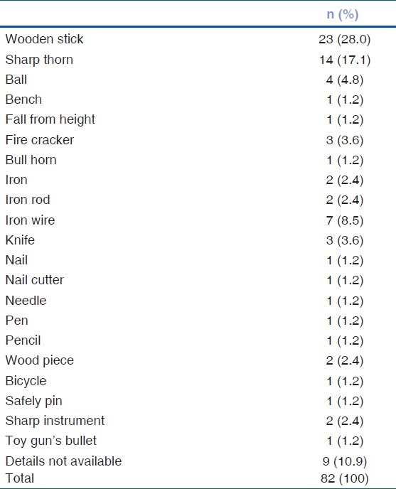
The causes of trauma are enumerated in Table 1. Injury by wooden stick and sharp thorns were the most common cause of injury.
Table 1.
Cause of trauma

Eight (9.8%) had undergone only lens aspiration, 2 (2.4%) cataract + anterior vitrectomy (AV) + primary posterior capsulotomy (PPC), 31 (37.8%) had undergone manual small incision cataract surgery (SICS) with intraocular lens (IOL) implantation, and 29 (35.4%) had undergone phaco emulsification with IOL implantation. Most surgeries were irrigation and aspiration. They were termed manual small incision cataract surgery if the phacoemulsification machine was not used. One eye had small incision cataract surgery (SICS) + AV + PPC + posterior chamber intraocular lens (PCIOL) and another phaco AV + PPC + PCIOL.74/82 (90.2%) eyes had an intra-ocular lens implanted.
Table 2 shows the aided visual acuity before surgery, unaided and aided visual acuity at the 6 weeks and 3–7 year follow-up. The preoperative and 6 weeks data were collected from chart review while the follow-up data were collected prospectively at the follow-up appointment. The preoperative visual acuity mean was 0.059 (SD 0.073) by decimal notation and postoperative visual acuity mean was 0.483 (SD 0.417), P<0.001on doing a paired t-test.
Table 2.
Visual acuity before and after traumatic cataract surgery in children

Of the 82, 6 (7.3%) eyes had intraocular pressure >20mm of Hg in the operated eye while 8 (9.8%) eyes had postoperative uveitis. Of the 82, 53 (64.7%) had more than grade I posterior capsular opacification (PCO) after 5 years. Of these, 28 eyes underwent a Nd:YAG LASER capsulotomy and one eye membranectomy during the follow-up visit. Before the intervention, 25/53 (47.2%) of eyes with PCO had BCVA ≥ 6/18 compared with 12/29 (41.3%) had BCVA ≥ 6/18 in those that did not have PCO. Thirty-two children were dispensed new spectacles, two were given aphakic contact lens, and four were given a cosmetic contact lens for their corneal opacity. Four children underwent strabismus surgery.
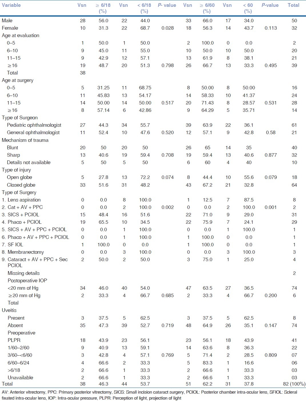
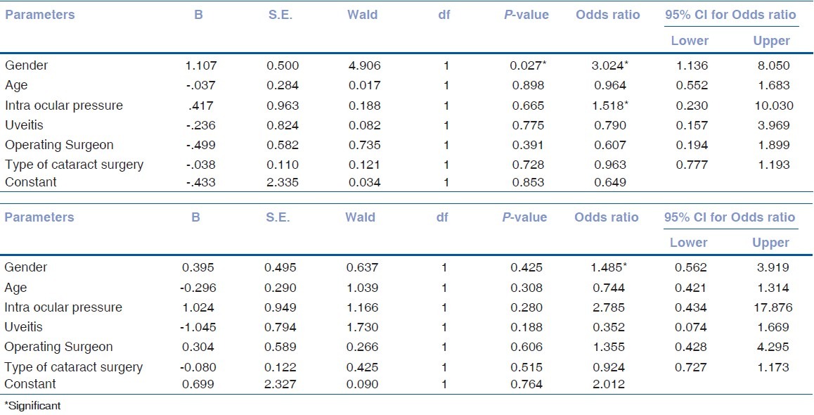
Table 3 demonstrates the correlation between various demographic and ocular parameters and visual acuity after surgery. Table 4 demonstrates the multivariable analysis using binary logistic regression with ≥ 6/18 and ≥ 6/60 as cut-off.
Table 3.
Factors affecting visual acuity after traumatic cataract surgery

Table 4.
Multivariable analysis: By using binary logistic regression, (A) Binary logistic regression with ≥6/18 as cut-off

Discussion
Our study is limited by the fact that the surgical details were as recorded 3–7 years ago and there may have been a recall bias in parents reporting what they spent for the surgery. But the >3 years outcome was reported after prospective evaluation and standardized clinical examination. There were no significant differences in the visual outcome depending on age and type of operating surgeon. As with other studies, boys were more commonly affected than girls; no doubt due to their outdoor habits and more chances of playing rough and contact and projectile sports.[1,2,4,8,9] But a large study reporting traumatic cataracts from tribal regions of India had young adults with traumatic cataracts equally common in both the genders.[6]
Age was not a significant variable affecting visual acuity unlike some studies from India and the USA.[6,10] But very young children had poorer outcome. Majority of children were of the school-going age group though traumatic cataracts were seen in preschoolers too, one child being an infant at the time of injury.
Injury by wooden sticks and sharp thorns were the most common causes of traumatic cataract in this rural community. Wooden sticks were used as firewood and many children, boys and girls, helped their parents in collecting them. This was similar to reports from tribal belt in India and Nepal.[6,8] Playing with a cricket ball, toy guns, and fire-crackers (during the festival of Diwali) were the common causes of trauma while playing. Most of the children came from lower socio-economic strata and were thus more likely to be participating in some kind of agricultural activities and playing more outdoor sports. Except in one child with a history of fall from height, the traumatic cataracts were invariably unilateral.
The median time interval between injury and presentation to the hospital was only 4 days, which was heartening, unlike the delay seen in congenital or developmental cataracts.[4,11] Most children were brought to the hospital by their parents who did not brook delay, indicating the seriousness with which the injury was taken. It also explains why age of patient and preoperative vision did not affect visual outcome as amblyopia could not develop. Congenital and developmental cataract surgery results depend on age of the child and visual acuity before surgery.[12]
Gradin et al. reported that 64.7% had vision better than 20/60 after surgery for traumatic cataract, compared with 38 (46.3%) in this series.[13] Aldakaf et al. and Sternberg et al. reported that initial vision and mechanism of injury were predictors of final outcome.[10,14] Eyes with sharp trauma had poorer visual results, as did eyes that needed multiple surgeries due to coexisting ocular morbidity, commonly corneal tears. Eyes that had postoperative uveitis and raised intra-ocular pressure had a poorer visual outcome.
In this study the only significant variable affecting visual outcome (≥6/18) on univariable analysis was gender and the type of surgery which in turn depended on the type of trauma and the age of presentation. On multivariable analysis only gender was significant. Girls had poorer outcomes, even though injuries were similar. Use of IOLs was a norm and only 8/82 underwent a plain cataract extraction at the time of surgery. This was in line with other series of traumatic cataracts globally.[15–18] Nonuse of IOLs was associated with poor outcome in our study, but IOLs were not used only in circumstances where the eye was badly damaged.
A series of traumatic cataracts from south India more than a decade ago had 92% of children developing posterior capsular opacification if primary posterior capsulotomy was not performed.[15] Another study from India comparing traumatic cataract surgery with and without posterior capsule management had demonstrated that AV + PPC had a beneficial effect on visual acuity (P = 0.001) and prevented further intervention.[19] This study showed that a significant number of children with intact posterior capsule may develop visually significant PCO even after placement of square-edge IOL in the capsular bag. Eyes with PCO in our study had similar vision than those without, as PPC + AV was done in very young children who had poorer vision. Ocular co-morbidity also was a confounding factor.
A lot of these injuries could have been prevented had proper protective wear being used, but it would be difficult to convince rural communities the advantages of protective wear to their children. Many house-hold items like pins, scissors, knives, pens, pencils, and nail cutters were responsible for the injury. Local government schools should inculcate the safety habits through the school curriculum as has been done in many urban private schools in India, along with lessons on first aid and safety precautions. It was heartening to note that bow and arrow injuries were not recorded as was the case more than a decade ago and there were only three instances of fire work injuries. These had been very common in the past decade and had been subjected to intense health education through television commercials.
In conclusion, even in developing country rural setting, satisfactory visual outcome is possible on long-term for children with traumatic cataract.
Acknowledgement
The clinical examination protocol was validated by Professor Clare Gilbert, (from the London School of Hygiene and Tropical Medicine/International Centre for Eye Health), Dr. Joan McLeod-Omnawale, ORBIS Director of Monitoring and Evaluation, Dr. Rupal Trivedi, Associate Professor, Storm Eye Institute, South Carolina, USA, Dr. H. Kishore, senior pediatric ophthalmologist, Al-Nahda Hospital, Muscat, Oman and Dr. Milind Killedar, pediatric ophthalmologist from Sangli, India. The final version was in concurrence of the authors with Dr. G.V. Rao, Rishi Raj Bora, Dr. Lutful Hussain (all from ORBIS India) and Dr. A.H. Mahadik (from Lions NAB Eye Hospital, Miraj). Faiz Mushrif and Poonam Shinde helped in data collection; Shrivallabh Sane in data analysis.
Footnotes
Source of Support: ORBIS International, New York through the USAID's AED Operational research project under the A2Z child micronutrient program
Conflict of Interest: No.
References
- 1.Reddy AK, Ray R, Yen YG. Surgical intervention of traumatic cataracts in children: Epidemiology, complications and outcomes. J AAPOS. 2009;13:170–4. doi: 10.1016/j.jaapos.2008.10.015. [DOI] [PubMed] [Google Scholar]
- 2.Thakur J, Reddy H, Wilson ME, Jr, Paudyal G, Gurung R, Thapa S, et al. Pediatric cataract surgery in Nepal. J Cataract Refract Surg. 2004;30:1629–35. doi: 10.1016/j.jcrs.2003.12.047. [DOI] [PubMed] [Google Scholar]
- 3.Khandekar R, Sudhan A, Jain BK, Shrivastav K, Sachan R. Pediatric cataract and surgery outcomes in central India: A hospital based study. Indian J Med Sci. 2007;61:15–22. [PubMed] [Google Scholar]
- 4.Gogate P, Khandekar R, Srisimal M, Dole K, Taras S, Kulkarni S, et al. Cataracts with delayed presentation- are they worth operating upon? Ophthalmic Epidemiol. 2010;17:25–33. doi: 10.3109/09286580903450338. [DOI] [PubMed] [Google Scholar]
- 5.Eckstein M, Vijayalakshmi P, Killedar M, Gilbert C, Foster A. Aetiology of childhood cataract in south India. Br J Ophthalmol. 1996;80:628–32. doi: 10.1136/bjo.80.7.628. [DOI] [PMC free article] [PubMed] [Google Scholar]
- 6.Shah M, Shah S, Shah S, Prasad V, Parikh A. Visual recovery and predictors of visual prognosis after managing traumatic cataracts in 555 patients. Ind J Ophthalmol. 2011;59:217–22. doi: 10.4103/0301-4738.81043. [DOI] [PMC free article] [PubMed] [Google Scholar]
- 7.Kumar N, Shekhar C, Kumar P, Kundu AS. Kuppuswamy's socioeconomic status scale-updating for 2007. Indian J Pediatr. 2007;74:1131–2. [PubMed] [Google Scholar]
- 8.Khatry SK, Lewis AE, Schein OD, Thapa MD, Pradhan EK, Katz J, et al. The epidemiology of ocular trauma in rural Nepal. Br J Ophthalmol. 2004;88:456–60. doi: 10.1136/bjo.2003.030700. [DOI] [PMC free article] [PubMed] [Google Scholar]
- 9.Wos M, Mirkiewicz-Sieradzka B. Traumatic cataract-treatment results. Klin Oczna. 2004;106:31–4. [PubMed] [Google Scholar]
- 10.Sternberg P, Jr, Juan E, Jr, Michel RG, Auer C. Multivariate analysis of prognostic factors in penetrating ocular injuries. Am J Ophthalmol. 1984;98:467–72. doi: 10.1016/0002-9394(84)90133-8. [DOI] [PubMed] [Google Scholar]
- 11.Mwende J, Bronsard A, Mosha M, Bowman R, Geneau R, Courtright P. Delay in presentation to hospital for surgery for congenital and developmental cataract in Tanzania. Br J Ophthalmol. 2005;89:1478–82. doi: 10.1136/bjo.2005.074146. [DOI] [PMC free article] [PubMed] [Google Scholar]
- 12.Bowman R, Joy K, Guy N, Wood M. Outcome of bilateral cataract surgery in Tanzanian children. Ophthalmology. 2007;114:2287–92. doi: 10.1016/j.ophtha.2007.01.030. [DOI] [PubMed] [Google Scholar]
- 13.Gradin D, Yorston D. Intraocular lens implantation for traumatic cataract in children in East Africa. J Cataract Refract Surg. 2001;27:2017–25. doi: 10.1016/s0886-3350(01)00823-9. [DOI] [PubMed] [Google Scholar]
- 14.Aldakaf A, Almogahed A, Bakir H, Carstocea B. Intraocular foreign bodies associated with traumatic cataract. Oftalmologia. 2006;50:90–4. [PubMed] [Google Scholar]
- 15.Eckstein M, Vijayalakshmi P, Killedar M, Gilbert C, Foster A. Use of intraocular lenses in children with traumatic cataract in south India. Br J Ophthalmol. 1998;82:911–5. doi: 10.1136/bjo.82.8.911. [DOI] [PMC free article] [PubMed] [Google Scholar]
- 16.Brar GS, Ram J, Pandav SS, Reddy GS, Singh U, Gupta A. Postoperative complications and visual results in uniocular pediatric traumatic cataract. Ophthalmic Surg Lasers. 2001;32:233–8. [PubMed] [Google Scholar]
- 17.Verma N, Ram J, Sukhija S, Pandav SS, Gupta A. Outcome of in-the-bag implanted square-edge poly-methyl-methacrylate intraocular lens with and without primary posterior capsulotomy in pediatric traumatic cataract. Indian J Ophthalmol. 2011;59:347–51. doi: 10.4103/0301-4738.83609. [DOI] [PMC free article] [PubMed] [Google Scholar]
- 18.Burke JP, Willshaw HE, Young JD. Intra-ocular lens implants for uniocular cataracts in childhood. Br J Ophthalmol. 1989;73:860–4. doi: 10.1136/bjo.73.11.860. [DOI] [PMC free article] [PubMed] [Google Scholar]
- 19.Rastogi A, Monga S, Khurana C, Anand K. Comparison of epilenticular IOL implantation vs technique of anterior and primary posterior capsulorhexis with anterior vitrectomy in pediatric cataract surgery. Eye (Lond) 2007;21:1367–74. doi: 10.1038/sj.eye.6702451. [DOI] [PubMed] [Google Scholar]


