

Abstract
Background:
Implant surgery requires local anesthesia and drilling. This surgery may affect the blood circulation of the adjacent teeth. In this study, we evaluated the blood oxygen saturation of the healthy adjacent tooth with a pulse oximeter, during implant surgery.
Materials and Methods:
In this clinical trial study, 15 healthy adult patients, who were candidates for anterior implant surgery and had healthy anterior adjacent teeth, were selected. Blood oxygen saturation of the adjacent tooth and index finger was measured with a pulse oximeter, before and after local anesthetic injection, and also immediately and one hour after completion of surgery. The collected data were analyzed with a Paired Samples Test and Spearman's Correlation Coefficient. (the significance level was at alpha P < 0.05).
Results:
The mean value of peripheral finger blood Spo2 before local anesthetic injection was 98.2% and remained stable during surgery. In the adjacent tooth, the mean values of the pulpal Spo2, before and after local anesthesia, were 87.73 and 79.27%, respectively; immediately after surgery it was 86.13% and one hour after surgery was 86.4%. The decrease in the value of pulpal Spo2 after local anesthesia compared to before the injection was significant. Also there was an inverse relationship between the numbers of utilized local anesthetic cartridges and the value of pulpal Spo2 after local anesthesia.
Conclusion:
After giving local anesthesia, the mean value of Spo2 in the adjacent tooth, because of the vasoconstructive effect of epinephrine, was decreased to about 8%. According to this study, the effect of the local anesthetic drug, containing epinephrine, on the blood circulation in the adjacent tooth was significantly more than the trauma from the implant surgery. We wonder if this temporary decrease in blood flow in the adjacent toot is clinically important or not. To answer this question more studies are required.
Keywords: Dental implants, dental pulp, oximetry
INTRODUCTION
Dental pulp is a loose connective tissue in the center of the teeth and supports its surrounding dentin.[1] Injuries to teeth, including caries or trauma, may cause pulpal diseases, therefore, it is important to evaluate the vitality of the dental pulp.[2,3]
There are different tests to assess pulp vitality. These tests fall into two groups. The sensibility tests including cold, thermal, and electrical pulp tests, and the pulpal blood flow evaluation tests including pulse oximetery, Laser Doppler flowmetery, and spectrophotometery.[1,4]
As sensibility tests are not able to define normal variations numerically, many false positive or false negative responses are expected. Also, different factors affect the results.[1,4–7] The major drawback of these tests is the assessment of pulp vitality only via stimulation of the sensory neurons, which is not an allocated evaluation in some cases, such as, dental trauma.[1,8]
Regarding the limitations of the first group of tests, study on the circulatory status of the dental pulp has been recently proposed, to assess pulp vitality. One of these methods is assessing the blood oxygen saturation of the pulp, using a pulse oximeter. Pulse oximetery is a digital, noninvasive, and inexpensive method to determine the oxygen saturation of the pulp, especially in cases such as dental trauma or pediatric dentistry, where other tests are not valid.[9,10]
Pulse oximetery applies a principle known as the Beer-Lambert law. The pulse oximeter has a probe containing two light-emitting diodes: One transmits red light (660 nm) and the other transmits an infrared light (900 – 940 nm). Blood hemoglobin (Hb) absorbs the red light, but oxygenated hemoglobin (Hbo2) absorbs the infrared light. The computer part of the device computes the ratio of the absorbed light by Hbo2 to the total light absorption by Hb and Hbo2. There is no specific probe for teeth in the market. Researchers use a finger probe, ear probe, modified ear probe, or a modified finger probe.[5–11]
Clail, et al.[12] reported a mean value of 95% for standard deviation (SD) (Spo2) in the finger and over 90% in the anterior teeth. In 2006, Gopi Krishna[13] studied 100 patients and used a custom-made probe on the teeth. The mean value of blood oxygen saturation in the finger was 97.58% and in the anterior teeth it was over 79%. He concluded that the blood oxygen shortage in the teeth was the cause of more refraction of infrared radiation by the dentin and enamel compared to the finger. Regarding the lack of sufficient information about the use of the pulse oximeter device as a reliable and noninvasive method to evaluate blood flow in dentistry, this study was conducted to evaluate the effect of a vasoconstructive agent in the local anesthetic drug, on reducing the pulpal blood flow during implant surgery, using pulse oximetry.
MATERIALS AND METHODS
The present study is a prospective clinical trial performed in the Operation Room of the School of Dentistry, Isfahan University of Medical Sciences, Isfahan, Iran during 2009 – 2010. Samples were 15 candidates for implant surgery, to replace one of their anterior teeth. Informed content was obtained from all patients.
The inclusion criteria were
Having at least one intact adjacent anterior tooth without any restoration, an age range of 19 to 49 years, being a nonsmoker, lack of any cyanotic cardiovascular disease, respiratory condition, such as, asthma, and any hematological or periodontal disease. The exclusion criteria were the patient's need for a bone graft and complicated procedures.
In this study convenience sampling was performed. A pulse oximeter (CRITICARE-Systems, Inc., 504, USA), with an ear and finger probe was used. The ear probe was applied to measure the blood oxygen saturation (Spo2) of the dental pulp and the finger probe for that of the finger (Spo2). Following this, comparison of the two measurements was done [Figures 1 and 2].
Figure 1.
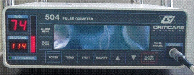
Pulse oximetery
Figure 2.

Pulse oximetry ear probe
The probes were cleaned and disinfected with 2.4% Glutaraldehyde for 45 minutes, according to the instruction of the Dolphin Corporation, 2001.[14]
The procedure had four steps, was performed in the morning, and was similar for all patients.
In the first step, in each patient, the blood oxygen saturation (Spo2) values of the finger and dental pulp were measured, before the anesthetic drug injection was given, using finger and ear probes, respectively. In the second step, the values were measured one minute after local anesthesia injection of two to four cartridges containing 1.8 ml of lidocaine 2% with 1/80000 of epinephrine were administered. In the third step, all the values were measured at the end of surgery, after cleaning the area. One hour after surgery, the values were measured again, as the fourth step.
The collected data from the four steps were analyzed using the Paired Samples Test and the Spearman's Correlation Coefficient.
RESULTS
The data were obtained from 15 patients (10 males, 5 females) with an age range of 19 – 49 years. The averages of age, number of cartridges, and duration of surgery were, 31 years, two cartridges, and 52 minutes, respectively.
In our study, the mean value of the peripheral Spo2 was over 97% in all four steps. The mean value of the pulpal Spo2 before local anesthesia was 87.73%, which decreased to 79.27% one minute after injection, then increased to 86.13% at the end of surgery, and to 86.4% one hour after surgery. [Table 1] [Figure 3].
Table 1.
Blood and pulpal Spo2% in two groups of study
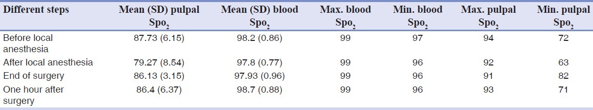
Figure 3.

Mean of Spo2 in four steps of the study in two groups
The paired samples test showed that the mean value of the pulpal Spo2 decreased significantly after anesthesia compared to before injection (P < 0.001), however, there was no significant difference between the mean value of the pulpal Spo2 at the end of surgery and one hour after the procedure (P = 0.267, P = 0.065), respectively.
A significant increase was found in the mean value of the pulpal Spo2 at the end of surgery relative to after anesthesia (P = 0.007), but there was no significant increase in the mean value of the pulpal Spo2 one hour after surgery compared to after the procedure. (P = 0.855).
According to the Spearman's correlation coefficient, the number of injections was inversely correlated with the mean value of the pulpal Spo2 after anesthesia (P = 0.041, r = 0.533) and also directly correlated with the duration of the procedure. (P = 0.035, r = 0.547).
DISCUSSION
The mean values of peripheral blood Spo2 in the four steps did not show significant changes; however, there were significant differences between the mean values of the pulpal Spo2 during the study. Therefore, the values of the pulpal Spo2 might have changed independently from the peripheral Spo2.
The mean value of the pulpal Spo2 was smaller than that of the peripheral Spo2. The reason was that thick dentin and enamel did not let much light pass through. This result is not consistent with the result of Gopi et al.[13]
The mean value of the pulpal Spo2 after anesthesia decreased compared to that before the injection, because the local anesthetic material included epinephrine, which constricted the blood vessels and caused regional blood flow reduction. As no significant decrease was found in the mean value of the pulpal Spo2 immediately and one hour after surgery in comparison with before anesthesia, this reduction in blood flow was temporary.
Finding a significant increase in the mean value of pulpal Spo2 after surgery compared to after the anesthetic material injection, indicated increased pulpal blood flow after surgery. As there was no remarkable decrease in the mean value of pulpal Spo2 immediately and one hour after surgery, compared to before anesthesia, surgical trauma was not the cause of decrease in the pulpal Spo2, and the change in pulpal Spo2 occurred only as the result of anesthesia.
Our study showed the effectiveness of the pulse oximeter probes to evaluate pulpal vitality. This result is consistent with the results of the studies of Goho,[5] Radhakrishnan,[6] Calil,[12] and Gopi Krishnan.[13]
The mean values of Spo2 in the finger and anterior teeth reported by Calil[12] were 95 and 90%, and by Gopi Krishnan et al.[13] were 97.85 and 79%, respectively. The results of this study accept the results of Kaviani et al.,[11] showing mean values of 97.5 and 92% for Spo2 in the ear and anterior teeth, respectively.
CONCLUSION
In conclusion, it can be stated that, injection of a local anesthetic drug (lidocaine, Epinephrine) causes a temporary decrease in pulpal Spo2, without any effect on the pulpal vitality, and also the surgical trauma does not have any influence on the vitality and Spo2 of the adjacent tooth pulp.
ACKNOWLEDGMENTS
We are very thankful to the staff of the Operation Room and the Research Deputy of the School of Dentistry, Isfahan University of Medical Sciences.
Footnotes
Source of Support: This report is based on a thesis which was submitted to the School of Dentistry, Isfahan University of Medical Sciences, Isfahan, Iran, in partial fulfillment of the requirements for the DDS degree in general dentistry, # 388361). The study was approved by the Medical Ethics and Research Office at the Isfahan University of Medical Sciences and financially supported by this University.
Conflict of Interest: The authors declare no conflicts of interest, real or perceived, financial or nonfinancial.
REFERENCES
- 1.Torabinejad M, Walton RE. Edodontics: Principles and practice. 4th ed. New York: Elsevier; 2008. [Google Scholar]
- 2.Arwill T, Edwall L, Lilja J, Olgart L, Svensson SE. Ultrastructure of nerves in the dentinal-pulp border zone after sensory and autonomic nerve transection in the cat. Acta Odontol Scand. 1973;31:273–81. doi: 10.3109/00016357309002514. [DOI] [PubMed] [Google Scholar]
- 3.Koling A, Rask–Andersen H. The blood capillaries in the subodondontoblastic region of the human dental pulp, as demonstrated by freeze- fracturing. Acta Odontol Scand. 1983;41:333–41. doi: 10.3109/00016358309162344. [DOI] [PubMed] [Google Scholar]
- 4.Rickoff B, Trowbridge H, Baker J, Fuss Z, Bender IB. Effects of thermal vitality tests on human dental pulp. J Endod. 1988;14:482–5. doi: 10.1016/S0099-2399(88)80104-3. [DOI] [PubMed] [Google Scholar]
- 5.Goho C. Pulse oximetry evaluation of vitality in primary and immature permanent teeth. Pediatr Dent. 1999;21:125–7. [PubMed] [Google Scholar]
- 6.Radhakrishnan S, Munshi AK, Hegde AM. Pulse oximetry: A diagnostic instrument in pulpal vitality testing. J Clin Pediatr Dent. 2002;26:141–5. doi: 10.17796/jcpd.26.2.2j25008jg6u86236. [DOI] [PubMed] [Google Scholar]
- 7.John IO. Endodontics. 5th ed. London: Hamilton; 2002. [Google Scholar]
- 8.Jafarzadeh H, Rosenberg PA. Pulse oximetry: Review of a potential aid in endodontic diagnosis. J Endod. 2009;35:329–33. doi: 10.1016/j.joen.2008.12.006. [DOI] [PubMed] [Google Scholar]
- 9.Noblett WC, Wilcox LR, Scamman F, Jaohnson WT, Diaz-Arnold A. Detection of pulpal circulation in vitro by pulse oximetry. J Endod. 1996;22:1–5. doi: 10.1016/S0099-2399(96)80226-3. [DOI] [PubMed] [Google Scholar]
- 10.Hilde Brand C, Fried k, Tuisku F, Johansson CS. Teeth and tooth nerves. Prog Neurobiol. 1995;45:165–222. doi: 10.1016/0301-0082(94)00045-j. [DOI] [PubMed] [Google Scholar]
- 11.Kaviani N, Mosavi B, Vahedi H. Comparative evaluation of blood oxygen saturation in healthy anterior teeth and ear with pulse oximetry. Isfahan Dent Sch J. 2007;312:53–7. [Google Scholar]
- 12.Calil E, Caldeira Cl, Gavini G, Lemos EM. Determination of pulp vitality in vivo with pulse oximetry. Int Endod J. 2008;41:741–6. doi: 10.1111/j.1365-2591.2008.01421.x. [DOI] [PubMed] [Google Scholar]
- 13.Gopi Krishna V, Kandaswamy D, Gupta T. Assesment of the efficacy of an indigenously developed pulse oximeter dental sensor holder for pulp vitality testing. Indian J Dent Res. 2006;17:111–3. doi: 10.4103/0970-9290.29880. [DOI] [PubMed] [Google Scholar]
- 14.Scharf JE, Shah B. high-level disinfection and reusable pulse oximetry sensors. Dolphin Medical Company. 2001 [Google Scholar]