Abstract
Background:
Several studies have shown that laser-etching of enamel for bonding orthodontic brackets could be an appropriate alternative for acid conditioning, since a potential advantage of laser could or might be caries prevention. This study compared enamel resistance to demineralization following etching with acid phosphoric or Er:YAG laser for bonding orthodontic brackets.
Materials and Methods:
Fifty sound human premolars were divided into two equal groups. In the first group, enamel was etched with 37% phosphoric acid for 15 seconds. In the second group, Er:YAG laser (wavelength, 2 940 nm; 300 mJ/pulse, 10 pulses per second, 10 seconds) was used for tooth conditioning. The teeth were subjected to 4-day PH-cycling process to induce caries-like lesions. The teeth were then sectioned and the surface area of the lesion was calculated in each microphotographs and expressed in pixel. The total surface of each specimen was 196 608 pixels.
Results:
Mean lesion areas were 7 171 and 7532 pixels for Laser-etched and Acid-etched groups, respectively. The two sample t-test showed that there was no significant difference in lesion area between the two groups (P = 0.914).
Conclusion:
Although Er:YAG laser seems promising for etching enamel before bonding orthodontic brackets, it does not reduce enamel demineralization when exposed to acid challenge.
Keywords: Demineralization, enamel, Er:YAG laser, etching, caries resistance
INTRODUCTION
Today, acid etching is the most common method of preparing enamel for bonding orthodontic brackets. Although this method results in high bond strength, its great disadvantage is the potential for caries formation. Acid etching removes and demineralizes the most superficial layer of enamel and makes the teeth more susceptible to long-term acid attack, especially when resin monomers cannot sufficiently fill the demineralized area due to saliva contamination or air bubbles.[1] Considering the high prevalence of white spot lesions in orthodontic patients,[2] and with respect to the fact that white spot lesions can form in as early as 4 weeks in the presence of inadequate oral hygiene,[3] prevention of enamel demineralization is of great importance during treatment. There have been ample attempts to find a method to decrease the incidence of white spot lesions in orthodontic patients. Fluoride mouth rinses have been shown to significantly reduce caries formation,[4] but excellent cooperation in using a mouth rinse can be achieved in only 13% of the patients.[5] Another method to prevent demineralization is to use fluoride-releasing composite resins and conventional or resin-modified glass ionomer cements, but the lower bond strengths of these materials compared with conventional acid etching[6–8] have raised questions on their clinical efficiency.
Recently, great attention is given to conditioning dental surfaces with laser light. The physical and chemical changes in laser-irradiated surfaces hold promise for prevention of enamel demineralization. Although different laser systems have been used in previous studies for conditioning dental hard tissues,[9–14] it is well clear that with available technology, only erbium family lasers (Er:YAG and Er,Cr:YSGG) are suitable for this purpose. The wavelength of Er:YAG laser is highly absorbed by water and hydroxyapatite,[15,16] making it suitable for both hard and soft tissue ablation. Laser irradiation removes smear layer and creates surface roughness;[11,13] both of them are favorable for monomer infiltration and adhesion process.[17]
There are contradictory reports about caries prevention effect of Er:YAG laser. Hossain et al. reported an increase in the calcium to phosphorus ratio during laser irradiation, which resulted in caries inhibition.[18] In the study of Kim et al., laser-treated enamels showed improvement in crystalline structure and had the lowest mineral dissolution compared to control and phosphoric acid-etched specimens.[19] Another study showed that Er:YAG laser treatment reduced the carbonate content and modified the organic matrix, thus providing caries-preventive effect on enamel.[20] However, some studies did not find any significant difference between Er:YAG-lased and non-lased groups with respect to the enamel demineralization.[21,22] Apel et al. observed that Er:YAG laser was unable to achieve any notable reduction in acid solubility of dental enamel.[23] Some authors concluded that the application of sub-ablative erbium lasers solely for preventive caries treatment does not seem to be sensible under the conditions they studied.[23,24]
If it was possible to reduce the prevalence of demineralization, while using Er:YAG laser for etching the enamel surface, this would be of great advantage for orthodontic patients. Therefore, the objective of this study was to evaluate the caries resistance potential of Er:YAG laser-etched surfaces after exposure to an in vitro caries challenge, compared to acid-etched specimens.
MATERIALS AND METHODS
In this in vitro study, 50 intact and caries-free human premolars were used. Any remaining soft tissue was removed with a scaler and the teeth were kept in 0.1% thymol solution to prevent bacterial growth. The teeth were randomly assigned into two equal groups based on the etching procedure. Prior to the experiment, the roots were sectioned 2 mm below the cementoenamel junction using a high-speed drill and a diamond bur, to facilitate further microscopic sample preparation. The enamel surfaces of the teeth were cleaned with pumice slurry and rubber prophylactic cups, then rinsed with running water and dried with a moisture-free air syringe. To limit and standardize the etching area, the facial surfaces of the teeth were painted with a thin coat of nail varnish except a 4 × 4 mm window which was left exposed on the center of the clinical crown.
The enamel surfaces of the teeth in group 1 were etched with a 37% phosphoric acid gel for 15 seconds, rinsed for 15 seconds with copious amount of water, and dried with oil-free air for another 15 seconds. The frosty white appearance was observed on all the teeth.
The teeth in group 2 were etched with an Er:YAG laser device (Fidelis Plus II, Fotona, Slovenia) emitting a wavelength of 2.94 mm. The laser was irradiated using short pulse mode (180 μs), 300 mJ of energy per pulse, and 10 pulses per second under air and water spray (5 ml/min).[25] The spot size of the laser was 1 mm. The laser beam was directed manually at 1 mm distance and was delivered with a sweeping motion perpendicular to the enamel surface for 10 seconds with the use of RO7 handpiece. After laser etching, the teeth were washed and dried with an oil-free air source. The frosty white appearance was noticed on all the specimens similar to the acid-etched group.
To compare the acid resistance of the teeth exposed to Er:YAG laser with acid-etched group, the teeth in both groups were subjected to 4-day pH cycling process to form caries-like lesions. In this process, the teeth were immersed in demineralization and remineralization solutions for 18 hours and 6 hours per day, respectively.[26] The demineralization solution (pH = 4.6) consisted of 0.05M acetic acid, 2.2 mM calcium, and 2.2mM phosphate ions, and the remineralization solution (pH = 7.0) contained 0.15 M potassium chloride, 1.5mM calcium, and 0.9 mM phosphate ions.[25] This process started with the demineralization solution. Between the two phases and at the end of the process, the teeth were washed with running water for 5 minutes. The groups were cycled separately in individual glass containers throughout the 4-day procedure. Then, the teeth were stored in normal saline solution until sectioning.
Slide preparation and microscopic observation
The samples were embedded in epoxy resin (Struers, Copenhagen, Denmark) using plastic tubes (2 cm diameter and 1.5 cm height) as molds. The crowns were mounted in such a way that the facial surface of each tooth was parallel to the bottom of the mold. The microscopic sections were obtained via trimming the teeth by a ground section apparatus from both mesial and distal aspects in a buccolingual direction. To do this, the tooth was first trimmed from one side until it reached the middle of the treatment window. Then, the tooth was fixed on a glass slab with a transparent stone adhesive and the other half was trimmed until the thickness of the specimen reached approximately 100 μ. For thickness evaluation, another slab was placed on top of the specimen, and the specimen was compared with a matrix strap (thickness, 100 μ), which was placed next to it on the slab. One microscopic section was obtained from each tooth. The sections were evaluated under a polarized light microscope (Olympus, model BH2, Dualmont Corporation, Minnepolis, Minn) and the photograph of each section was taken with maximum illumination at ×40 magnificence. The resulting pictures were analyzed with the aid of Adobe Photoshop CS software (Adobe System Incorporated, USA). Using this software, the demineralized zone on the enamel surface of each sample was captured with Magic wand tool (Photoshop software) and the surface area of the lesion was calculated and expressed in pixel. The total surface of all pictures was 196 608 pixels. One examiner assessed all the photographs, blinded to the treatment groups.
The lesion areas were compared between the laser-etched and acid-etched groups by student t-test. Statistical calculation was performed with SPSS software (Version 11.5, SPSS Inc, Chicago, Ill), and P value less than 0.05 was considered significant.
RESULTS
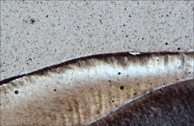
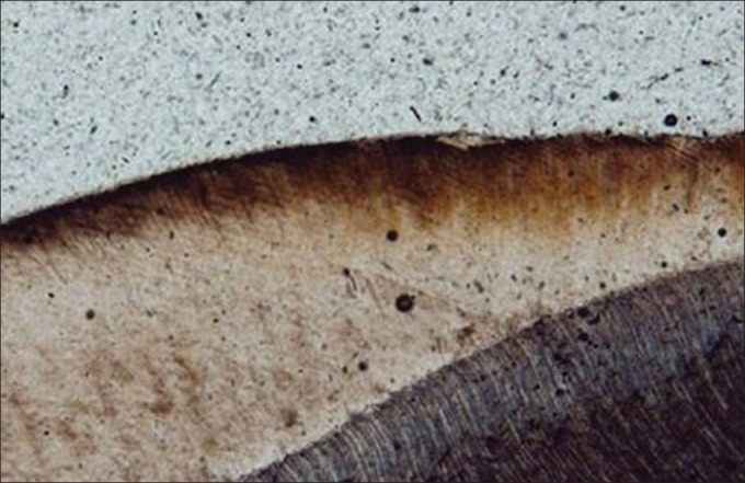
Photomicrographs of representative lesions in the laser-etched and acid-etched groups are demonstrated in Figures 1 and 2, respectively. In general, the demineralized tissue seems dark under polarized light microscope, while the intact enamel shows a yellowish or bluish appearance. Lesion area measurements for the two study groups are presented in Table 1. Student t test revealed no significant difference between the laser-etched and acid-etched groups regarding the area of demineralization (P = 0.914).
Figure 1.

Photomicrograph of enamel lesion representing the Er:YAG laser-etched group at ×40 magnification under polarized light microscope
Figure 2.

Photomicrograph of enamel lesion representing the acid-etched group at ×40 magnification under polarized light microscope
Table 1.
Lesion area measurements in the study groups

DISCUSSION
This in vitro study evaluated the acid resistance of Er:YAG laser-etched specimens, compared with those prepared by conventional acid-etching technique. The main disadvantage of phosphoric acid etching, i.e., the potential for producing decalcification,[19] has prompted many clinicians to find an alternative conditioning method. Enamel pretreatment with polyacrylic acid has been suggested by some authors to reduce the rate of enamel loss during etching,[27] but this method failed to achieve adequate bond strength to resist intraoral forces.[28] The application of laser for tooth conditioning may be a suitable alternative for acid etching because it can save chair time and provide caries prevention effect. The bond strengths of adhesive resins bonded to laser-etched enamel surfaces have been a matter of concern for many clinicians. Some studies have found no significant differences between bond strengths of the teeth etched with erbium lasers or phosphoric acid,[29–32] while others reported the reverse finding.[1,17,33] However, it seems reasonable that with suitable laser parameters, bond strengths of laser-etched surfaces should be comparable to those prepared by conventional acid etching.
The caries protective effect of laser light has been attributed to the heat produced during laser irradiation, which can cause changes in the chemical and crystalline structure of the enamel.[19,23,24,34] Previous studies have reported a minimum solubility of enamel and smallest lesion depths after heating the dental enamel to between 300 and 400°C.[23,24,35] Most authors believe that carbonate decomposition and loss of water are important contributing factors to caries prevention.[23,24,34,36,37] Removal of carbonate improves the crystalline structure, and thus decreases the enamel susceptibility to demineralization, because carbonate fit less well in the lattice and produce more acid-soluble apatite-phase.[20,34,38] Zuerlein et al.[39] indicated that decomposition of carbonate begins from 400°C onward, but Fowler ad Kuroda[37] reported substantial loss of carbonate and water at temperatures between 100 and 400°C. Another theory for the protective effect of laser light is based on the blocking of enamel diffusion pathway. In lased enamel, the decomposed organic materials can block the interprismatic and intraprismatic spaces that act as ion diffusion channels during the demineralization process, making the enamel less vulnerable to mineral loss.[19,23,34,40,41] One other explanation for the laser-induced caries prevention is the formation of microspaces and microfissures in lased enamel. These spaces are believed to trap the calcium, phosphorus, and fluoride ions released from the tooth during the demineralization process.[19,36] The size of these spaces can vary with the laser energy; higher energies leading to the formation of deeper or more extensive spaces in the enamel.[36] However, some authors suggest that these spaces can also act as open channels, facilitating the acid attack to subsurface, thus enhancing the mineral loss.[19,24]
The present study indicates that Er:YAG laser parameters for enamel conditioning cannot simultaneously prevent demineralization when compared to phosphoric acid etching. This finding is in agreement with the studies of Apel et al.[23] and Chimello et al.[22] who found no significant differences between Er:YAG-lased and unlased groups with respect to the enamel demineralization. Apel et al. found fine cracks in the enamel surface after sub-ablative erbium laser irradiation which can act as starting points for acid attack and demineralization, therefore counteracting the positive effect of laser light in caries prevention.[24] The results of this study are in contrast with the study of Hossain et al.[18] who reported significant caries resistance in Er:YAG laser-irradiated specimens, compared to control group. Liu et al. exposed molars to Er:YAG laser irradiation using 100, 200, and 300 mJ pulse energies and found that laser treatments of 100 and 200 mJ provided significant protection of enamel demineralization, but not the treatment with 300 mJ.[34] Cecchini et al. evaluated the different settings of Er:YAG laser on enamel acid resistance and reported that lower energies (60-80 mJ) caused a significant reduction in enamel solubility.[36] The difference between the findings of this study with those of Cecchini et al.[36] can be attributed to the higher energy per pulse (300 mJ) we employed for enamel etching.
Some previous studies[24,34] did not use water cooling during Er:YAG laser treatment, while in the present study, water-cooling was employed to closely simulate the clinical condition used for etching the enamel surface. A previous study indicated that Er:YAG laser is effective for increasing the caries resistance either used with or without water cooling, but the degree of caries prevention was considerably higher without water mist than with water mist.[18] It is apparent that the application of Er:YAG laser without water cooling may increase the enamel surface temperature higher than that achieved with water application. Therefore, the difference between the results of this study compared to those of Liu et al.[34] may be related to the different surface temperature of the enamel, achieved with different laser parameters. However, it should be noted that the time of laser exposure in the studies that did not use water cooling[24,34] was shorter than that needed for effective enamel conditioning.
The time of phosphoric acid etching in the clinical condition may vary between 15 to 60 seconds.[28,31] In the present study, 15 seconds of acid etching was selected in order to minimize the adverse effect of acid etching on the enamel surface. It is expected that longer etching times, as are used commonly by many clinicians, may make the teeth more susceptible to demineralization, and in this case, the difference between the caries resistance of acid-etched and laser-etched surfaces would be more remarkable. The limitations of the present study were sample preparation for microscopic evaluation. Further research should focus on finding suitable range of Er:YAG laser parameters to achieve both optimal caries resistance and high bond strength in the lased enamel.
CONCLUSION
The present study indicates that Er:YAG laser cannot increase the enamel resistance to demineralization when using for enamel conditioning.
ACKNOWLEDGEMENTS
The authors would like to thank Vice Chancellor of Research of Mashhad University of Medical Sciences who financially supported this research. Sincere thanks are also expressed to Dr. Matin Basiri for his assistance in performing this project.
Footnotes
Source of Support: Vice Chancellor of Research of Mashhad University of Medical Sciences.
Conflict of Interest: None declared.
REFERENCES
- 1.Martinez-Insua A, Da Silva Dominguez L, Rivera FG, Santana-Penin UA. Differences in bonding to acid-etched or Er:YAG-laser-treated enamel and dentin surfaces. J Prosthet Dent. 2000;84:280–8. doi: 10.1067/mpr.2000.108600. [DOI] [PubMed] [Google Scholar]
- 2.Gorelick L, Geiger AM, Gwinnett AJ. Incidence of white spot formation after bonding and banding. Am J Orthod. 1982;81:93–8. doi: 10.1016/0002-9416(82)90032-x. [DOI] [PubMed] [Google Scholar]
- 3.Ogaard B, Rolla G, Arends J. Orthodontic appliances and enamel demineralization. Part 1. Lesion development. Am J Orthod Dentofacial Orthop. 1988;94:68–73. doi: 10.1016/0889-5406(88)90453-2. [DOI] [PubMed] [Google Scholar]
- 4.Benson PE, Parkin N, Millett DT, Dyer FE, Vine S, Shah A. Fluorides for the prevention of white spots on teeth during fixed brace treatment. Cochrane Database Syst Rev. 2004;3:CD003809. doi: 10.1002/14651858.CD003809.pub2. [DOI] [PubMed] [Google Scholar]
- 5.Geiger AM, Gorelick L, Gwinnett AJ, Benson BJ. Reducing white spot lesions in orthodontic populations with fluoride rinsing. Am J Orthod Dentofacial Orthop. 1992;101:403–7. doi: 10.1016/0889-5406(92)70112-N. [DOI] [PubMed] [Google Scholar]
- 6.Cook PA, Youngson CC. An in vitro study of the bond strength of a glass ionomer cement in the direct bonding of orthodontic brackets. Br J Orthod. 1988;15:247–53. doi: 10.1179/bjo.15.4.247. [DOI] [PubMed] [Google Scholar]
- 7.Fox NA, McCabe JF, Gordon PH. Bond strengths of orthodontic bonding materials: An in-vitro study. Br J Orthod. 1991;18:125–30. doi: 10.1179/bjo.18.2.125. [DOI] [PubMed] [Google Scholar]
- 8.Klockowski R, Davis EL, Joynt RB, Wieczkowski G, Jr, MacDonald A. Bond strength and durability of glass ionomer cements used as bonding agents in the placement of orthodontic brackets. Am J Orthod Dentofacial Orthop. 1989;96:60–4. doi: 10.1016/0889-5406(89)90230-8. [DOI] [PubMed] [Google Scholar]
- 9.Ariyaratnam MT, Wilson MA, Mackie IC, Blinkhorn AS. A comparison of surface roughness and composite/enamel bond strength of human enamel following the application of the Nd:YAG laser and etching with phosphoric acid. Dent Mater. 1997;13:51–5. doi: 10.1016/s0109-5641(97)80008-5. [DOI] [PubMed] [Google Scholar]
- 10.Drummond JL, Wigdor HA, Walsh JT, Jr, Fadavi S, Punwani I. Sealant bond strengths of CO(2) laser-etched versus acid-etched bovine enamel. Lasers Surg Med. 2000;27:111–8. doi: 10.1002/1096-9101(2000)27:2<111::aid-lsm2>3.0.co;2-l. [DOI] [PubMed] [Google Scholar]
- 11.Kwon YH, Kwon OW, Kim HI, Kim KH. Nd:YAG laser ablation of enamel for orthodontic use: Tensile bond strength and surface modification. Dent Mater J. 2003;22:397–403. doi: 10.4012/dmj.22.397. [DOI] [PubMed] [Google Scholar]
- 12.Shahabi S, Brockhurst PJ, Walsh LJ. Effect of tooth-related factors on the shear bond strengths obtained with CO2 laser conditioning of enamel. Aust Dent J. 1997;42:81–4. doi: 10.1111/j.1834-7819.1997.tb00101.x. [DOI] [PubMed] [Google Scholar]
- 13.Walsh LJ, Abood D, Brockhurst PJ. Bonding of resin composite to carbon dioxide laser-modified human enamel. Dent Mater. 1994;10:162–6. doi: 10.1016/0109-5641(94)90026-4. [DOI] [PubMed] [Google Scholar]
- 14.Whitters CJ, Strang R. Preliminary investigation of a novel carbon dioxide laser for applications in dentistry. Lasers Surg Med. 2000;26:262–9. doi: 10.1002/(sici)1096-9101(2000)26:3<262::aid-lsm3>3.0.co;2-7. [DOI] [PubMed] [Google Scholar]
- 15.Hale GM, Querry MR. Optical constants of water in the 200-nm to 200-microm wavelength region. Appl Opt. 1973;12:555–63. doi: 10.1364/AO.12.000555. [DOI] [PubMed] [Google Scholar]
- 16.Wigdor HA, Walsh JT, Jr, Featherstone JD, Visuri SR, Fried D, Waldvogel JL. Lasers in dentistry. Lasers Surg Med. 1995;16:103–33. doi: 10.1002/lsm.1900160202. [DOI] [PubMed] [Google Scholar]
- 17.Chimello-Sousa DT, de Souza AE, Chinelatti MA, Pecora JD, Palma-Dibb RG, Milori Corona SA. Influence of Er:YAG laser irradiation distance on the bond strength of a restorative system to enamel. J Dent. 2006;34:245–51. doi: 10.1016/j.jdent.2005.06.009. [DOI] [PubMed] [Google Scholar]
- 18.Hossain M, Nakamura Y, Kimura Y, Yamada Y, Ito M, Matsumoto K. Caries-preventive effect of Er:YAG laser irradiation with or without water mist. J Clin Laser Med Surg. 2000;18:61–5. doi: 10.1089/clm.2000.18.61. [DOI] [PubMed] [Google Scholar]
- 19.Kim JH, Kwon OW, Kim HI, Kwon YH. Acid resistance of erbium-doped yttrium aluminum garnet laser-treated and phosphoric acid-etched enamels. Angle Orthod. 2006;76:1052–6. doi: 10.2319/11405-398. [DOI] [PubMed] [Google Scholar]
- 20.Liu Y, Hsu CY. Laser-induced compositional changes on enamel: A FT-Raman study. J Dent. 2007;35:226–30. doi: 10.1016/j.jdent.2006.08.006. [DOI] [PubMed] [Google Scholar]
- 21.Apel C, Birker L, Meister J, Weiss C, Gutknecht N. The caries-preventive potential of subablative Er:YAG and Er:YSGG laser radiation in an intraoral model: A pilot study. Photomed Laser Surg. 2004;22:312–7. doi: 10.1089/pho.2004.22.312. [DOI] [PubMed] [Google Scholar]
- 22.Chimello DT, Serra MC, Rodrigues AL, Jr, Pecora JD, Corona SA. Influence of cavity preparation with Er:YAG Laser on enamel adjacent to restorations submitted to cariogenic challenge in situ: A polarized light microscopic analysis. Lasers Surg Med. 2008;40:634–43. doi: 10.1002/lsm.20684. [DOI] [PubMed] [Google Scholar]
- 23.Apel C, Meister J, Schmitt N, Graber HG, Gutknecht N. Calcium solubility of dental enamel following sub-ablative Er:YAG and Er:YSGG laser irradiation in vitro. Lasers Surg Med. 2002;30:337–41. doi: 10.1002/lsm.10058. [DOI] [PubMed] [Google Scholar]
- 24.Apel C, Meister J, Gotz H, Duschner H, Gutknecht N. Structural changes in human dental enamel after subablative erbium laser irradiation and its potential use for caries prevention. Caries Res. 2005;39:65–70. doi: 10.1159/000081659. [DOI] [PubMed] [Google Scholar]
- 25.Lee BS, Hsieh TT, Lee YL, Lan WH, Hsu YJ, Wen PH, et al. Bond strengths of orthodontic bracket after acid-etched, Er:YAG laser-irradiated and combined treatment on enamel surface. Angle Orthod. 2003;73:565–70. doi: 10.1043/0003-3219(2003)073<0565:BSOOBA>2.0.CO;2. [DOI] [PubMed] [Google Scholar]
- 26.Hsu CY, Jordan TH, Dederich DN, Wefel JS. Laser-matrix-fluoride effects on enamel demineralization. J Dent Res. 2001;80:1797–801. doi: 10.1177/00220345010800090501. [DOI] [PubMed] [Google Scholar]
- 27.Maijer R, Smith DC. A new surface treatment for bonding. J Biomed Mater Res. 1979;13:975–85. doi: 10.1002/jbm.820130614. [DOI] [PubMed] [Google Scholar]
- 28.Jones SP, Gledhill JR, Davies EH. The crystal growth technique–a laboratory evaluation of bond strengths. Eur J Orthod. 1999;21:89–93. doi: 10.1093/ejo/21.1.89. [DOI] [PubMed] [Google Scholar]
- 29.Basaran G, Ozer T, Berk N, Hamamci O. Etching enamel for orthodontics with an erbium, chromium:yttrium-scandium-gallium-garnet laser system. Angle Orthod. 2007;77:117–23. doi: 10.2319/120605-426R.1. [DOI] [PubMed] [Google Scholar]
- 30.Kim JH, Kwon OW, Kim HI, Kwon YH. Effectiveness of an Er:YAG laser in etching the enamel surface for orthodontic bracket retention. Dent Mater J. 2005;24:596–602. doi: 10.4012/dmj.24.596. [DOI] [PubMed] [Google Scholar]
- 31.Ozer T, Basaran G, Berk N. Laser etching of enamel for orthodontic bonding. Am J Orthod Dentofacial Orthop. 2008;134:193–7. doi: 10.1016/j.ajodo.2006.04.055. [DOI] [PubMed] [Google Scholar]
- 32.Usumez S, Orhan M, Usumez A. Laser etching of enamel for direct bonding with an Er,Cr:YSGG hydrokinetic laser system. Am J Orthod Dentofacial Orthop. 2002;122:649–56. doi: 10.1067/mod.2002.127294. [DOI] [PubMed] [Google Scholar]
- 33.De Munck J, van Meerbeek B, Yudhira R, Lambrechts P, Vanherle G. Micro-tensile bond strength of two adhesives to Erbium:YAG-lased vs.bur-cut enamel and dentin. Eur J Oral Sci. 2002;110:322–9. doi: 10.1034/j.1600-0722.2002.21281.x. [DOI] [PubMed] [Google Scholar]
- 34.Liu JF, Liu Y, Stephen HC. Optimal Er:YAG laser energy for preventing enamel demineralization. J Dent. 2006;34:62–6. doi: 10.1016/j.jdent.2005.03.005. [DOI] [PubMed] [Google Scholar]
- 35.Sato K. Relation between acid dissolution and histological alteration of heated tooth enamel. Caries Res. 1983;17:490–5. doi: 10.1159/000260708. [DOI] [PubMed] [Google Scholar]
- 36.Cecchini RC, Zezell DM, de Oliveira E, de Freitas PM, Eduardo Cde P. Effect of Er:YAG laser on enamel acid resistance: Morphological and atomic spectrometry analysis. Lasers Surg Med. 2005;37:366–72. doi: 10.1002/lsm.20247. [DOI] [PubMed] [Google Scholar]
- 37.Fowler BO, Kuroda S. Changes in heated and in laser-irradiated human tooth enamel and their probable effects on solubility. Calcif Tissue Int. 1986;38:197–208. doi: 10.1007/BF02556711. [DOI] [PubMed] [Google Scholar]
- 38.Bachra BN, Trautz OR, Simon SL. Precipitation of calcium carbonates and phosphates. 3. The effect of magnesium and fluoride ions on the spontaneous precipitation of calcium carbonates and phosphates. Arch Oral Biol. 1965;10:731–8. doi: 10.1016/0003-9969(65)90126-3. [DOI] [PubMed] [Google Scholar]
- 39.Zuerlein MJ, Fried D, Featherstone JD. Modeling the modification depth of carbon dioxide laser-treated dental enamel. Lasers Surg Med. 1999;25:335–47. doi: 10.1002/(sici)1096-9101(1999)25:4<335::aid-lsm8>3.0.co;2-f. [DOI] [PubMed] [Google Scholar]
- 40.Hsu CY, Jordan TH, Dederich DN, Wefel JS. Effects of low-energy CO2 laser irradiation and the organic matrix on inhibition of enamel demineralization. J Dent Res. 2000;79:1725–30. doi: 10.1177/00220345000790091401. [DOI] [PubMed] [Google Scholar]
- 41.Stern RH, Sognnaes RF, Goodman F. Laser effect on in vitro enamel permeability and solubility. J Am Dent Assoc. 1966;73:838–43. doi: 10.14219/jada.archive.1966.0319. [DOI] [PubMed] [Google Scholar]


