Abstract
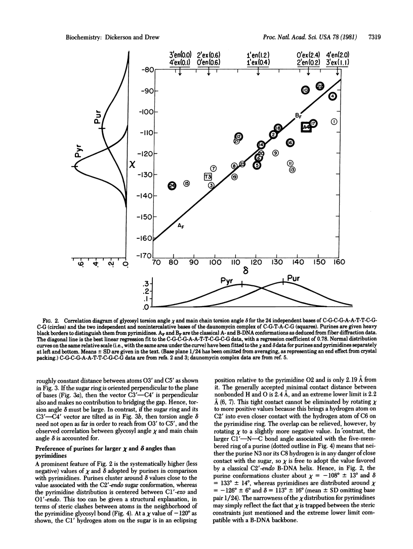
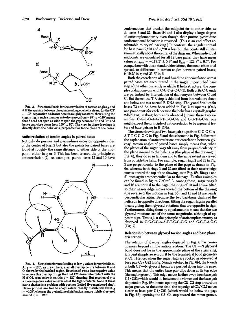
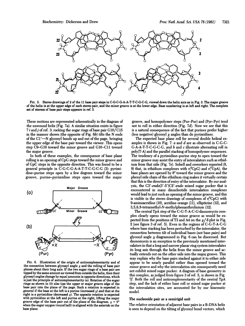
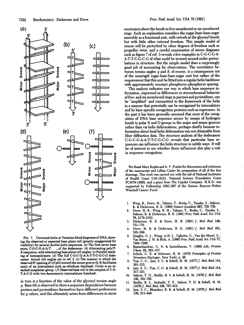
X-ray structure analysis of B-DNA double helix with sequence C-G-C-G-A-A-T-T-C-G-C-G has revealed several sequence-dependent structural features. Four of these are shown in this paper to be related to one another by simple structural or kinematic principles: (i) the correlation between glycosyl torsion and chi and main chain C4'--C3' torsion angle delta, (ii) the observations that purines prefer larger phi and delta angles than do pyrimidines, (iii) the anticorrelation of phi or of delta angles between sugars associated with one base pair, and (iv) the observation that successive base planes in purine-pyrimidine steps open up the angle between them toward the major groove, whereas pyrimidine-purine steps open toward the minor groove. These features offer the beginning of an understanding of the way in which specific base sequences can perturb the structure of a B-DNA double helix so as to be "read" by intercalating drugs, repressors, and other recognition proteins.
Full text
PDF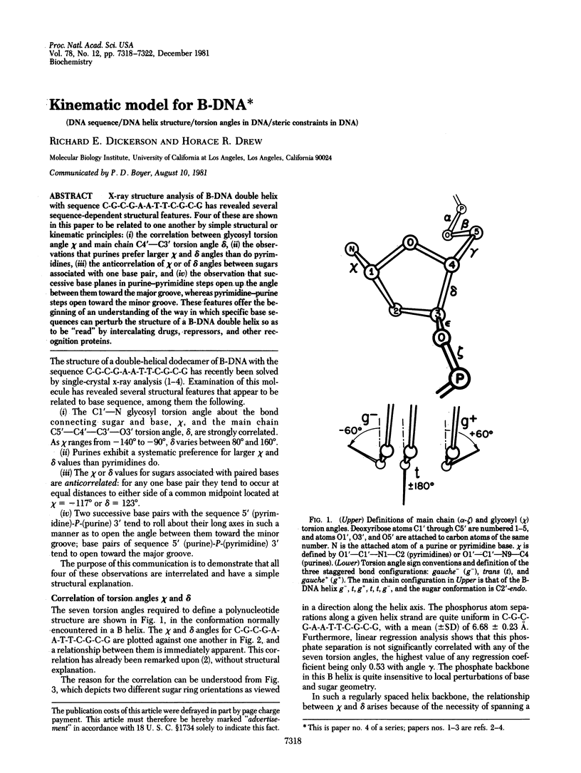
Selected References
These references are in PubMed. This may not be the complete list of references from this article.
- Drew H. R., Dickerson R. E. Structure of a B-DNA dodecamer. III. Geometry of hydration. J Mol Biol. 1981 Sep 25;151(3):535–556. doi: 10.1016/0022-2836(81)90009-7. [DOI] [PubMed] [Google Scholar]
- Drew H. R., Wing R. M., Takano T., Broka C., Tanaka S., Itakura K., Dickerson R. E. Structure of a B-DNA dodecamer: conformation and dynamics. Proc Natl Acad Sci U S A. 1981 Apr;78(4):2179–2183. doi: 10.1073/pnas.78.4.2179. [DOI] [PMC free article] [PubMed] [Google Scholar]
- Jain S. C., Bhandary K. K., Sobell H. M. Visualization of drug--nucleic acid interactions at atomic resolution. VI. Structure of two drug--dinucleoside monophosphate crystalline complexes, ellipticine--5-iodocytidylyy (3'-5') guanosine and 3,5,6,8-tetramethyl-N-methyl phenanthrolinium--5-iodocytidylyl (3'-5') guanosine. J Mol Biol. 1979 Dec 25;135(4):813–840. [PubMed] [Google Scholar]
- Jain S. C., Tsai C. C., Sobell H. M. Visualization of drug-nucleic acid interactions at atomic resolution. II. Structure of an ethidium/dinucleoside monophosphate crystalline complex, ethidium:5-iodocytidylyl (3'-5') guanosine. J Mol Biol. 1977 Aug 15;114(3):317–331. doi: 10.1016/0022-2836(77)90253-4. [DOI] [PubMed] [Google Scholar]
- Quigley G. J., Wang A. H., Ughetto G., van der Marel G., van Boom J. H., Rich A. Molecular structure of an anticancer drug-DNA complex: daunomycin plus d(CpGpTpApCpG). Proc Natl Acad Sci U S A. 1980 Dec;77(12):7204–7208. doi: 10.1073/pnas.77.12.7204. [DOI] [PMC free article] [PubMed] [Google Scholar]
- Ramachandran G. N., Sasisekharan V. Conformation of polypeptides and proteins. Adv Protein Chem. 1968;23:283–438. doi: 10.1016/s0065-3233(08)60402-7. [DOI] [PubMed] [Google Scholar]
- Reddy B. S., Seshadri T. P., Sakore T. D., Sobell H. M. Visualization of drug--nucleic acid interactions at atomic resolution. V. Structure of two aminoacridine--dinucleoside monophosphate crystalline complexes, proflavine--5-iodocytidylyl (3'-5') guanosine and acridine orange--5-iodocytidylyl (3'-5') guanosine. J Mol Biol. 1979 Dec 25;135(4):787–812. doi: 10.1016/0022-2836(79)90513-8. [DOI] [PubMed] [Google Scholar]
- Sakore T. D., Reddy B. S., Sobell H. M. Visualization of drug-nucleic acid interactions at atomic resolution. IV. Structure of an aminoacridine--dinucleoside monophosphate crystalline complex, 9-aminoacridine--5-iodocytidylyl (3'--5') guanosine. J Mol Biol. 1979 Dec 25;135(4):763–785. doi: 10.1016/0022-2836(79)90512-6. [DOI] [PubMed] [Google Scholar]
- Tsai C. C., Jain S. C., Sobell H. M. Visualization of drug-nucleic acid interactions at atomic resolution. I. Structure of an ethidium/dinucleoside monophosphate crystalline complex, ethidium:5-iodouridylyl (3'-5') adenosine. J Mol Biol. 1977 Aug 15;114(3):301–315. doi: 10.1016/0022-2836(77)90252-2. [DOI] [PubMed] [Google Scholar]
- Wing R., Drew H., Takano T., Broka C., Tanaka S., Itakura K., Dickerson R. E. Crystal structure analysis of a complete turn of B-DNA. Nature. 1980 Oct 23;287(5784):755–758. doi: 10.1038/287755a0. [DOI] [PubMed] [Google Scholar]


