



Abstract
Persistent mullerian duct syndrome (PMDS) is a rare form of male pseudohermaphroditism in which mullerian duct derivatives are present in an otherwise normally differentiated 46 XY male. We report a case of a 33-year-old male with PMDS operated for postchemotherapy seminoma. A diagnosis of PMDS was made on subsequent histopathological evaluation.
KEY WORDS: Bilateral cryptorchidism, PMDS, seminoma
INTRODUCTION
Persistent mullerian duct syndrome (PMDS) is a rare form of male pseudohermaphroditism, characterized by the presence of a uterus and fallopian tubes owing to failure of mullerian duct regression in genotypically normal males.[1] The syndrome is caused either by an insufficient amount of mullerian inhibiting factor or due to the insensitivity of the target organ to the MIF. The diagnosis of PMDS is often established during operative treatment of associated abnormalities such as inguinal hernia and undescended testis, when a uterus and/or fallopian tube is found along with undescended testis in a genotypically and phenotypically normal male.[2]
We present a case of PMDS in a case of postchemotherapy seminoma.
CASE REPORT
A 33-year-old married male patient came to our hospital for postchemotherapy surgical removal of seminoma. The patient was a known case of bilateral undescended testis. Patient developed a mass in the abdomen before four months, which on investigation by CT scan and a needle biopsy was confirmed as seminoma.
The CT scan showed a heterogenous lesion between the rectum and urinary bladder measuring 13 × 12 × 17 (AP × RL × CC) in size with multiple enlarged retroperitoneal, preaortic and mediastinum lymph nodes.
Needle biopsy [Figure 1] showed nests of malignant epithelial cells separated by fibrous stroma. The cells are large with clear cytoplasm and distinct cytoplasmic borders. The findings were suggestive of seminoma.
Figure 1.
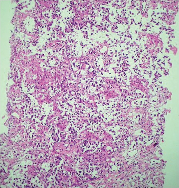
Needle biopsy of semioma (H and E, stain ×10)
Subsequently two cycles of chemotherapy were given.
Patient was referred to our hospital for the surgical management. On exploration, a mass was seen adherent to the posterior vesical wall, along with it on the right side was a uterine-like structure with bilateral fallopian tube. The mass surrounded the left iliac vessels and the sigmoid colon. The structures were dissected in total and were submitted for further histopathological evaluation. Grossly, [Figure 2] the tumor mass was welldefined, measuring 7 × 6.5 × 4 cm. The cut surface was white-tan in color, and was firm. The uterus measured about 6.5 × 5 cm in size, with bilateral fallopian tubes. The right fallopian tube measured 7 cm and left 9 cm.
Figure 2.

Gross appearance of the tumor with uterus and B/L testes
To the right of the uterus is tube like and cord-like structure, and at the end is testis measuring 2.5 × 2.5 cm.
Microscopically, the left testis showed seminoma with postchemotherapy changes [Figures 3 and 4] with individual cell necrosis, dense fibrosis with focal areas of lymphocytic infiltration.
Figure 3.

High-power view showing postchemotherapy changes of seminoma (H and E, stain ×40)
Figure 4.

Postchemotherapy changes of seminoma (H and E, stain ×10)
The section from the uterus showed atrophic endometrium [Figure 5] and measured 0.1 cm in thickness. The myometrium and the cervix were unremarkable. The right tube showed normal tubal histology [Figure 6] while the left tube showed changes of hydrosalpinx. No ovarian tissue was found on either of the side. Right undescended testis was atrophic with tubular hyalinization and Leydig cell hyperplasia [Figure 7]. The right spermatic cord was identified and was unremarkable.
Figure 5.

Atrophic endometrium from the uterus (H and E, stain ×10)
Figure 6.

Section showing histology of fallopian tube (H and E, stain ×10)
Figure 7.

Right atrophic testis with Leydig cell hyperplasia (H and E, stain ×10)
A chromosome analysis revealed a normal male karyotype of 46 XY.
DISCUSSION
PMDS is a rare form of male pseudohermaphroditism, characterized by the presence of a uterus and fallopian tubes owing to failure of mullerian duct regression in genotypically normal males.[1]
In a human foetus, both mullerian and wolffian ducts, the anlagen of the female and male reproductive tracts, respectively, are present at 7-week gestation.
The normal sex differentiation in males is controlled by testosterone and MIF. Testosterone has a direct local effect on the wolffian ducts, including differentiation into the epididymides, vas deferens, and seminal vesicles. Also the formation of the urogenital sinus and male external genitalia requires in situ conversion of testosterone into dihydrotestosterone.[2] Despite the normal male genotype and the subsequent normal development of foetal testis, if there is a failure in production of MIF or insensitivity of the target organ to MIF, Mullerian structures do not regress.[3]
Since the secretion and action of testosterone is not affected, the Wolffian (mesonephric) duct derivatives and the external genitalia of the foetus progress in the normal male direction. An intersex condition is therefore not usually suspected. But the malformation is incidentally detected during operative treatment of associated abnormalities such as an inguinal hernia or an undescended testis, generally in the first year of life. Henceforth, the diagnosis of PMDS is often established when a uterus and/or fallopian tube is found along with undescended testis in a genotypically and phenotypically normal male child.[2]
In PMDS, the testes are usually histologically normal, apart from lesions due to longstanding cryptorchidism. The overall incidence of malignant transformation in these testes is 18%, similar to the rate in abdominal testes in otherwise healthy men.[4]
There are three anatomic variants of PMDS, the most common is the male type in which one testis is usually found within the scrotum; the ipsilateral uterus and fallopian tube are either in the inguinal canal or can be brought into it by gentle traction on the presenting testis. The second type is crossed testicular ectopia, which is characterized by herniation of both the testes, the entire uterus and both the fallopian tubes. The least common form, or female type, is characterized by bilateral cryptorchidism with testes embedded in the broad ligaments in an ovarian position with respect to the uterus, which is fixed in the pelvis. It is seen in only 10-20% of the cases.[5]
In our case, the PMDS was of the female type, with bilateral cryptorchid testis and uterus and bilateral fallopian tubes.
To conclude, the possibility of PMDS should be kept in mind in a case of bilateral cryptorchid testis.
Footnotes
Source of Support: Nil
Conflict of Interest: None declared.
REFERENCES
- 1.Gutte AA, Pendharkar PS, Sorte SZ. Transverse testicular ectopia associated with persistent Mullerian Duct syndrome - the role of imaging. Br J Radiol. 2008;81:176–8. doi: 10.1259/bjr/76916885. [DOI] [PubMed] [Google Scholar]
- 2.Dekker HM, De Jong IJ, Sanders J, Wolf RF. Persistent mullerian duct syndrome. Radiographics. 2003;23:309–13. doi: 10.1148/rg.232025141. [DOI] [PubMed] [Google Scholar]
- 3.Tadmouri GO. Persistent mullerian duct syndrome, Types I and II. Dubai, United Arab Emirates: Centre for Arab Genomic Studies; 2005. p. 2. OMIM Number 261550. [Google Scholar]
- 4.Sichani MM, Heidarpour M, Dadkhah A, Rezvani M. Persistent mullerian duct syndrome with anIrreducible inguinal hernia. Urol J. 2009;6:298–300. [PubMed] [Google Scholar]
- 5.Renu D, Rao BG, Ranganathan K, Namitha Persistent mullerian duct syndrome. Indian J Radiol Imaging. 2010;20:72–4. doi: 10.4103/0971-3026.59761. [DOI] [PMC free article] [PubMed] [Google Scholar]