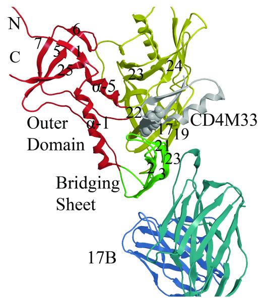
Figure 1. Structural details of gp120 core.
Ribbon diagram of the crystal structure (2I5Y) from the YU2 strain showing gp120 inner, outer and bridging sheet colored red, yellow and green, respectively. The mini-protein CD4 mimetic (CD4M33) containing a biphenyl group (purple) binds in the cavity formed at the junction of the three domains. The D1 domain of the 17b Fab is shown in blue. (Rendered with MOE70 )

