Abstract
There is a need for time-efficient, valid measures of distal paretic upper extremity (UE) movement. The purposes of this study were to: (a) determine the psychometric properties of the wrist stability and mobility and hand items of the upper extremity scale of the Fugl-Meyer (w/h UE FM) as a “stand alone” measure of distal UE movement; and (b) provide detailed instructions on w/h UE FM administration and scoring. The UE FM and Action Research Arm Test (ARAT) were administered on 2 separate occasions to each of 29 subjects exhibiting stable, mild, UE hemiparesis (23 males; age (mean (sd)) 60.8 (12.3) years; mean time since stroke onset for subjects in the sample: 36.0 months). Fifty-eight observations were collected on each measure. w/h UE FM internal consistency levels (measured by Cronbach’s alpha) were high (0.90 and 0.88 for first and second testing sessions, respectively). The intraclass correlation coefficients for the UE FM was 0.98, while the intraclass correlation coefficient for the w/h UE FM was 0.97. Concurrent validity measured by Spearman’s correlation was moderately high between the w/h UE FM and ARAT (.72 p < .0001). From these data, it appears that the w/h UE FM is a promising tool to measure distal UE movement in minimally impaired stroke, although more research with a larger sample is needed. A standardized approach to UE test administration is critical to accurate score interpretation across patients and trials. Thus, the article also provides instructions and pictures for w/h UE FM administration and scoring.
Upper extremity (UE) hemiparesis constitutes a common stroke-induced impairment. To measure UE hemiparesis, researchers have frequently administered the UE section of the Fugl-Meyer Assessment (UE FM).1 Developed in 1975, the UE FM is the most established stroke motor measure, is recommended for use in stroke rehabilitative trials,2 and, unlike other measures of paretic UE dysfunction,3,4,5 only requires a few household items to administer. Thus, because no special equipment is needed, the UE_FM is relatively easy to administer.
UE FM items are hierarchically organized in proximal to distal fashion (i.e., shoulder items are followed by elbow, wrist, and then finger items), while their sequencing is based on the principle that recovery of reflexes is followed by paretic UE movement recovery within and then outside of flexor and extensor synergies.6,7 However, these premises have recently been challenged,8 and this item sequencing can make administration of the initial UE FM scales superfluous in patients exhibiting minimal UE impairment. This is because individuals with this level of movement usually exhibit intact reflexes, and can usually perform proximal UE movements outside of synergy. Additionally, several therapy regimens have been developed that are most efficacious in patients with stroke exhibiting minimal UE impairment.9,10,11,12 These facets make a quickly-performed, distal paretic UE motor measure an unmet need, especially given the time required to properly set up and administer other UE measures.3–5
To address these shortfalls, we wondered whether the UE FM wrist stability and mobility and hand scale could be viably used as a brief, “stand-alone,” outcome measure of distal movement in minimally impaired stroke. We also recognized the need for formalized UE FM scoring rules, due to: (a) the diverse UE movement abilities and sequelae exhibited by patients with stroke. These presentations often make outcome measure item scoring more challenging; (b) a paucity of detailed UE FM administration and scoring instructions in the literature. Such sparse descriptions allow for multiple interpretations. This may cause only minor scoring variances, yet a slight score change may significantly affect the outcome of a clinical intervention or research trial; (c) because no formalized instructions have been available, it has been our experience that researchers and clinicians have used divergent UE FM testing and scoring methods in stroke rehabilitative clinical trials. Such inconsistencies challenge the ability to interpret and compare the outcomes of clinical interventions and research reports using the UE FM. A recent attempt to address some of the above challenges was made,13 by suggesting standardized instructions for the UE FM, as well as providing psychometrics for the measure when using these instructions. However, that study only included 15 subjects who were heterogenous in their UE impairment levels (UE FM scores ranged from 5 to 63). Pictorial depictions of UE FM positions and verbal instructions that enable consistent directions to the client have also not been provided in previous papers,13,14 with the latter paper14 only providing a UE FM description and review of its psychometric properties. Thus, several gaps remain unmet in regards to assuring consistent UE FM administration, and facilitating its clinical use.
This paper addresses the above challenges through the following actions: (a) This study determined the psychometric properties of the wrist stability and mobility and hand items of the upper extremity scale of the Fugl-Meyer (w/h UE FM) as a “stand alone” measure of distal UE movement. This study also reports the intra-rater reliability and the concurrent validity of the w/h UE FM with an established, stroke-specific measure of distal paretic UE movement (i.e., the Action Research Arm Test), which constitutes an advancement over previous work.13 (b) This paper provides detailed instructions on w/h UE FM administration and scoring. Data were collected in a well-defined cohort of chronic subjects with stable, minimal UE impairment. To our knowledge, this was the first study assessing the psychometric characteristics of the w/h UE FM.
Method
Study Design and Subjects
Subjects had been recruited for an outpatient, randomized, controlled, multiple baseline trial approved by the local ethics board. They were recruited from local, outpatient rehabilitation clinics and stroke support groups in the Midwestern United States, and met the following inclusion criteria: (1) 10° of active flexion in the paretic wrist, as well as 2 digits in the more affected hand; (2) stroke experienced > 12 months prior to study enrollment; (3) a score ≥ 70 on the Modified Mini Mental Status Examination;15 (4) age > 18 ≤ 75; (5) only had experienced one stroke; (6) discharged from all forms of physical rehabilitation. Exclusion criteria were: (1) excessive spasticity in the paretic UE, as defined as a score of ≥ 3 in the paretic elbow, wrist, or fingers as determined by the Modified Ashworth Spasticity Scale;16 (2) excessive pain in the paretic UE, as measured by a score ≥ 5 on a 10-point visual analog scale; and (3) participating in any experimental rehabilitation or drug studies.
The current study was a secondary analysis of pre-intervention scores from the aforementioned trial. Information on the study’s power considerations, the interventions administered, and study outcomes are published elsewhere.17
Instruments
For over three decades, the UE FM1 has been used to assess changes in upper extremity impairment. UE FM items are organized into scales that discern isolated movements at increasingly distal UE regions. Data arise from 33 items that are scored using a 3-point ordinal scale (0=cannot perform; 1= partially performed; 2=can perform fully), for a total score of 66. The UE FM has been shown to have high intra-rater reliability, interrater reliability, and construct validity.18,19 As stated earlier, the w/h UE FM was comprised of the 12 most distal UE FM items, and collectively made up the UE FM wrist stability and mobility and hand scales for a total possible score of 24. Greater description on these items – including their scoring and administration - is provided in Table 2. The w/h UE FM requires no more than 8–10 minutes to administer.
Table 2.
| Wrist Stability and Mobility | Starting Position | Ending Position | Scoring Criteria | Special Instructions | Verbal instructions |
|---|---|---|---|---|---|
Wrist stability elbow at 90°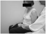
|
Shoulder at 0° in all planes, elbow at 90° with the forearm at 10° less than full pronation and wrist extended at least 15° beyond neutral. | NA | 0 = Correct starting position cannot be obtained. 1 = Wrist extension can be obtained but slight forward pressure to the dorsal surface of the hand causes movement towards wrist flexion or loss of any other aspect of starting position. 2 = Can retain wrist extension even with slight pressure to the dorsal side of the hand. |
Support: If the elbow cannot maintain 90° of flexion the RT is allowed to assist elbow flexion. The RT may use an open hand to provide support midway between the elbow and wrist on the anterior (palmer / volar) portion of the forearm. Assistance into elbow flexion requires the following caveats: If the Pt. is unable to extend the elbow to 90° the RT should not assist the elbow into extension. The RT should be careful not to provide pressure that aids proper positioning of shoulder (0° in all planes). Shoulder positioning should be maintained by the strength and coordination of the Pt. Pressure applied to the dorsal aspect of the Pt’s hand should be directly perpendicular to the angle of the hand. |
Starting Position: “Hold your arm at you side with your elbow at 90, as in grasping a bicycle handlebar, with your hand facing down.” Evaluation: “Raise your hand at the wrist. I am going to apply pressure to the back of your hand. Hold your hand firm, don’t let me move you.” |
Wrist flexion / extension, elbow at 90°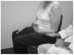
|
Shoulder at 0° in all planes, elbow at 90° with the forearm at 10° less than full pronation. | End ranges of full wrist flexion and full extension of the wrist are evaluated | 0 = Correct starting position cannot be obtained or movement toward wrist extension or flexion or cannot be achieved 1 = Partial active range of motion achieved. 2 = full active flexion and extension of the wrist with proper stating position maintained throughout movement as much or more than the unaffected side. |
Support: If the elbow cannot maintain 90° of flexion the RT is allowed to assist elbow flexion with the RT using an open hand midway between the elbow and wrist on the anterior (palmer / volar) portion of the forearm. Assistance into elbow flexion requires the following caveats: The RT should be careful not to provide pressure that aids proper positioning of shoulder (0° in all planes). Shoulder positioning should be maintained by the strength and coordination of the Pt. Wrist flexion may be attributable to force of gravity; score to the point at which the Pt. actively flexes wrist. Provide anti gravity support to the palmer surface of the hand to determine the end of the active range of motion. |
Starting Position “Hold your arm at your side with your elbow at 90° and the palm of your hand facing down, as if grasping a bicycle handlebar.” Evaluation “Raise and lower the hand, at the wrist, as much as possible.” |
Wrist stability with elbow at 0°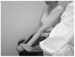
|
Wrist in 15° of flexion and shoulder in at least 25° of anywhere in the arc from flexion to abduction, elbow at 0 and 10° less than full pronation. | Same as starting position. | 0 = Correct starting position cannot be obtained. 1 = Wrist extension can be obtained but slight forward pressure to the dorsal surface of the hand causes movement towards wrist flexion or loss of any other aspect of starting position. 2 = Can retain wrist extension even with slight pressure to the dorsal side of the hand. |
Elbow extension may be lost either upon wrist extension or upon pressure against hand. The shoulder can be postured anywhere in the arc between flexion, through scaption and into pure abduction. Support: If the shoulder cannot be maintained in 25° of shoulder flexion, the RT is allowed to assist to position using an open hand, positioned just proximal to the elbow joint. |
Starting Position “Hold your arm at you side with your elbow straight, and the palm of your hand facing down. Evaluation “Raise your hand at the wrist. I am going to apply pressure to the back of your hand. Hold your hand firm, don’t let me move you.” |
Wrist flexion / extension, With elbow at 0°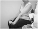
|
Shoulder in at least 25° of anywhere in the arc from flexion to abduction, elbow at 0 and 10° less than full pronation. | End ranges of full wrist flexion and full extension of the wrist are evaluated | 0 = Correct starting position cannot be obtained or movement toward wrist extension or flexion or cannot be achieved 1 = Partial active range of motion achieved or loss of any other aspect of starting position occurs during movement. 2 = full active flexion and extension of the wrist with proper stating position maintained throughout movement as much or more than the unaffected side without loss of any other aspect of starting position. |
Look for full elbow extension to cease when subject attempts wrist extension. The shoulder can be postured anywhere in the arc between flexion, trough scaption and into pure abduction. Support: If the shoulder cannot be maintained in 25° of shoulder flexion, the RT is allowed to assist to position using an open hand just proximal to the elbow joint. |
Starting Position “Hold your arm at your side with your elbow straight, and the palm of your hand facing down. Evaluation Raise and lower the hand, at the wrist, as much as possible.” |
| Starting Position | Ending Position | Scoring Criteria | Special Instructions | Verbal instructions | |
Circumduction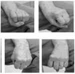
|
Shoulder at 0° in all planes, elbow at 90° with the forearm at 10° less than full pronation. | Full complete circumduction equal or greater than unaffected side. | 0 = Correct starting position cannot be obtained or no element of a circular motion at wrist available 1 = Partial circumduction achieved as evidenced of a full but smaller circle than unaffected side or movement is accomplished but the performed circular movement is jerky and/or broken and/or incomplete. |
Support: If the elbow cannot maintain 90° of flexion the RT is allowed to assist elbow flexion with the RT using an open hand midway between the elbow and wrist on the anterior portion of the forearm. Assistance into elbow flexion requires the following caveats: The RT should be careful not to provide pressure that aids proper positioning of shoulder (0° in all planes). Shoulder positioning should be maintained by the strength and coordination of the Pt. Circumduction is performed by mixing elements of wrist flexion, ulnar deviation, wrist extension and radial deviation in such a way that a complete and unbroken circle is formed. Verbally cuing the Pt. with an explanation of these four elements of Circumduction can be useful. Circumduction can be completed clockwise or counterclockwise. |
Starting Position “Hold your arm at your side with your elbow at 90° and the palm of your hand facing down, as if you are grasping a bicycle handlebar” Evaluation “Wth the back of your hand facing the ceiling the entire time, and without moving your forearm, make a circle with your hand by moving the wrist” |
| Hand | |||||
| Starting Position | Ending Position | Scoring Criteria | Special Instructions | Verbal instructions | |
Mass Flexion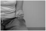
|
Forearms on lap in a resting posture. Hand and fingers as “open” as possible with as much finger and thumb extension as possible. |
Full fisted posture equal or greater than the unaffected side. Thumb on the outside of the fingers postured between the PIP and DIP of index and long fingers. | 0 = no movement towards flexion of any of the joints of any of the digits 1 = Some flexion at any or all joints of the digits 2 = full flexion of all digits at all joints equal or greater than the unaffected side |
Passive stretching of the fingers by the patient is allowed prior to testing. Forearm can be postured anywhere from full supination to full pronation |
Starting Position “Hold your arm at your side with your elbow at 90° and the palm of your hand as open as possible. Evaluation Make as much of a fist as you can, with the thumb on the outside of the fingers. |
Mass Extension
|
Forearms on lap in a resting posture. Hand in full fisted posture with all digits flexed as possible at all joints. |
Full extension of all joints of all digits | 0 = cannot release from mass flexion 1 = can release a mass posture 1 = can extend fingers partially 2=Full extension of all digits at all joints |
Starting position may involve the hand being placed by Pt. or RT into maximum possible flexion of all MCP and IP joints of all fingers and the thumb. |
Starting Position “Hold your arm at your side with your elbow at 90° and your hand in as much of a fist as possible Evaluation Open the hand as much as possible” |
| ALL GRASPING TASKS | The following position applies to all of the grasping tasks: Shoulder at 0° in all planes and the elbow at 90°. Forearm neutral pronation / supination. When possible, the wrist is maintained in 0°. However, if wrist cannot be held in 0 (i.e. it is held in flexion) the score is not affected. |
This rule applies to all of the hand (C) movements, grasps and details: Support: If the elbow cannot maintain 90° of flexion the RT is allowed to assist elbow flexion with the RT using an open hand midway between the elbow and wrist. |
|||
| Starting Position | Ending Position | Scoring Criteria | Special Instructions | Verbal instructions | |
Grasp A: Hook Grasp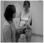
|
Hand in hook grasp position: Digits 2–5 MCP fully extended = or > unaffected side and fully flexed PIP and DIP |
Hand in hook grasp position: Digits 2–5 MCP fully extended = or > unaffected side and fully flexed PIP and DIP |
0 = Starting position cannot be attained 1 = Starting position is broken upon application of pressure 1 = Grasp is broken upon application of pressure 2 = Hook grasp starting position is maintained throughout performance of task. |
The starting position is extraordinarily hard for neurological conditions involving hemiparesis. Pressure is provided with the RT using his/her own hook grasp in direct opposition to that of the Pt. Pressure provided should be approximate to the weight of the RT forearm with gravity. |
Starting Position “Hold your arm at your side with your elbow at 90° and your hand straight at all of your knuckles. Evaluation I am going to apply pressure away from you at your fingers. Hold your hand firm, don’t let me move you” |
| Starting Position | Ending Position | Scoring Criteria | Special Instructions | Verbal instructions | |
Grasp B: Radial Grasp (Thumb Flexion)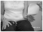
|
Hand and fingers as “open” as possible. All thumb joints and all interphalangeal joints must be postured at 0°. Thumb can be postured at any degree of extension such that it is separated from the lateral side of 2nd digit’s MCP. |
Hand and fingers as “open” as possible. All thumb joints at 0° grasping a piece of paper between the DIP of the thumb and the lateral surface of the metacarpal and proximal phalanx of the 2nd digit. | 0 = Starting position cannot be attained 1 = Starting position is broken upon onset of tug by RT of paper 1 = Pt is unable to hold paper against tug 2 = Hook grasp and starting position are maintained throughout performance of task. |
The RT may be able to tell from the “mass extension” movement, that the Pt. Is unable to achieve the starting position of this task. As the thumb is flexed (adducted) towards the 2nd digit there will be tendency to flex all the fingers at all joints. Verbally cue the Pt not do this. If, however, the Pt is not able to keep the fingers straight, score as is applicable. |
Starting Position “Hold your arm at your side with your elbow at 90° and your hand as open as possible.” Evaluation “Grasp this paper between your thumb and the side of your index finger. Keep your fingers as straight as possible. I am going to gently tug the paper, but try to hold the paper in place.” |
Grasp C: Pincer Grasp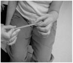
|
Thumb and index finger open in anticipation | As the pencil is pulled horizontally away from the midline of the Pt. the grasp and the position of the extremity are maintained | 0 = Starting position cannot be attained 1 = Starting position is broken upon onset of tug by RT of pencil 1 = Pt is unable to hold pencil against tug 2 = Pincer Grasp and starting position are maintained throughout performance of task. |
Pts. with hemiparesis may show an ability to complete a pinching grasp in which contact is maintained at a variety of points on the index finger and thumb. A pincer grasp specifically requires opposition between the pad of the thumb and the pad of the index finger. |
Starting Position “Hold your arm at your side with your elbow at 90°. Now, grasp this paper between the pads of your thumb and index finger.” Evaluation “I am going to gently tug the pencil, but try to hold the pencil in place.” |
| Starting Position | Ending Position | Scoring Criteria | Special Instructions | Verbal instructions | |
Grasp D: Cylinder Grasp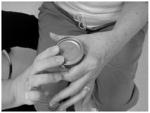
|
Thumb as extended as possible | As the can is pulled horizontally away from the midline of the Pt. the grasp and the position of the extremity are maintained | 0 = Starting position cannot be attained 1 = Starting position is broken upon onset of tug by RT of can 1 = Pt is unable to hold can against tug 2 =Cylindrical Grasp and starting position are maintained throughout performance of task. |
The can should meet the area between the thumb and the index finger in a way at least equal to the unaffected side. There is a tendency for patients with hemiparesis to not be able to extend the thumb wide enough to accept the can without the RT pushing the can into place. To eliminate this possibility, have the can meet the hand at the web space prior to touching any other part of the hand or fingers. |
Starting Position “Hold your arm at your side with your elbow at 90°. Now, grasp this can between your thumb and index finger.” Evaluation “I am going to gently tug the can, but try to hold the can in place.” |
Grasp E: Spherical Grasp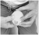
|
Fingers as actively extended as possible | As the ball is pulled horizontally away from the midline of the Pt. as the grasp and the position of the extremity are maintained | 0 = Starting position cannot be attained 1 = Starting position is broken upon onset of tug by RT of ball 1 = Pt is unable to hold ball against tug 2 = Spherical Grasp and starting position are maintained throughout performance of task. |
A tennis ball is used. All 5 fingers should be in contact with the ball. There is a tendency for patients with hemiparesis to not be able to extend one or more of the fingers enough to accept the ball without the RT pushing the ball into place. To eliminate this possibility, have the ball meet the palm surface prior to touching any other part of the hand. |
Starting Position “Hold your arm at your side with your elbow at 90°.” Evaluation “Grasp this in your hand and fingers as if you want to throw it. Now, I am going to gently tug the ball, but try to hold the ball in place.” |
Note: “RT” denotes rater; “Pt” denotes patient; “MCP” denotes meta carpal phalangeal; “DIP” denotes distal interphalangeal; “PIP” denotes proximal interphalengeal.
To discern concurrent validity of the w/h UE FM, we also administered the Action Research Arm Test, (ARAT);3 a measure of paretic UE functional limitation, and the second-oldest, stroke-specific, motor outcome measure (second only to the UE FM). It is a 19-item test divided into four categories (grasp, grip, pinch, and gross movement), with 16 of the nineteen ARAT items measuring distal regions of the arm (e.g., pinching a ball bearing or marble between the thumb and each finger of the affected hand), making it an ideal comparator to the wrist/hand scale of the UE FM. Each ARAT item is graded on a 4-point ordinal scale (0=can perform no part of the test; 1=performs test partially; 2=completes test but takes abnormally long time or has great difficulty; 3=performs test normally) for a total possible score of 57. For this test, subjects were seated in a comfortable chair with a straight back, while the ARAT items that they had to grasp were placed on an adjustable table in front of them. Table height was adjusted according to the needs of each subject. The ARAT has high intrarater (r = .99) and retest (r = .98) reliability and validity,20,21 all in stroke-induced hemiparesis.
Each of the above measures was administered to each subject on two occasions prior to the study intervention phase by a single rater with > 8 years experience with the measures. He was blinded in that he had no knowledge of the intent of the current study. The tests were administered approximately 2 weeks and one week before study intervention, respectively. As noted in the study criteria, subjects were chronic (i.e., past the point of spontaneous neurological recovery), and stable as verified by medical records and clinicians.
Statistical Analyses
Intra-rater reliability of the ARAT, UE FM, and w/h UE FM was assessed using the intraclass correlation coefficient (ICC). High reliability was indicated by a test ICC greater than 0.80.22 Minimal detectable change, the minimum amount of change not due to measurement error, was calculated based on the intrarater reliability (ICC) as: 1.96 * σpretest 1 * (2*(1-ICC))1/2, where σpretest 1 is the standard deviation at the first pretest.23 Cronbach’s alpha and the associated bootstrap confidence intervals (CI) were calculated to determine the internal consistency of items comprising the w/h UE FM at each administration. A Cronbach’s alpha between 0.70 and 0.95 was considered satisfactory.24 Concurrent validity was established between the w/h UE FM and ARAT using the Spearman’s rank correlation coefficient. A value > 0.70 represented a high association between the measures. Analyses were performed in Stata (StataCorp. 2007. Stata Statistical Software: Release 10. College Station, TX: StataCorp LP) and R software.25
Results
Data were collected from 29 subjects (23 males; mean age of subjects in sample = 60.8 (sd: 12.3) years, age range of subjects in sample = 21 – 76 years; mean time since stroke onset for subjects in sample = 36.0 months, range of time since stroke = 12 – 169 months; 23 subjects with ischemic stroke; 15 subjects with hemiparesis affecting their right arms; 22 were White;; 7 were black), with a total of 58 test administrations collected on the sample (2 per subject). Average ARAT, UE FM and w/h UE FM scores were 22.1 (sd: 15.5), 29.7 (sd: 11.4), and 7.2 (sd: 5.7) respectively.
ICC’s for the w/h UE FM and ARAT were 0.97 and 0.71, respectively (Table 1). w/h UE FM intra-rater reliability and internal consistency (measured by Cronbach’s alpha) were each high (Table 1). Concurrent validity was also above the aforementioned 0.70 criterion, with w/h UE FM and ARAT scores showing a high correlation (Table 1).
Table 1.
Psychometrics of w/h UE FM
| Intra-rater Reliability
| ||||
|---|---|---|---|---|
| Mean(sd) | ICC (95% CI) | MDC95 | ||
| PreTest 1 | PreTest 2 | |||
| ARAT | 22.4 (15.1) | 21.9 (16.1) | 0.71 (0.53–0.89) | 22.54 |
| UE FM | 29.8 (12.1) | 29.7 (10.8) | 0.98 (0.96–0.99) | 4.74 |
| w/h UE FM | 7.1 (5.9) | 7.2 (5.5) | 0.97 (0.95–0.99) | 1.64 |
| w/h UE FM Internal Consistency | ||
|---|---|---|
|
| ||
| Cronbach’s Alpha | 95% CI | |
| Pre test 1 | 0.90 | 0.84–0.94 |
| Pre test 2 | 0.88 | 0.81–0.92 |
|
| ||
| Concurrent Validity: ARAT and w/h UE FM | ||
|
| ||
| 0.72 (p<0.001) | ||
Discussion
Commonly-used stroke scales that quantify disability (e.g., Barthel Index26) or the global picture of neurologic status (e.g., National Institutes of Health Stroke Scale {NIHSS}27) insufficiently quantify a patient’s degree of motor recovery. For example, a patient may be considered “recovered” by scoring a “0” on the NIHSS, yet may still exhibit a sizable paretic UE motor deficit. Additionally, since minimally-impaired individuals are most likely to respond to paretic UE regimens,28,29 subtle motor changes in these patients may not be detected by the above measures. Such challenges with conventionally-used stroke measures and the UE FM’s specificity to paretic limb recovery underscore its utility and importance. Thus, the current paper focused on the psychometric properties of the w/h UE FM as well as providing detailed instructions on its clinical use.
Based on its high intrarater reliability, item consistency, and concurrent validity values (Table 1), the w/h UE FM appears to be appropriate for use as a “stand alone” scale. Indeed, the values reported herein compare favorably with values obtained on the scale when it was administered as part of the entire UE FM in terms of reliability (e.g., reliability coefficients between 0.96 and 0.99 reported by Duncan and colleagues;18,0.97 when administered by Sanford and colleagues30). Our results also compare somewhat favorably with evidence of concurrent validity between the UE FM and ARAT reported by others. For example, DeWeerdt and colleagues31 found that the UE FM and ARAT were highly correlated both at 2 weeks (r = 0.91) and at 8 weeks poststroke (r = 0.94). Moreover, comparison between the entire UE FM and the w/h UE FM in the present study revealed no detriment to reliability when using the shortened test, as evidenced by similar ICC’s. Given aforementioned shortfalls of existing stroke measures, we encourage w/h UE FM use as a bedside measure of paretic limb movement. Since deployment of many UE therapies hinges on active distal UE movement,9–11 use of such a measure would assist clinicians in identifying candidates for these regimens.
As with other medical sub-disciplines, stroke motor rehabilitation stands to benefit from use of standardized measures and procedures to discern the efficacy of its techniques. For example, such standardization is important given the variety of UE pathologies and patient heterogeneity that can obfuscate paretic UE testing.32 Standardized, consistent measurement is also fundamental to cost-effective, appropriate rehabilitative care. Despite these needs, the original UE FM report1 offered no detailed procedures on how to administer its items. Since UE FM administration and scoring is human rater-based, consistent procedures are needed, and are expected to reduce variability among raters. Such consistency is of particular importance given the proliferation of stroke clinical trials using the UE FM as an outcome measure. To address this need, Table 2 is a “clinical reference tool” that can be used by the therapist to administer the w/h UE FM. Both are based on our extensive experiences with the measure, which include over a decade of research and clinically-based experiences. The instructions are the first of which we are aware that provide pictoral representations of each position, descriptions of the position that the subject is trying to attain, and verbal instructions to be used by the rater. This is important for standardization, and is expected to facilitate straightforward w/h UE FM clinical use and measurement for individuals exhibiting active distal movement.
Limitations and Future Directions
Study limitations and next steps underway in this line of research include: (a) The current study enrolled 29 subjects. While findings are likely valid, verifying the current findings with larger numbers of patients exhibiting minimal UE impairment is desirable; (b) The current study measured psychometrics of the w/h UE FM in people with active paretic wrist and finger flexion. An important next step is determining psychometrics of the w/h UE FM in patients exhibiting higher degrees of paretic UE impairment, but still with some measurable degree of active distal movement.9–11 This is important since some clinical interventions use these criteria as entry criteria. (c) Finally, given that this article suggests a standardized method for administering the w/h UE FM, there remains a need to determine inter-rater reliability. Such research would allow researchers to determine whether the use of such instructions results in high reliability scores among different raters, as would be the intention of the current work.
Conclusions
The w/h UE FM appears to be valid and reliable when administered as a “stand alone” measure to stroke survivors exhibiting minimal UE impairment. Next steps include its testing in a larger sample, and with individuals exhibiting moderate levels of impairment, and testing its inter-rater reliability and its concurrent validity with other UE measures.
Acknowledgments
This work was supported by grants from the National Institutes of Health (R21AT002110-02; R01AT004454-02). The authors gratefully acknowledge the contributions of Pierce Boyne, DPT, who assisted in the review of this manuscript.
ABBREVIATIONS
- UE
Upper extremity
- UE FM
upper extremity Fugl Meyer
- w/h UE FM
wrist hand scale of the upper extremity Fugl Meyer
- ARAT
Action Research Arm Test
- ICC
Interclass correlation coefficient
Footnotes
The authors certify that no party having a direct interest in the results of the research supporting this article has or will confer a benefit on them or on any organization with which they are associated AND, if applicable, certify that all financial and material support for this research (e.g., NIH or NHS grants) and work are clearly identified in the title page of the manuscript. There is no drug or device tested herein.
Contributor Information
Stephen J. Page, School of Health and Rehabilitation Sciences and Director of the Neuromotor Recovery and Rehabilitation Laboratory (the “RehabLab”®) at the Ohio State University Medical Center (OSUMC) Columbus, OH.
Peter Levine, OSUMC.
Erinn Hade, Center for Biostatistics at OSUMC.
References
- 1.Fugl-Meyer AR, Jaasko L, Leyman I, Olsson S, Steglind S. The post-stroke hemiplegic patient. I. A method for evaluation of physical performance. Scand J Rehabil Med. 1975;7:13–31. [PubMed] [Google Scholar]
- 2.Gladstone DJ, Daniells CJ, Black SE. The Fugl-Meyer Assessment of motor recovery after stroke: A critical review of its measurement properties. Neurorehabil Neural Repair. 2002;16:232–240. doi: 10.1177/154596802401105171. [DOI] [PubMed] [Google Scholar]
- 3.Lyle RC. A performance test for assessment of upper limb function in physical rehabilitation treatment and research. Int J Rehabil Res. 1981;4:483–492. doi: 10.1097/00004356-198112000-00001. [DOI] [PubMed] [Google Scholar]
- 4.Kopp B, Kunkel A, Flor H, Platz T, Rose U, Mauritz K, et al. The Arm Motor Ability Test: Reliability, validity, and sensitivity to change of an instrument for assessing disabilities in activities of daily living. Arch Phys Med Rehabil. 1997;78:615–620. doi: 10.1016/s0003-9993(97)90427-5. [DOI] [PubMed] [Google Scholar]
- 5.Morris DM, Uswatte G, Crago JE, Cook EW, Taub E. The reliability of the Wolf motor function test for assessing upper extremity function after stroke. Arch Phys Med Rehabil. 2001;82:750–755. doi: 10.1053/apmr.2001.23183. [DOI] [PubMed] [Google Scholar]
- 6.Twitchell TE. The restoration of motor function following hemiplegia in man. Brain. 1951;74:443. doi: 10.1093/brain/74.4.443. [DOI] [PubMed] [Google Scholar]
- 7.Brunnstrom S. Movement Therapy in Hemiplegia: A Neurophysiological Approach. New York: Harper and Row, Publishers, Inc; 1970. [Google Scholar]
- 8.Woodbury ML, Velozo CA, Richards LG, Duncan PW, Studenski S, Lai SM. Dimensionality and construct validity of the Fugl-Meyer Assessment of the upper extremity. Arch Phys Med Rehabil. 2007;88(6):715–723. doi: 10.1016/j.apmr.2007.02.036. [DOI] [PubMed] [Google Scholar]
- 9.Wolf SL, Winstein CJ, Miller JP, Taub E, Uswatte G, Morris D, Giuliani C, Light KE, Nichols-Larsen D EXCITE Investigators. Effect of constraint-induced movement therapy on upper extremity function 3 to 9 months after stroke: the EXCITE randomized clinical trial. JAMA. 2006;296:2095–2104. doi: 10.1001/jama.296.17.2095. [DOI] [PubMed] [Google Scholar]
- 10.Page SJ, Levine P, Leonard A. Mental practice in chronic stroke: Results of a randomized, placebo controlled trial. Stroke. 2007 Apr;38:1293–1297. doi: 10.1161/01.STR.0000260205.67348.2b. [DOI] [PubMed] [Google Scholar]
- 11.Page SJ, Levine P, Leonard AC, Szaflarski J, Kissela B. Modified constraint-induced therapy in stroke: Results of a single blinded, randomized controlled trial. Phys Ther. 2008;88:333–340. doi: 10.2522/ptj.20060029. [DOI] [PubMed] [Google Scholar]
- 12.De Kroon JR, IJzerman MJ, Lankhorst GJ, Zilvold G. Electrical stimulation of the upper limb in stroke: stimulation of the hand vs. alternate stimulation of flexors and extensors. Am J Phys Med Rehabil. 2004;83:592–600. doi: 10.1097/01.phm.0000133435.61610.55. [DOI] [PubMed] [Google Scholar]
- 13.Sullivan KJ, Tilson JK, Cen SY, Rose DK, Hershberg J, Correa A, Gallichio J, McLeod M, Moore C, Wu SS, Duncan PW. Fugl-Meyer assessment of sensorimotor function after stroke: standardized training procedure for clinical practice and clinical trials. Stroke. 2011 Feb;42(2):427–432. doi: 10.1161/STROKEAHA.110.592766. [DOI] [PubMed] [Google Scholar]
- 14.Deakin A, Hill H, Pomeroy V. A rough guide to the Fugl-Meyer Assessment, upper limb section. Physiother. 2003;89:751–763. [Google Scholar]
- 15.Teng EL, Chui HC. The Modified Mini-Mental State Exam. J Clin Psychiatry. 1987;48 (8):314–318. [PubMed] [Google Scholar]
- 16.Bohannon RW, Smith MB. Interrater reliability of a modified Ashworth scale of muscle spasticity. Phys Ther. 1987;67:206–207. doi: 10.1093/ptj/67.2.206. [DOI] [PubMed] [Google Scholar]
- 17.Page SJ, Dunning K, Hermann V, Leonard A, Levine P. Longer versus shorter mental practice sessions for affected upper extremity movement after stroke: a randomized controlled trial. Clin Rehabil. 2011 Jul;25(7):627–637. doi: 10.1177/0269215510395793. [DOI] [PMC free article] [PubMed] [Google Scholar]
- 18.Duncan PW, Propst M, Nelson SG. Reliability of the Fugl-Meyer assessment of sensorimotor recovery following cerebrovascular accident. Phys Ther. 1983;63:1606–1610. doi: 10.1093/ptj/63.10.1606. [DOI] [PubMed] [Google Scholar]
- 19.DiFabio RP, Badke RB. Relationship of sensory organization to balance function in patients with hemiplegia. Phys Ther. 1990;70 (9):542–548. doi: 10.1093/ptj/70.9.542. [DOI] [PubMed] [Google Scholar]
- 20.Van der Lee JH, De Groot V, Beckerman H, Wagenaar RC, Lankhorst GJ, Bouter LM. The intra- and interrater reliability of the action research arm test: a practical test of upper extremity function in patients with stroke. Arch Phys Med Rehabil. 2001 Jan;82(1):14–19. doi: 10.1053/apmr.2001.18668. [DOI] [PubMed] [Google Scholar]
- 21.Hsieh CL, Hsueh IP, Chiang FM, Lin PH. Inter-rater reliability and validity of the action research arm test in stroke patients. Age Ageing. 1998;27:107–113. doi: 10.1093/ageing/27.2.107. [DOI] [PubMed] [Google Scholar]
- 22.Munro BH. Statistical methods for health care research. 5. Philadelphia: Lippincott Williams & Wilkins; 2005. [Google Scholar]
- 23.Brennan RL. Elements of Generalizability Theory. Iowa City, Iowa: ACT Publications; 1983. [Google Scholar]
- 24.Terwee CB, Bot SD, de Boer MR, van der Windt DA, Knol DL, Dekker J, et al. Quality criteria were proposed for measurement properties of health status questionnaires. J Clin Epidemiol. 2007;60:34–42. doi: 10.1016/j.jclinepi.2006.03.012. [DOI] [PubMed] [Google Scholar]
- 25.R Development Core Team. R. A language and environment for statistical computing. R Foundation for statistical computing; Vienna, Austria: 2011. [Google Scholar]
- 26.Mahoney FI, Barthel D. Functional evaluation: the Barthel Index. Maryland State Med J. 1965;14:56–61. [PubMed] [Google Scholar]
- 27.Brott TG, Adams HP, Olinger CP, Marler JR, Barsan WG, et al. Measurements of acute cerebral infarction: a clinical examination scale. Stroke. 1989;20:864–870. doi: 10.1161/01.str.20.7.864. [DOI] [PubMed] [Google Scholar]
- 28.Hendricks HT, van Limbeek J, Geurts AC, Zwarts MJ. Motor recovery after stroke: a systematic review of the literature. Arch Phys Med Rehabil. 2002 Nov;83(11):1629–1637. doi: 10.1053/apmr.2002.35473. [DOI] [PubMed] [Google Scholar]
- 29.Kwakkel G, Kollen BJ, van der Grond J, Prevo AJ. Probability of regaining dexterity in the flaccid upper limb: impact of severity of paresis and time since onset in acute stroke. Stroke. 2003 Sep;34(9):2181–2186. doi: 10.1161/01.STR.0000087172.16305.CD. [DOI] [PubMed] [Google Scholar]
- 30.Sanford J, Moreland J, Swanson LR, Stratford PW, Gowland C. Reliability of the Fugl-Meyer assessment for testing motor performance in patients following stroke. Phys Ther. 1993;73:447–454. doi: 10.1093/ptj/73.7.447. [DOI] [PubMed] [Google Scholar]
- 31.De Weerdt WJG, Harrison MA. Measuring recovery of armhand function in stroke patients: a comparison of the Brunnstrom-Fugl-Meyer test and Action Research Arm Test. Physiother Can. 1985;37:65–70. [Google Scholar]
- 32.Duncan PW, Lai SM, Keighley J. Defining post-stroke recovery: implications for design and interpretation of drug trials. Neuropharmacol. 2000;39:835–841. doi: 10.1016/s0028-3908(00)00003-4. [DOI] [PubMed] [Google Scholar]


