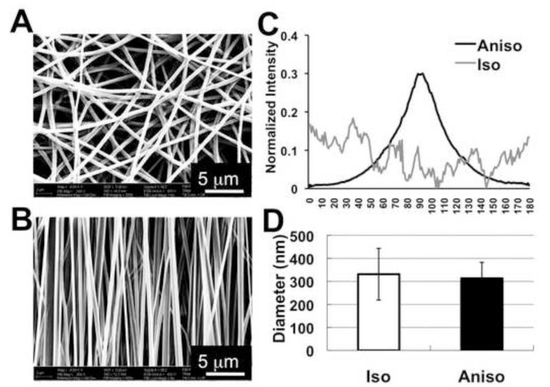
Figure 1.
Characterization of isotropic and anisotropic electrospun PCL/Coll nanofibrous matrices. SEM image of nanofibrous matrices with two types of fiber spatial organizations: isotropic (A) and anisotropic (B). (C) Normalized intensity plots against the angle of acquisition for isotropic and anisotropic nanofibrous matrices. (D) The measured average fiber diameter based on SEM images. Scale bar: 5μm.

